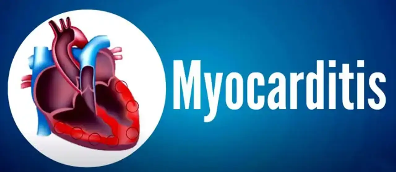
Myocarditis
Myocarditis is an underdiagnosed cause of acute heart failure, sudden death, and persistent dilated cardiomyopathy, and it refers to the clinical and histological symptoms of a wide variety of pathogenic immunological processes in the heart.
Patients with acute and chronic myocarditis generally have changes in the quantity and function of lymphocyte subsets and macrophages, as well as antibody-mediated damage. The immunological response in the heart produces structural and functional abnormalities in cardiomyocytes, which leads to regional or global contractile dysfunction, chamber stiffness, or conduction system illness. The inflammation can impair the heart's capacity to pump blood and produce rapid or irregular heartbeats (arrhythmias).
Viral infections are the most common cause of Myocarditis in the United States and other industrialized countries. Severe myocarditis weakens the myocardium, causing the rest of the body to get insufficient blood. Clots in the heart can cause a stroke or heart attack.
Epidemiology
The exact incidence is unknown. It is assumed that mild or asymptomatic cases of myocarditis may go unrecognized and resolve spontaneously.
Myocarditis affects around 1.5 million cases worldwide per year. The incidence is often considered to be between 10 and 20 cases per 100 000. The total incidence is unclear and most likely underdiagnosed.
In the United States, the frequency of myocarditis is difficult to ascertain as many cases are subclinical. In community-based populations The incidence and consequences of myocarditis remain unclear since epidemiologic studies indicate that the majority of Coxsackie B virus infections, a major cause of myocarditis, are subclinical and thus have a benign course. 1% to 5% of all patients with acute viral infections may involve the myocardium.
The majority of patients are young and healthy. Individuals who are susceptible include children, pregnant women and those who are immunocompromised. In ∼ 10% of sudden deaths in young adults, myocarditis is diagnosed in the post-mortem examination.
Myocarditis pathophysiology
Myocarditis begins with the direct invasion of an infectious agent and its subsequent replication within or around the myocardium causing myonecrosis.
As a result of the infectious agent's invasion and reproduction, the heart tissue is destroyed. Later, the offending substance activates the cytotoxic effects of host immunity, and the host cellular immune reacts. There may also be a toxic effect of exogenous or endogenous chemicals produced by the systemic pathogen directly on the myocyte.
Three stages of the disease process:
- Acute: defined by direct viral cytotoxicity and focal or diffuse necrosis of the myocardium
- Subacute: defined as an increase in autoimmune-mediated damage with activated T and B cells, followed by antibody formation, resulting in cardiac autoantibodies as well as inflammatory proteins. Anti-b-myosin antibody concentrations are greater in patients with myocarditis and dilated cardiomyopathy than in control groups
- Chronic: defined by diffuse myocardial fibrosis and cardiac dysfunction that may lead to dilated cardiomyopathy and its consequences, such as CHF, ventricular dysrhythmias, and ECG abnormalities.
Histopathology
Classic histologic examination of the endomyocardial biopsy will reveal cellular infiltrates, which are usually histiocytic and mononuclear with or without associated myocyte damage. Specific findings include eosinophilic, granulomatous, and giant cell myocarditis. The infiltrates are highly variable, often associated with varying degrees myonecrosis. With subacute and chronic myocarditis, interstitial fibrosis may result from the previous insult of the myocardial cytoskeleton.
Myocarditis causes
Myocarditis can result from a wide spectrum of infectious pathogens, including viruses, bacteria, chlamydia, rickettsia, fungi, and protozoans, as well as toxic and hypersensitivity reactions and can be classified as:
Infectious causes:
Viral: Up to 50% of myocarditis cases are classified as idiopathic. In these cases, a viral etiology is suspected, but cannot be sufficiently proven.
Viruses are the infectious pathogens most frequently implicated in reports of acute myocarditis including adenovirus, arborvirus, Cytomegalovirus, echovirus, Enterovirus (Coxsackie B), Epstein-Barr virus, hepatitis B virus, hepatitis C virus, herpes viruses (human herpesvirus-6), HIV/AIDS, influenza A and B viruses, Parvovirus (parvovirus B-19), mumps virus, poliovirus, rabies virus, respiratory syncytial virus, rubeola virus, rubella virus and varicella virus. Myocarditis is the most common cardiac pathological finding at autopsy of patients infected with the human immunodeficiency virus (HIV), with a prevalence of 50% or more.
Bacterial: Corynebacterium diphtheriae can cause myocarditis associated with bradycardia in nonimmunized children. Many other bacterial causes are identified including Brucella, Chlamydia (especially Chlamydia pneumonia and Chlamydia psittacosis), Francisella tularensis, Haemophilus influenzae, gonococcus, Clostridium, Legionella pneumophila (Legionnaire disease), Mycobacterium (tuberculosis), Neisseria meningitidis, Salmonella, Staphylococcus, Streptococcus A (rheumatic fever) and Streptococcus pneumoniae.
Fungal: Invasive fungal illness has become much more common in recent decades, owing to a rise in the number of immunocompromised individuals. Broad-spectrum antibiotics, corticosteroids, and cytotoxic drugs, invasive medical procedures, and human immunodeficiency virus (HIV) infection are all important risk factors for severe fungal illness. Fungal myocarditis generally occurs in the setting of a disseminated fungal infection . Candida is the most prevalent cause of fungal myocarditis along with other fungal causes such as Actinomyces, Aspergillus, Blastomyces, Cryptococcus and Histoplasma.
Parasitic: Trypanosoma cruzi, the cause of Chagas disease, has been a leading cause of myocarditis in parts of rural South and Central America. Entamoeba histolytica (amebiasis), Leishmania, Plasmodium falciparum (malaria), Trypanosoma brucei (African sleeping sickness), Toxoplasma gondii (toxoplasmosis) are all involved in developing severe myocarditis.
Non-infectious causes:
Autoimmune diseases: Dermatomyositis, inflammatory bowel disease, Sjögren syndrome, systemic lupus erythematosus, Takayasu’s arteritis, Churg-Strauss syndrome, collagen-vascular diseases, hypereosinophilic syndrome with eosinophilic endomyocardial disease and Kawasaki disease are the main autoimmune disorder involved in the pathogenesis of myocarditis. Patients with systemic inflammatory diseases such as rheumatoid arthritis have an increased cardiovascular mortality rate compared with the general population. the incidence of non-ischaemic cardiomyopathy, such as myocarditis, was estimated to be as high as 39% in those patients.
Drug induced: drug hypersensitivity reaction—including non-specific skin rash, malaise, fever, and eosinophilia are absent in most cases. Clozapine, sulfonamide antibiotics, methyldopa, and certain anti-seizure medications have all been associated to hypersensitivity myocarditis.
Myocarditis symptoms
Acute myocarditis is frequently first diagnosed as nonischemic dilated cardiomyopathy in a patient with symptoms that have been present for a few weeks to several months. However, symptoms vary from preclinical illness to sudden death, with new-onset atrial or ventricular arrhythmias, total heart block, or an acute myocardial infarction–like syndrome.
Patients may present with chest pain, dyspnea, palpitations, fatigue, decreased exercise tolerance, or syncope. Chest pain in acute myocarditis may mimic typical angina and be associated with electrocardiographic changes, including ST-segment elevation. Alternatively, Chest pain in acute myocarditis can result from an associated pericarditis or occasionally from coronary-artery spasm.
A viral prodrome including fever, rash, myalgias, arthralgias, fatigue, and respiratory or gastrointestinal symptoms frequently precedes the onset of myocarditis by several days to a few weeks. In the European Study of the Epidemiology and Treatment of Inflammatory Heart Disease, 72 % of the 3055 patients with suspected myocarditis reported dyspnea, 32% had chest discomfort, and 18% had arrhythmias.
Most studies of acute myocarditis report a slight preponderance in male patients, which may be due to a protective effect of natural hormone variations on immune responses in women.
The clinical presentation of myocarditis in children differs from that in adults. children often have a more fulminant presentation and may require advanced circulatory and respiratory support in early stages of their disease. The most common findings during initial presentation to the emergency department were respiratory distress, tachycardia, lethargy, fever, hepatomegaly and abnormal heart sounds including murmur. Because of the wide range of clinical manifestations, physicians must include myocarditis in the differential diagnosis of numerous cardiac disorders.
Rash, peripheral eosinophilia, fever or a temporal relation with recently initiated medications suggest a possible hypersensitivity myocarditis. Patients with acute dilated cardiomyopathy associated with thymoma, autoimmune diseases, ventricular tachycardia, or high-grade heart block should be evaluated for giant-cell myocarditis.
Furthermore, myocarditis, as well as childhood cardiomyopathy, are major causes of sudden death. A new long-term research of pediatric myocarditis found that the biggest impact of myocarditis may not be apparent until 6 to 12 years after diagnosis, when children die or require heart transplantation for chronic dilated cardiomyopathy.
Common clinical scenarios associated with myocarditis:
- Possible subclinical acute myocarditis: Transient elevations in troponin (a biomarker of cardiac injury) or ECG changes after an acute viral illness, adverse drug reaction, or rarely vaccination. 5·5 per 10000 patients may develop clinical myocarditis. The long-term risk of developing heart failure or arrhythmias is not known.
- Acute heart failure with dilated cardiomyopathy: Patients can present with shortness of breath, fatigue, paroxysmal nocturnal dyspnoea, orthopnoea and the inability to tolerate exercise. Patients typically have a dilated ventricle, with symptoms that occur about 2 weeks to a few months after gastrointestinal or upper respiratory infections.
- Myocarditis resembling a heart attack: Myocarditis can mimic an acute coronary syndrome, While the heart function is largely preserved. When inflammation occurs in the pericardium, the presentation can mimic an acute myocardial infarction but without significant coronary artery disease on angiography and the pain is worsen with leaning backward and better leaning forward.
- Syncope from ventricular arrhythmias or heart block: 6% of patients with unexplained atrioventricular heart block had myocarditis.
- Heart failure associated with progressive or chronic dilated cardio myopathy: Up to 40% of individuals with persistent DCM who have symptoms of heart failure despite normal medical treatment have myocarditis as determined by immunohistological criteria.
Myocarditis diagnosis
When myocarditis is suspected, more common causes of cardiovascular disease, such as valvular heart disease, should be excluded.
Patients with clinically suspected acute myocarditis should have the following diagnostic tests:
Electrocardiogram:
Although widely used as a screening tool, the sensitivity of electrocardiogram (ECG) for myocarditis is not very high. The ECG in those with myocarditis show a variety of changes including:
- Nonspecific T-wave change (most common).
- Sinus tachycardia.
- Arrhythmias: atrial or ventricular ectopic beat, atrial tachycardia and complex ventricular arrhythmias.
- Repolarization abnormalities: non-specific ST segment, T wave changes and ST segment elevation mimics myocardial infarction.
- Heart block: right bundle branch block and AV block.
- Low R-wave with poor progression which is more prominent when pericardium is involved.
Laboratory tests:
- Nonspecific serum markers of inflammation, including high ESR, high CRP and leucocytosis.
- Increased serum concentrations of troponin I and T are more common than increased levels of CK or CK-MB . Higher levels of troponin T is a prognostic marker for poor outcome
Chest x-ray:
May show cardiomegaly, pericardial effusion, or both. Additional findings may include interstitial infiltrates, and pleural effusions.
Echocardiography:
Echocardiography can be used to assess heart chamber size, myocardial thickness, systolic and diastolic function. Its primary function is to rule out alternative causes of heart failure. The patient may have one or more of the following radiological findings:
- Ventricles: dilation, increased ventricular sphericity, reduced ejection fraction, impaired contractility, ventricular thrombi and regional wall motion abnormalities.
- Pericardial effusion: localized or circumferential fluid surrounding the ventricles.
Cardiac MRI:
Cardiac MRI can detect intracellular and interstitial edema, hyperemia and capillary leakage, pericardial effusion and myocardial necrosis and fibrosis.
Myocardial biopsy:
Histological evidence of myocardial inflammation with or without myocyte damage is the gold standard for the diagnosis of myocarditis and can show focal necrosis with lymphocytic infiltration most often has viral etiology.
Can myocarditis be cured?
In many people, myocarditis improves on its own or with treatment, leading to a complete recovery. Myocarditis treatment focuses on the management of the underlying cause and the presenting symptoms.
Heart failure therapy:
Standard heart failure medication, which includes diuretics, angiotensin-converting enzyme inhibitors or angiotensin receptor antagonists, and the addition of -blockers, works effectively for most patients with acute myocarditis who present with dilated cardiomyopathy.
Intravenous vasodilators may be used to support patients with high filling pressures, whereas intravenous inotropic drugs may be used to treat patients with severe symptoms.
Antiviral treatment:
Most patients with acute myocarditis are identified many weeks after viral infection, thus antiviral treatment given after myocarditis has been established is unlikely to be of much use.
On the other hand, Treatment with interferon, resulted in the eradication of viral genomes and improved left ventricular function in individuals with chronic dilated cardiomyopathy and persisting viral genomes.
Immunosuppressive agents:
Long-term survival in patients with giant cell or eosinophilic myocarditis is enhanced by therapy with cyclosporin and corticosteroids, and withdrawal of immunosuppression in this group has occasionally resulted in an increased risk of recurrent, often fatal outcome.
Antiarrhythmic treatment:
High-grade AV block and tachyarrhythmias can be treated with suitable medications and, if necessary, the placement of a temporary or permanent pacemaker. Arrhythmias generally go away after a few weeks. If arrhythmias continue after the initial inflammatory phase, patients with symptomatic or persistent ventricular tachycardia may require antiarrhythmic treatment, an implanted cardioverter-defibrillator, or heart transplantation.
Conclusion
Myocarditis is a rare, possibly fatal illness that causes a variety of symptoms in both children and adults. In developed countries, viral infection is the most common cause of myocarditis, but other causes include bacterial and protozoal infections, toxins, drug reactions, autoimmune diseases, giant cell myocarditis, and sarcoidosis.
Acute injury damages myocytes, which stimulates the innate and humeral immune systems, resulting in acute inflammation. The immune response is gradually down-regulated in the majority of patients, and the myocardium heals.
The symptoms vary from preclinical illness to sudden death, with new-onset atrial or ventricular arrhythmias, total heart block, or an acute myocardial infarction–like syndrome. On the other hand, persistent myocardial inflammation causes continuing myocyte destruction and chronic symptomatic heart failure, or even death.
The diagnosis is generally determined based on the clinical presentation, laboratory test such as cardiac markers and noninvasive imaging including ECG and Cardiac MRI.
Most patients react well to conventional heart failure treatment, but in extreme situations, mechanical circulatory assistance or heart transplantation may be required. Except in individuals with giant cell myocarditis, the prognosis in acute myocarditis is typically favourable. Persistent, chronic myocarditis typically progresses, however it may respond to immunosuppression.
