Neuritis of the facial nerve

Last updated date: 25-Aug-2023

Originally Written in English

Neuritis of the facial nerve

Overview

The facial nerve is a nerve that governs the side muscles of the face. It lets us to express ourselves through smiling, crying, and winking. A facial nerve injury can result in a socially and psychologically debilitating physical abnormality; while most instances cure spontaneously, therapy may eventually need considerable rehabilitation or numerous operations.

The motor and parasympathetic functions of the facial nerve, as well as taste to the anterior two-thirds of the tongue, are all carried out by it. It also regulates the salivary and lacrimal glands. The upper and lower face muscles are controlled by the motor function of the peripheral facial nerve. As a result, Bell palsy diagnosis necessitates paying close attention to forehead muscle strength. If the strength of the forehead is retained, a primary source of weakness should be addressed.
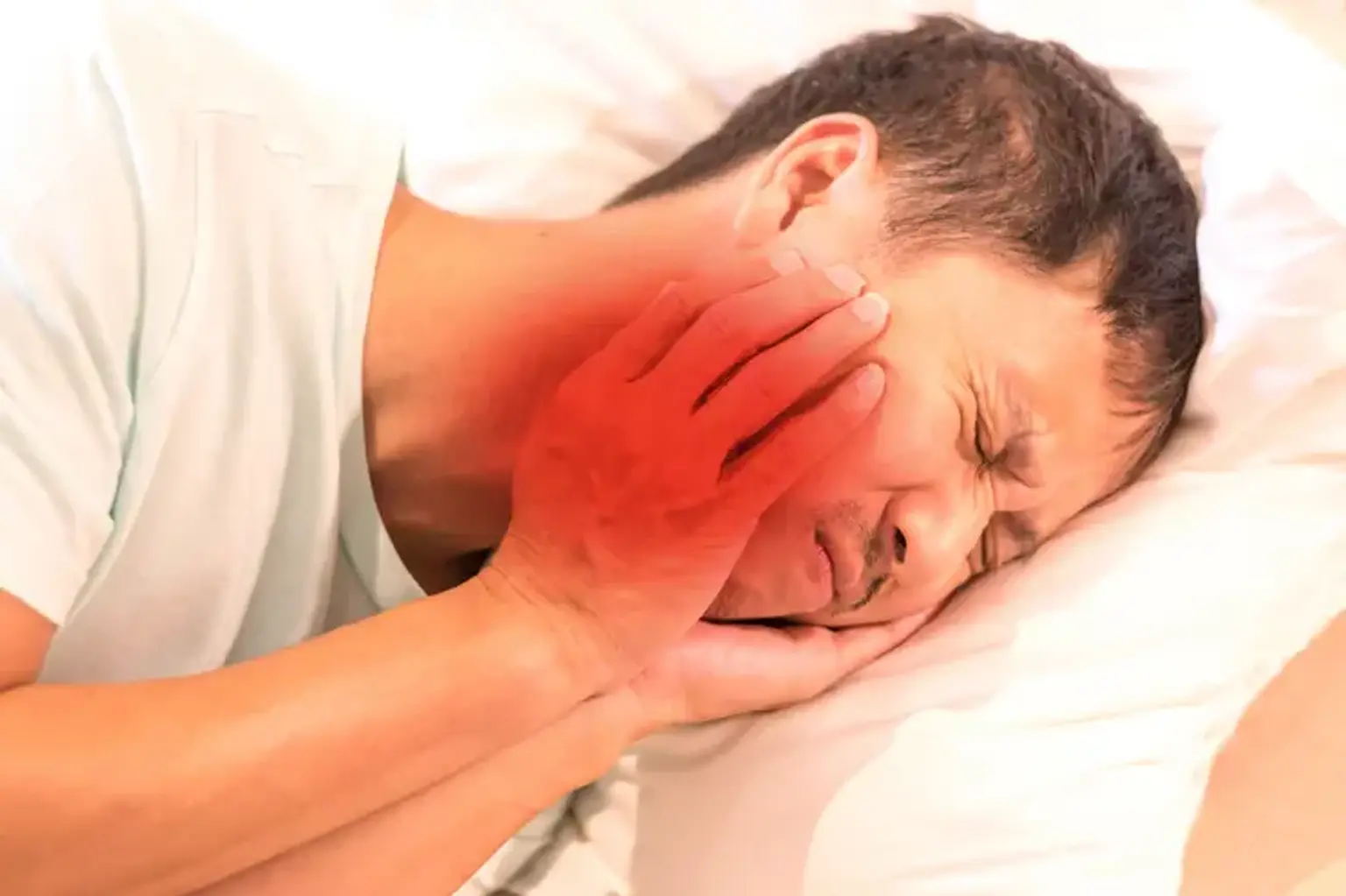
 

Neuritis of the facial nerve definition

facial nerve definition

The face nerve is the seventh of the twelve cranial nerves. Each individual has two facial nerves, one on each side of the face. The face nerve travels through and around the middle ear structures with the hearing nerve (the eighth cranial nerve). It leaves the front of the ear through the stylomastoid foramen (a hole at the base of the skull) and goes into the parotid gland. It separates into multiple branches in the parotid gland, which supply motor activity to the different muscles and glands of the head and neck.

Neuritis of the facial nerve, commonly known as Bell's palsy, is a medical disorder that causes weakening and/or paralysis of the facial muscles when the nerve controlling the muscles of the face becomes inflamed, swollen, and/or compressed. The condition is named after the Scottish anatomist Charles Bell. He was the first to describe the illness.

The Bell's syndrome causes one side of the face to droop and occasionally stiffen. Patients on the afflicted side may have trouble moving, smiling, or shutting their eyes. Bell's palsy is generally quite transitory, with symptoms disappearing within a few weeks.


Epidemiology

The yearly incidence is 15 to 20 instances per 100,000 people, with 40,000 new cases reported each year, and the lifetime risk is one in sixty. The recurrence rate ranges from 8% and 12%. Even without therapy, 70% of patients will recover completely. Palsy has no gender or racial preference, and it can develop at any age, but it is more common in middle and late life, with the median age of onset at 40 years. Diabetes, pregnancy, preeclampsia, obesity, and hypertension are all risk factors.


Neuritis of facial nerve causes

Neuritis of facial nerve causes

  • Bell palsy

A growing body of data in the literature reveals a variety of probable clinical diseases and pathologies that appear, at least in part, with a period of unilateral facial paralysis. Several viral infections have been emphasized in the literature, including herpes simplex virus, varicella-zoster virus, and Epstein-Barr virus. In the context of a presumably known etiologic process, providers may ambiguously allude to a BP diagnosis. This can happen, for example, in the context of well-known relationships (e.g. Ramsay-Hunt syndrome and Lyme disease).

While there are other probable causes, including idiopathic, traumatic, neoplastic, congenital, and autoimmune, BP is responsible for around 70% of facial nerve palsies.

Compression of the seventh cranial nerve at the geniculate ganglion is hypothesized to cause BP. The labyrinthine region of the facial canal is the narrowest, and it is here that most occurrences of compression occur. Because of the limited aperture of the facial canal, inflammation promotes nerve compression and ischemia. A unilateral facial paralysis that involves the muscles of the forehead is the most typical finding.


Other cases of facial nerve palsy include:

  • Ramsay Hunt syndrome

Ramsay Hunt documented facial paralysis, herpetiform vesicular eruptions, and vestibulocochlear dysfunction as a syndromic event. People with Ramsay Hunt syndrome have a higher chance of hearing loss than patients with Bell palsy, and the condition progresses more painfully. Furthermore, these individuals have a worse recovery rate.

Medical therapy is similar to that for Bell palsy, with a combination of steroids and antiviral medicines most commonly employed.

  • Lyme disease

Infection with Borrelia burgdorferi through tick bites shows another cause of facial paralysis, presenting with all of the symptoms of Lyme disease. Ten percent of Lyme disease patients develop facial paralysis, with a quarter of these individuals presenting with bilateral palsy.

  • Bacterial infection

Bacterial infection can also cause facial nerve paralysis, which is most commonly associated with acute otitis media or externa. In cholesteatoma patients, facial nerve palsy develops slowly.

  • Noninfectious causes

Head trauma affecting the intracranial intratemporal path of the facial nerve or, less typically, the infratemporal course, as observed in face blunt or sharp damage, are noninfectious causes of facial nerve palsy.

Iatrogenic facial nerve damage is most common following cervicofacial rhytidectomies, parotid gland operations, auditory neuroma resection, or tumor excision at any point along the facial nerve's journey. As a result, if facial paralysis arises following surgery, surgical investigation is required if the facial nerve is not intact. The facial nerve must occasionally be sacrificed voluntarily as part of appropriate oncologic therapy due to topographic relationships and/or tumor extension.

Facial nerve palsy is frequently associated with malignancies of the face nerve (e.g., hemangioma, neuroma) or tumors in the immediate neighborhood of the facial nerve. In general, the slow onset of paralysis may raise the possibility of a tumor as the reason. 


How does a person get Bell's palsy?

Bell's palsy

While the exact process of facial nerve damage in Bell's palsy is uncertain, one potential mechanism of injury includes:

  • Previously, you had a primary viral infection (herpes).
  • From months to years, the virus might be found in the nerve (geniculate ganglion).
  • The virus reactivates at a later time.
  • The virus multiplies and spreads down the nerve.
  • The virus affects the Schwann cells that surround the nerve, causing inflammation.
  • The immune system reacts to the injured Schwann cells, resulting in nerve inflammation and consequent weakening or paralysis of the face.
  • The severity and extent of the nerve injury will determine the course of the paralysis and recovery.


What are symptoms of a facial nerve problems?

symptoms of a facial nerve problems

Patients often arrive with quick and escalating symptoms that worsen over the course of a day to a week, reaching a peak in severity after 72 hours. Weakening will be limited to one-half of the face, resulting in weakness of the brows, forehead, and mouth angle. Patients may complain of being unable to shut the afflicted eyelid or lip on the affected side.

A partial or total weakening of the forehead is the most important physical exam result. If the strength of the forehead is retained, a core reason should be examined. Patients may also experience changes in taste, sensitivity to hearing, otalgia, and tears and salivation.

Ocular features include:  

  • Corneal exposure
  • Lagophthalmos
  • Brow droop
  • Paralytic ectropion of the lower lid
  • Upper eyelid retraction
  • Decreased tear output
  • Loss of nasolabial fold


Depending on the damage to the nerve following degrees of paralysis of the nerve can be formed:

  • Normal - Normal coordinated movements of the face
  • Slight - Deformation and slight asymmetry with preserved forehead mimics
  • Moderate - Obvious facial asymmetry, Bell symptom
  • Significant - movements of the forehead are dropped
  • Severe – Asymmetry, impossibility of closing the eye, speech impairment
  • Total - Impossible speech


Diagnosis of Neuritis of the facial nerve

Diagnosis of Neuritis of the facial nerve

The evaluation is guided by the history and physical examination. To define the degree of facial nerve weakening, the Facial Nerve Grading System might be employed. This grading system ranges from I (no weakness) to VI (complete weakness). If the appearance is consistent with BP, no lab or radiographic investigations are necessary. If there are any uncommon traits, individuals should be investigated for an underlying cause of their symptoms.

Similarly, Lyme disease testing is predicated on a history of tick-borne illness. Routine Lyme disease testing is not advised in the absence of additional symptoms of the disease, such as a history of a tick bite, skin rash, or arthritis. Diabetic testing should not be done since facial nerve palsy does not qualify as diabetic neuropathy.

There is little agreement on the ideal time of Lyme disease imaging, however most sources advocate after two months of no improvement in the facial palsy. Magnetic Resonance Imaging (MRI) is the imaging modality of choice. MRI of the facial nerve can reveal inflammation while also ruling out other disorders such as schwannoma, hemangioma, or a space-occupying lesion.

In patients with severe Bells Palsy, nerve conduction tests and electromyography (EMG) may aid in determining outcomes. EMG is used in electroneurography to measure the difference in potentials generated by the face muscles on both sides.

Auditory evoked potentials and audiography should be done if hearing loss is suspected. For clinical examination of blood pressure, there is a grading system. The method grades dysfunction from light to severe. Saliva flow, tear function, and nerve excitability are among the other tests.

  • Imaging

CT scanning and magnetic resonance imaging (MRI) are effective in the diagnosis of intratemporal and/or intracranial facial nerve injuries because they can identify temporal fracture patterns (vertical, transversal, mixed) and edema development. The facial nerve can be visualized under specific conditions, and swelling or disruption might be noticed.

  • Electrophysiologic studies

Electrophysiology can help identify the level of nerve damage, potential outcomes, and treatment alternatives. The minimum and maximum stimulation tests (MST) and electroneuronography (ENog) are the most commonly employed. Percutaneous stimulation of the facial nerve is used in these examinations.

ENog tests are essential to establish the appropriateness and timing of surgical intervention (decompression or microneurorrhaphy). electroneuronography measures the compound action potential (CAP) as well as the delay following nerve stimulation. Without surgical intervention, degeneration of 90% or greater has been proven to indicate a bad prognosis.


Management Neuritis of the facial nerve

Management Neuritis of the facial nerve

It is critical to understand that spontaneous healing does occur, and so the function of therapy remains debatable.

Corticosteroids are the primary therapy, with a typical regimen of 60 mg to 80 mg per day for roughly one week. There is also evidence that combining corticosteroids and antivirals improves BP outcomes when compared to corticosteroids alone. 

Patients with severe facial nerve palsy may benefit from a combination of steroids and antivirals. There was no statistically significant increase in adverse events when antivirals were compared to placebo or corticosteroids. To lessen the probability of a corneal abrasion, patients should be told to use eye lubricant and patch the afflicted eye at sleep.

When symptoms do not improve after many weeks or months, surgical treatments may be considered. Eyelid weights and muscle transfers are two methods for presenting ocular desiccation. Facial nerve decompression is not a suggested therapeutic option and is examined on an individual basis. Prior research on facial nerve compression was of poor quality. If no progress has been noticed in 4 weeks, it is advised to consult a specialist (plastic surgery, neurology, otolaryngology) as soon as possible to investigate more severe therapies.


Surgery for Facial Nerve Palsy

Surgery for Facial Nerve Palsy

Surgical reconstruction options

Patients with facial muscular weakness or paralysis may benefit from one or more of the following reconstructive procedures:

  • Nerve repair or nerve grafts: Facial nerve regeneration happens at a one-millimeter-per-day rate. Direct microscopic repair is the best option if a nerve has been cut or removed.
  • Nerve transposition: The tongue nerve (hypoglossal nerve) or another face nerve is frequently linked to the existing facial nerve. The patient, for example, can then train themselves to move their face by moving their tongue.
  • Muscle transposition or sling procedures: To move the face, the temporalis or masseter muscles (the only muscles on the face not supplied by the facial nerve) can be pushed down and attached to the corner of the mouth.
  • Muscle transfers: Leg free muscles (gracilis) can be employed to produce muscular hypertrophy as well as function. A cross facial nerve transposition is frequently performed to provide comparable nerve supply to the donor muscle flap.
  • Ancillary eyelid or oral procedures: In addition to one of the aforementioned, a brow lift or facelift, partial lip excision, eyelid relocation, lower eyelid shortening, upper eyelid weights, or eyelid springs are frequently required in reconstructive surgery following severe facial nerve palsies.


Treatment options for eye problems

Patients with facial nerve paralysis have trouble closing their eyes because the muscles that shut the eye are paralyzed. If the cornea of the eye becomes excessively dry, serious issues might arise. Treatment includes:

  • Eye protection glasses that keep dust out of the eyes;
  • Manual closure of the eye with a finger to keep it moist — patients should use the back of their finger rather than the tip to avoid injury to the eye;
  • Artificial tears or ointments to assist moisten the eyes;
  • When sleeping, taping or patching the eye closed with paper tape; and
  • If healing is not complete, a temporary or permanent constriction of the eye opening (tarsorrhaphy) may be required.


Differential Diagnosis

Lyme illness and Ramsey Hunt syndrome should be ruled out as causes of peripheral seventh nerve palsy. Tuberculosis, HIV, trauma, sarcoidosis, vasculitis, and tumor are also less prevalent causes of facial palsy. Misdiagnosis is found to be 10.8 percent in speciality referral facilities.

In addition, if there are recurrent occurrences, practitioners should examine Melkersson-Rosenthal syndrome. This is an uncommon neurocutaneous condition characterized by recurrent facial palsy, orofacial edema, and a fissured tongue. Females are more likely to be diagnosed with Melkersson-Rosenthal syndrome.


Prognosis

Bell Palsy Prognosis

Bell palsy (BP) disappears entirely without therapy in 71% of untreated cases. Corticosteroid treatment has been shown to boost the chance of nerve healing. Recurrence does occur, and one research discovered a recurrence incidence of 13%. Another study found that up to 10% of BP patients would develop symptomatic recurrence after a mean of ten years.

Risk factors associated with poor outcomes include:

  1. Complete paralysis 
  2. Age more than 60 and
  3. Decreased salivation or taste on the ipsilateral side.

The longer the recovery, the more likely that residual sequelae may develop. A recurrence rate of 5-15% has been reported.


Conclusion 

Neuritis of the facial nerve

The facial nerve, also known as the seventh cranial nerve, is a cranial nerve that exits from the pons of the brainstem and regulates facial expression muscles as well as conveying taste sensations from the anterior two-thirds of the tongue. The most frequent peripheral paralysis of the seventh cranial nerve is facial nerve neuritis, often known as Bell palsy (BP). The diagnosis is one of exclusion and is usually determined on physical examination. Corticosteroid therapy has been demonstrated to increase the chances of nerve recovery.