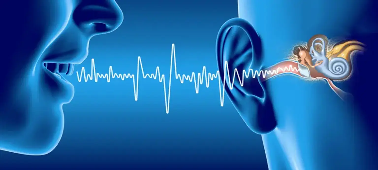
What Are Neuro-Otology Tests?
Neuro-otology tests are specialized diagnostic procedures used to evaluate the function of the inner ear, balance system, and auditory pathways. These tests are essential for diagnosing disorders that affect hearing and balance, such as vestibular disorders and auditory dysfunction. The field of neuro-otology bridges neurology and otology (ear science), focusing on how the nervous system and ear interact to control both hearing and balance.
These tests are particularly valuable for patients experiencing dizziness, vertigo, tinnitus, or hearing loss that cannot be attributed to more common causes. They help identify issues such as vestibular dysfunction, inner ear diseases, and neurological disorders, enabling healthcare providers to recommend effective treatments like vestibular rehabilitation or hearing aids.
Common Neuro-Otology Tests
Several neuro-otology tests are commonly used to assess balance and hearing functions:
Vestibular Testing: A set of tests to evaluate the vestibular system. These tests check how well the inner ear and brain work together to control balance.
Audiometry Tests: These tests measure the ability to hear different sounds and frequencies, helping to diagnose hearing loss and its severity.
Electronystagmography (ENG): A test used to assess eye movements in response to specific stimuli. This helps evaluate how the inner ear and balance system are functioning, often used to diagnose vertigo and other balance disorders.
Rotational Chair Test: This test evaluates the vestibular system by observing how the body responds to controlled rotations in a chair, helping diagnose balance disorders like vestibular neuritis or labyrinthitis.
Vestibular Evoked Myogenic Potentials (VEMP): A test that measures the response of muscles to sound stimuli. It helps evaluate the vestibular system and detect any dysfunction in the inner ear’s balance mechanisms.
Each of these tests plays a unique role in diagnosing the cause of a patient's symptoms and can be used in combination to provide a comprehensive diagnosis.
Electronystagmography (ENG)
Electronystagmography (ENG) is a key diagnostic tool used in neuro-otology to evaluate the function of the vestibular system. ENG measures nystagmus, which is the involuntary movement of the eyes, typically caused by disruptions in the balance system.
During ENG, electrodes are placed around the eyes to detect eye movements in response to various stimuli. These tests are particularly useful in diagnosing vestibular disorders such as vertigo, labyrinthitis, or vestibular neuritis, conditions that cause dizziness and imbalance.
ENG can assess how well the inner ear responds to changes in head position or caloric stimulation, helping doctors identify if the balance system is functioning properly. This test is typically non-invasive and can be performed in an outpatient setting, providing valuable insights into the cause of dizziness or balance issues.
Rotational Chair Test
The rotational chair test is another essential tool in neuro-otology for diagnosing vestibular disorders. This test evaluates how the vestibular system responds to rotational movements and is often used to identify dizziness, vertigo, or other balance issues related to inner ear dysfunction.
During the test, the patient sits in a special chair that is rotated back and forth while the movements are carefully controlled. Small sensors are placed on the patient’s eyes to track involuntary eye movements (nystagmus), which are a key indicator of balance dysfunction. The rotational chair test helps determine whether the vestibular system is responding appropriately to movement, and can identify issues like vestibular neuritis or benign paroxysmal positional vertigo (BPPV).
This test is particularly useful in assessing central vestibular disorders, which involve the brain’s processing of balance signals, and is often performed when other vestibular testing methods are inconclusive. It provides valuable information for creating an effective treatment plan, including vestibular rehabilitation or medication.
Vestibular Evoked Myogenic Potentials (VEMP)
Vestibular Evoked Myogenic Potentials (VEMP) is a specialized test used in neuro-otology to evaluate the function of the vestibular system, specifically the saccule and inferior vestibular nerve. VEMP measures the response of the neck or eye muscles to sound stimuli, which can indicate how well the vestibular system is working.
During the VEMP test, electrodes are placed on the patient’s neck or around the eyes, and a loud sound is played through earphones. The muscle responses to these sounds are measured to assess the integrity of the vestibular pathway. Abnormal responses can indicate a vestibular dysfunction or other issues in the inner ear that affect balance.
VEMP is particularly useful in diagnosing vestibular disorders like Ménière’s disease, vestibular neuritis, or labyrinthitis. It can also help identify asymmetries in the vestibular function, which is useful in cases of unexplained dizziness or vertigo. VEMP testing is non-invasive and can often provide additional information when other tests do not yield definitive results.
Role of Neuro-Otology in Diagnosing Balance and Hearing Disorders
The primary role of neuro-otology tests is to evaluate the health of the vestibular system (responsible for balance) and the auditory system (responsible for hearing). These tests are used to diagnose and treat conditions such as:
Vestibular Disorders: Disorders that affect balance, causing symptoms like dizziness and vertigo. Vestibular testing is used to determine if the inner ear or brain is responsible for these issues.
Auditory Dysfunction: Problems with hearing, whether from the inner ear, nerve pathways, or the brain. Audiometry tests assess the severity of hearing loss and its potential causes.
Neuro-otology specialists use a combination of diagnostic tests and clinical assessment to understand the cause of the symptoms and create personalized treatment plans. These tests help to identify underlying conditions such as meniere's disease, labyrinthitis, or vestibular neuritis, all of which can impact balance and hearing.
Postural Stability Testing
Postural stability testing is an important part of neuro-otology assessments for diagnosing balance disorders. This test evaluates a person’s ability to maintain balance while standing, walking, or in different positions, which is crucial for identifying issues in the vestibular system or other areas of balance control.
During the test, patients may be asked to stand on a platform or walk along a path while their posture and ability to maintain balance are measured. Specialized equipment often tracks movements and measures how much a person sways or loses balance under different conditions, such as with eyes open or closed, or when visual and auditory input is altered.
This test helps identify vestibular dysfunction, neurological disorders, or musculoskeletal issues that may be affecting a person's balance. It’s also used in evaluating patients with conditions like Parkinson’s disease, stroke, or diabetes, which can affect postural control. Postural stability testing is often used in conjunction with other neuro-otology tests to create a comprehensive picture of a patient’s balance health.
Audiometry Tests: Standard Hearing Assessment
Audiometry tests are fundamental in neuro-otology for evaluating hearing function and diagnosing auditory dysfunction. These tests measure the patient’s ability to hear sounds at different frequencies and intensities, helping doctors understand the nature and extent of hearing loss.
During audiometry testing, the patient listens to a series of sounds through headphones and indicates when they can hear the sound. The test typically involves measuring air conduction (sound traveling through the ear canal) and bone conduction (sound traveling through the skull to the inner ear), allowing doctors to differentiate between conductive and sensorineural hearing loss.
Audiometry tests are key for diagnosing conditions such as sensorineural hearing loss, conductive hearing loss, and tinnitus. They also help in monitoring hearing health in patients who are at risk for progressive hearing loss, such as those with ototoxic medications, age-related hearing loss, or noise-induced hearing damage.
These tests are often performed as part of a broader diagnostic approach to neuro-otology issues, providing essential information to guide treatment options like hearing aids, cochlear implants, or surgical interventions.
Otoacoustic Emissions (OAE)
Otoacoustic emissions (OAE) are another critical test in neuro-otology used to assess hearing function. This test evaluates the health of the cochlea, particularly the outer hair cells responsible for amplifying sound. OAE testing is a non-invasive procedure that plays a vital role in detecting early signs of sensorineural hearing loss.
During an OAE test, a small probe is placed in the ear canal, emitting sounds into the ear. The cochlea responds by producing faint sounds, or emissions, which are detected by the probe. If the emissions are absent or abnormal, it may indicate damage to the outer hair cells, which are essential for normal hearing.
OAE testing is valuable for newborn hearing screening, as well as for adults who may have subtle or early-stage hearing loss. It can help diagnose conditions like early-stage noise-induced hearing loss or ototoxicity, and can also be used to monitor hearing health over time, especially in patients with risk factors for hearing loss.
Auditory Brainstem Response (ABR)
Auditory Brainstem Response (ABR) is a test that evaluates the auditory nerve and brainstem’s response to sound stimuli. It is particularly useful in diagnosing auditory dysfunction and neurological hearing issues, especially in individuals who may not be able to participate in traditional audiometry tests (such as infants or individuals with certain disabilities).
During the test, electrodes are placed on the scalp, and sounds are played through earphones. The ABR test measures the timing of electrical responses from the auditory pathway from the ear to the brainstem. By analyzing the speed and clarity of these responses, doctors can determine whether there are issues with the auditory nerve or the neural pathways responsible for hearing.
ABR is crucial for diagnosing hearing loss related to issues in the nerve pathway, such as acoustic neuroma, auditory neuropathy, or other neuro-otology disorders. It can also be used to assess hearing in newborns, particularly those who fail newborn hearing screening tests.
How Neuro-Otology Tests Diagnose Hearing and Balance Disorders
Neuro-otology tests are essential for diagnosing both hearing and balance disorders as they help determine the cause and extent of the symptoms. These tests evaluate how well the inner ear, auditory nerve, and vestibular system are functioning, enabling healthcare providers to pinpoint the source of problems like dizziness, vertigo, or hearing loss.
By combining different types of tests, such as vestibular testing, audiometry, and ABR, doctors can determine whether the issue lies with the inner ear, vestibular nerve, or brainstem. For example, ENG and rotational chair tests help assess balance issues, while audiometry and OAE focus on hearing problems. Comprehensive testing can diagnose conditions such as:
Vestibular neuritis or labyrinthitis (inner ear infections causing balance issues).
Meniere’s disease (a condition involving both balance and hearing problems).
Sensorineural hearing loss (due to damage in the auditory nerve or cochlea).
Benign paroxysmal positional vertigo (BPPV) (causing dizziness with specific head movements).
These tests not only identify the root cause of the symptoms but also provide the information necessary for treatment planning, such as vestibular rehabilitation therapy, hearing aids, or even surgery.
Role of Neuro-Otology Tests in Treatment Planning
The results from neuro-otology tests are essential for creating personalized treatment plans for patients with balance or hearing disorders. These tests allow doctors to pinpoint whether the issues are related to the vestibular system, auditory pathways, or central nervous system. Based on the diagnosis, a variety of treatments may be recommended.
For Balance Disorders: Tests like vestibular rehabilitation testing or the rotational chair test help assess the severity of balance issues. If the problem is related to vestibular dysfunction, treatments such as vestibular rehabilitation therapy (VRT) can help retrain the brain to compensate for balance problems, while medication may be prescribed for conditions like Meniere’s disease or vestibular neuritis.
For Hearing Loss: Audiometry and OAE tests guide treatment for hearing problems. Hearing aids, cochlear implants, or auditory brainstem implants (ABI) may be recommended depending on the severity and type of hearing loss. In some cases, surgical interventions may be required to address conductive hearing loss caused by issues like ear infections or ear canal blockages.
By combining the results of these tests, neuro-otology specialists can create a holistic treatment plan that addresses both the symptoms and underlying causes of hearing and balance disorders, significantly improving the patient's quality of life.
Neuro-Otology Tests for Vertigo Diagnosis
Vertigo, a sensation of spinning or dizziness, is one of the most common symptoms evaluated through neuro-otology tests. Various vestibular tests help determine the cause of vertigo, whether it's related to the inner ear or other parts of the nervous system.
Electronystagmography (ENG) and rotational chair tests are key in diagnosing vertigo. These tests evaluate the vestibular system, which controls balance, by monitoring involuntary eye movements (nystagmus) that occur when the balance system is disrupted.
Vestibular Evoked Myogenic Potentials (VEMP) testing is another important diagnostic tool, helping to identify abnormalities in the vestibular system and its contribution to vertigo.
Postural stability testing assesses how well a patient can maintain balance during specific movements or under varying conditions, helping pinpoint if the vertigo is due to issues with the inner ear or brain.
By combining these tests, doctors can determine whether vertigo is caused by conditions like BPPV, vestibular neuritis, Ménière’s disease, or other vestibular disorders. Accurate diagnosis through neuro-otology tests is essential for effective treatment, which may involve medications, rehabilitation, or other interventions.
Dizziness and Balance Issues: How Tests Help
Dizziness and balance issues are frequently seen in patients with vestibular dysfunction. These symptoms can be caused by a variety of conditions, ranging from inner ear infections to neurological disorders. Neuro-otology tests play a vital role in diagnosing the root causes.
Postural stability testing helps evaluate how the body responds to changes in posture or movement, which is crucial for detecting balance issues.
Vestibular testing like ENG and rotational chair tests measure the effectiveness of the balance system and help identify disorders such as vestibular neuritis, labyrinthitis, or BPPV.
Audiometry tests can rule out auditory causes of dizziness, such as Ménière’s disease, where both hearing loss and balance issues are present.
These tests allow specialists to understand the specific causes of dizziness and balance disorders, ensuring that treatment, such as vestibular rehabilitation, is tailored to the patient’s needs. Whether the issue lies with the inner ear, nervous system, or muscles, accurate testing is crucial for proper management.
Neuro-Otology Testing in Korea: A Leading Destination
Korea is quickly becoming a global hub for neuro-otology testing, particularly for balance disorders and hearing dysfunction. The country offers cutting-edge technology and highly skilled specialists in vestibular and auditory health.
State-of-the-art equipment: Korea’s medical centers are equipped with the latest diagnostic technologies for neuro-otology tests like ENG, VEMP, rotational chair tests, and audiometry.
Expert Neuro-Otology Specialists: Korea is home to some of the world’s leading neuro-otology specialists who provide comprehensive diagnostic services for balance and hearing issues. Many of these specialists have extensive experience in diagnosing complex disorders and offering personalized treatment plans.
Medical Tourism: For international patients seeking high-quality and affordable neuro-otology tests, Korea is an attractive destination. Korean hospitals offer competitive pricing without compromising care quality, making it a popular choice for medical tourism.
Korea’s expertise in neuro-otology testing and rehabilitation provides patients with the best possible diagnostic outcomes and treatment options for hearing and balance disorders.
Neuro-Otology Test Costs and Options in Korea
When considering neuro-otology testing in Korea, patients can expect competitive costs, especially in comparison to other countries offering similar services. Neuro-otology tests like ENG, VEMP, rotational chair tests, and audiometry are highly advanced but still affordable in Korea.
Affordable pricing: The cost of neuro-otology tests in Korea can be significantly lower than in many Western countries, with high-quality care and modern equipment available at top hospitals and clinics.
Comprehensive care: Korean hospitals offer all-inclusive packages that cover diagnostic tests, follow-up care, and even rehabilitation, providing patients with a seamless treatment experience. This makes it particularly appealing for international patients who seek top-tier care at a fraction of the cost.
Insurance and Payment: Many hospitals in Korea accept international insurance or offer payment plans, making it easier for patients to access these tests. Patients should consult with the hospital to understand the exact costs and available options.
With its state-of-the-art technology and highly skilled specialists, Korea offers excellent value for those seeking neuro-otology testing and treatment, making it a leading destination for patients worldwide.
Conclusion
Neuro-otology tests are crucial for diagnosing and treating balance disorders and auditory dysfunction. These specialized tests, including vestibular testing, audiometry, and VEMP, offer invaluable insights into the functioning of the vestibular system and auditory pathways, helping healthcare providers accurately diagnose conditions like vertigo, hearing loss, and other related disorders.
By combining these diagnostic tools, specialists can craft personalized treatment plans that may include vestibular rehabilitation, hearing aids, or surgical interventions, ultimately restoring balance and hearing function and improving the patient's quality of life.
With Korea emerging as a global leader in neuro-otology testing, patients seeking advanced, affordable care can find world-class medical services in the country. Whether dealing with vertigo, dizziness, or hearing loss, neuro-otology tests provide the foundation for effective diagnosis and treatment, offering hope for those affected by these often-debilitating conditions.
If you or a loved one is experiencing balance or hearing issues, consider consulting with a neuro-otology specialist to explore the diagnostic options and begin a path toward recovery and improved well-being.