Neurofibromatosis type 2 (NF2)

Last updated date: 15-May-2023

Originally Written in English

Neurofibromatosis type 2 (NF2)

Overview

Neurofibromatosis type 2 (NF2) is a hereditary disorder in which tumors form along your nerves to cause either peripheral nerves tumors or brain tumors or both. The tumors are mostly non-cancerous (benign), although they can produce a variety of symptoms.

Almost everyone who has NF2 develops tumors along the nerves that control hearing and balance. Hearing loss that progressively worsens over time, ringing or buzzing in the ears (tinnitus), and balance issues – especially when moving in the dark or walking on uneven ground – are common symptoms.

Other tumors can develop inside the brain or spinal cord, or along the nerves leading to the arms and legs. This might result in symptoms such as arm and leg weakness and chronic headaches.
Treatment is determined by the size and kind of tumor, as well as whether or not the tumor is producing symptoms. If the tumor is asymptomatic, diligent monitoring with imaging investigations may be advised instead of surgical surgery.

The usual therapy for most NF2-related brain tumors is brain tumor surgery to remove the tumor. A craniotomy (one of many surgical options) is a treatment in which a neurosurgeon temporarily removes a part of the skull to enable access to the tumor during surgery.


What is Neurofibromatosis?

Neurofibromatosis (NF) is a hereditary neurological condition that can damage the brain, spinal cord, nerves, and skin. Tumors, sometimes known as neurofibromas, develop along the body's nerves or on or beneath the skin. Neurofibromatosis (NF) is divided into two forms, according to scientists: NF1 and NF2. The most frequent variety is NF1, also known as von Recklinghausen's NF. It happens in around 1 in every 4,000 births.

NF2, also known as bilateral acoustic NF, central NF, or vestibular NF, is rare, occurring in 1 in 40,000 births. NF1 and NF2 are prevalent in all ethnic groupings and impact both sexes equally. Tumors are caused by alterations in nerve and skin cells. Tumors may also strain on important organs as they grow in size. NF can cause developmental anomalies and/or an increased risk of learning difficulties.

Other types of NF have been discovered in which the symptoms do not match those of NF1 or NF2. Schwannomatosis is an uncommon type of NF. However, the genetic etiology of this kind of NF has yet to be identified.


Causes of NF2

Causes of NF2

In certain cases, NF2 is inherited as an autosomal dominant trait. The faulty gene can be inherited from either parent, and the chance of transferring the gene to kids from either parent is 50%. There is no family history of NF2 in some individuals, and the condition is caused by a de novo mutation in the NF2 gene. NF2 is caused by mutations in the NF2 gene, which is located on the long arm of chromosome 22. (22q12.2). The NF2 gene encodes the protein merlin, which functions as a tumor suppressor gene. Merlin is located in the nerve system's Schwann cells.


Risk Factors

Neurofibromatosis Risk Factors

A family history of the condition is the most important risk factor for neurofibromatosis. Approximately half of those with NF1 and NF2 inherited the condition from an afflicted parent. People with NF1 and NF2 who have no afflicted relatives are likely to have a novel gene mutation.

Both NF1 and NF2 are autosomal dominant illnesses, which means that each kid of a parent with the disorder has a 50% chance of inheriting the genetic mutation.

The schwannomatosis inheritance pattern is less apparent. Currently, researchers estimate that the likelihood of inheriting schwannomatosis from an afflicted parent is roughly 15%.


How is NF2 Inherited?

NF2 Inherit

Normally, each cell has two copies of each gene: one from the mother and one from the father. NF2 has an autosomal dominant inheritance pattern, which means that a change, also known as a mutation or alteration, occurs in only one copy of the gene.

This implies that a parent with an NF2 mutation may pass on a copy of their normal gene or a copy of the altered gene to their children. As a result, a kid who has a parent who has a mutation has a 50% chance of acquiring that mutation. A person's sibling, sister, or father who has a mutation has a 50% chance of having the same mutation.

However, if the parents test negative for the mutation (meaning each person’s test results found no mutation), the risk to the siblings significantly decreases but their risk may still be higher than an average risk.

Options exist for people interested in having a child when a prospective parent carries a gene mutation that increases the risk for this hereditary cancer syndrome. 

Preimplantation genetic diagnosis (PGD) is a medical process performed in conjunction with in-vitro fertilization (IVF). It enables adults who have a certain known genetic mutation to lessen the chances that their children may inherit the ailment. In a laboratory, eggs are extracted from the ovaries and fertilized. When the embryos reach a particular size, 1 cell is taken and checked for the aforementioned inherited disease. The parents can then elect not to transfer embryos with the mutation.

PGD has been utilized for numerous genetic cancer risk disorders for over two decades. However, this is a complicated operation that must be preceded by financial, medical, and emotional considerations. Speak with an assisted reproduction specialist at a fertility clinic for further information.


NF Complications

NF Complications

Complications of NF include:

  1. Neurological issues: The most prevalent neurological disorders linked with NF are learning and thinking impairments. Epilepsy and the accumulation of extra fluid in the brain are uncommon consequences.
  2. Concerns about appearance: Visible neurofibromatosis symptoms, such as widespread cafe au lait patches, multiple neurofibromas in the face area, or huge neurofibromas, can induce worry and mental discomfort even if they are not medically significant.
  3. Vision issues: Optic glioma is a tumor that can form on the optic nerve and impair eyesight.
  4. Hormonal changes: Neurofibromas may be increased by hormonal changes associated with puberty or pregnancy. Most NF women have safe pregnancies, but they will need to be monitored by an obstetrician who is experienced with the illness.
  5. Cardiovascular issues: People with NF are more likely to have high blood pressure and blood vessel problems.
  6. Breathing difficulties: Plexiform neurofibromas can cause airway obstruction in rare cases.
  7. Cancer: Cancerous tumors are expected to affect 3% to 5% of patients with NF, and fewer with NF. These are often caused by neurofibromas beneath the skin or plexiform neurofibromas. People with NF are also more likely to develop other types of cancer, including breast cancer, leukaemia, colorectal cancer, brain tumors, and several types of soft tissue cancer. Women with NF should begin breast cancer screening earlier than the general population.
  8. Adrenal tumor (e.g. pheochromocytoma): This noncancerous tumor produces hormones that increase blood pressure. The pheochromocytoma is generally removed surgically.


Signs & Symptoms

Signs & Symptoms of Neurofibromatosis

Patients with NF2 often appear with tumor-related symptoms around the age of 20. Vestibular schwannoma is the most frequent intracranial tumor linked with NF2, and it is usually bilateral in these patients. Tinnitus, sensorineural hearing loss, and balance issues are common symptoms. The cutaneous characteristics of NF2 are less prevalent and more modest than those of NF1.

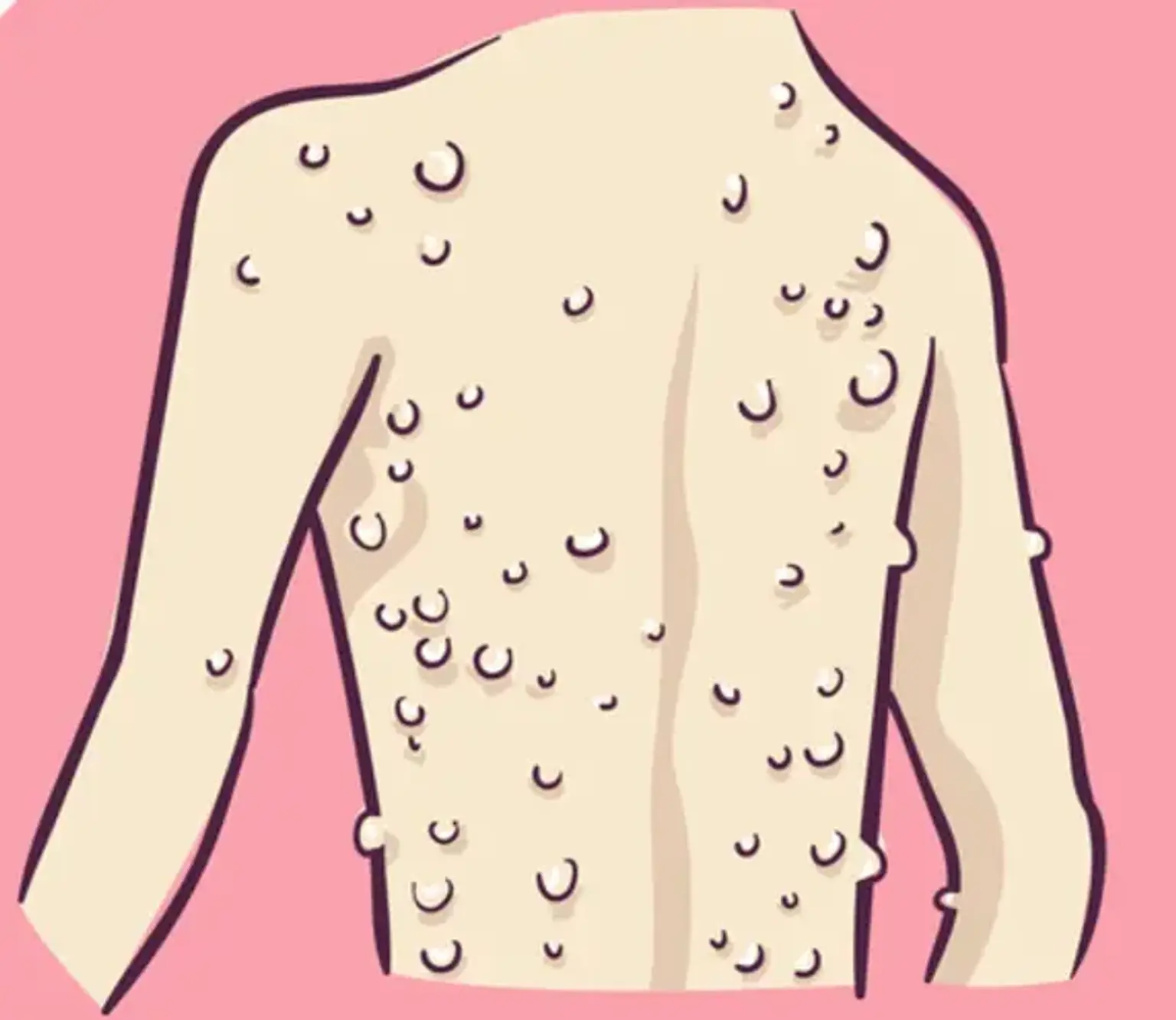
The most frequent skin finding is an elevated plaque-like lesion that is hyperpigmented in comparison to the surrounding skin. Other cutaneous abnormalities include subcutaneous nodules that signify nerve swelling and cutaneous tumors that are often schwannomas rather than neurofibromas. Meningiomas can cause headaches, seizures, or localized neurological symptoms, depending on where they are located.

NF2 patients are more likely to develop meningiomas than random meningiomas. NF2 is found in around 20% of children with meningiomas. Intraspinal tumors cause discomfort, muscle weakness, and paresthesia in patients.

The clinical diagnosis of NF2 is based on the presence of any one of the following criteria:

  1. Bilateral vestibular schwannomas less than 70 years of age
  2. Unilateral vestibular schwannoma before age 70 years and a first-degree relative with NF2
  3. Any two of the following: meningioma, schwannoma (non-vestibular), neurofibroma, glioma, cerebral calcification, cataract AND first-degree relative to NF2 OR unilateral vestibular schwannoma and negative LZTR1 testing
  4. Multiple meningiomas and unilateral vestibular schwannoma or any two of the following: schwannoma (non-vestibular), neurofibroma, glioma, cerebral calcification, cataract
  5. Constitutional or mosaic pathogenic NF2 gene mutation from the blood or by the identification of an identical mutation from two separate tumors in the same individual.


Diagnostic Work-up

Diagnostic Work-up

To evaluate symptoms, a neurological examination is frequently performed. This exam evaluates hearing, smell, eye movements, feeling, motor function, swallowing, balance, and coordination. Eye and auditory exams may also be conducted to assess eye health and hearing abilities.

To detect acoustic neuromas or other tumors and so diagnose NF2, imaging examinations are necessary. To see any tumor, computed tomography (CT) scans or magnetic resonance imaging (MRI) scans can be performed; nevertheless, MRI is the preferable technique. CT and MRI scans are available with and without contrast enhancement.

A tissue sample is required to determine the kind of tumor.

A blood sample may also be taken for genetic testing in order to discover a probable NF2 mutation.


Treatment

Neurofibromatosis Treatment

Treatment depends on the size and type of tumor and also whether the tumor is causing symptoms. If the tumor is asymptomatic, close observation using imaging studies may be recommended in lieu of surgical intervention.

For most brain tumors arising with NF2, the standard treatment is brain tumor surgery to resect the tumor. During surgery, a neurosurgeon temporarily removes a portion of the skull to gain access to the tumor, a procedure called a craniotomy. Our neurosurgeons rely on extensive experience to evaluate each tumor during surgery and to determine how much resection is safe. A neurosurgeon excises as much of the brain tumor as possible while maximally preserving the function of nearby healthy tissue.

In surgery to resect acoustic neuromas, removal of tumorous tissue and protecting the patient’s hearing are simultaneous priorities. Small acoustic neuromas may be treated by surgical resection along with stereotactic radiosurgery—a noninvasive technique in which a highly focused beam of radiation is used to destroy the tumor. Chemotherapy is sometimes also used.

For other brain tumors that may arise with NF2, brain tumor surgery, chemotherapy and/or radiation therapy may be used.

The benign peripheral nerve tumors associated with NF2 can be treated by surgical removal. Our experienced neurosurgeons meticulously dissect the tumor away from the fragile nerve, leaving the nerve intact and protecting its function.


Neurosurgical options for Neurofibromatosis Type 2

1. Computer-assisted surgery:

Computer-assisted surgery (also known as image-guided surgery) refers to any procedure in which imaging scans and computer technologies are combined to create a three-dimensional (3-D) model of an organ. The 3-D representation of the brain is used in neurosurgery. The model is used as a guide by neurosurgeons to safely and accurately route to and treat a tumor, vascular abnormality, or other lesion in the brain.

Prior to computer-assisted surgery, neurosurgeons had to create a huge incision in the scalp and an equally wide aperture in the skull to gain a complete view of the brain and other tissues and find the lesion. Our neurosurgeons can use computer-assisted surgery to:

  • Make much smaller incisions and openings
  • Perform minimally invasive procedures
  • Locate the lesion efficiently, resulting in a shorter length of operation
  • Know where critical structures are and avoid them
  • Plan, and even simulate, the best route to take

In the case of brain tumors, computer-assisted surgery has enabled certain tumors that were previously inoperable due to their location to become treatable, allowing more patients to be successfully treated with surgery. Furthermore, because this technology allows our neurosurgeons to see the tumor in 3-D, they may resect tumors more fully while limiting damage to healthy surrounding tissue, nerves, and blood vessels.


2. Craniotomy:

A craniotomy is the surgical removal of a tiny part of the skull bone for a short period of time. A craniotomy may be performed by a neurosurgeon to get access to the brain in order to treat one of several disorders. The skull bone is replaced at the end of whatever surgery is required. (Craniotomy should not be confused with craniectomy, which does not replace the bone.)

When addressing a specific craniotomy, clinicians may offer additional detailed information by include phrases indicating the location on the skull, the size of the opening, and the equipment and procedures employed.

When doing craniotomies, neurosurgeons employ the most advanced surgical methods and technologies. When an endoscope is employed, as is frequently the case with keyhole craniotomies, the operation is known as endoscopic craniotomy. When stereotactic procedures are employed, the process is known as stereotactic craniotomy. A craniotomy fenestration is a craniotomy done to fenestrate a cyst.

3. Microsurgery:

Microsurgery refers to a variety of techniques in which surgeons operate on small or delicate brain regions using a high-powered operating microscope and specialized tools.

Historically, massive skin incisions and bone holes were necessary to allow the neurosurgeon to examine the brain and detect the pertinent lesion. Some problems were just untreatable because the neurosurgeon could not view all of the delicate structures within the skull.

High-powered operating microscopes, which provide brilliant light and dramatically magnify the surgical area—including thin nerves and microscopic blood vessels—provided the cure. Most operating microscopes also provide a high-resolution image on a monitor that the surgical team may see.

neurosurgeons can repair or eliminate a brain abnormality using microsurgical procedures that allow them to traverse with incredible accuracy around the finest nerve endings and blood vessels. During a surgery, surgeons can also utilize the operating microscope to inspect the brain for persistent tumor, an inadequate blood artery repair, or any situation that demands care.

Microsurgery has eventually enabled neurosurgeons to do minimally invasive treatments, which provide patients with smaller incisions, less problems, and shorter recovery times.

4. Gamma Knife radiosurgery:

Gamma Knife radiosurgery is a type of radiation treatment that employs pinpoint precision to focus powerful beams of gamma rays on brain lesions.

Despite its name, Gamma Knife surgery does not entail traditional surgery or the use of a knife. Because the conclusion of Gamma Knife radiosurgery is comparable to that of a surgical treatment, it is referred to as "surgery."

Tumors, blood artery abnormalities, and nerve disorders can all benefit from Gamma Knife radiosurgery.


Outlook

Neurofibromatosis Outlook

The prognosis for these individuals is bleak. When cancers are discovered too late, the results are bad and the quality of life suffers.


Conclusion

Neurofibromatosis type 2

NF2 is a hereditary disease. The diagnosis is most prevalent in childhood and early adulthood (20–30 years); however, it can be made earlier. A bilateral vestibular schwannoma or an acoustic neuroma, which causes unilateral hearing loss, can be used to diagnose NF2.

If a patient does not match this requirement for diagnosis, they must have a family history of NF2, as well as a unilateral vestibular schwannoma and additional related tumors (cranial meningioma, cranial nerve schwannoma, spinal meningioma, spinal ependymomas, peripheral nerve tumor, spinal schwannoma, subcutaneous tumor, skin plaque).

Having said that, more than half of all NF2 patients have no family history of the disease. Peripheral neuropathy, or damage to the peripheral nerves, which commonly causes weakness, numbness, and discomfort in the hands and feet, may also lead to a diagnosis of NF2. NF2 can produce comparable symptoms in children, however it is more commonly associated with "visual abnormalities (cataracts, hamartomas), skin tumors, mononeuropathhy (facial paresis, drop foot), symptomatic spinal cord tumors, or non-vestibular intracranial tumors."

There is currently no treatment for NF2. Treatment include frequent monitoring and resolving any issues that arise.

Most tumors may be removed surgically, albeit there is a risk of complications such as total hearing or facial paralysis. As a result, the dangers and possible benefits must be carefully assessed before to therapy.

Most patients with NF2 ultimately have considerable hearing loss, and using a hearing aid or learning to lip read can help. To enhance a person's hearing, special implants are occasionally implanted.

NF2 tends to worsen with time, albeit the rate at which this occurs varies greatly. However, the majority of persons with NF2 eventually lose their hearing, and some require a wheelchair or other form of mobility aid.