Nucleoplasty
Overview
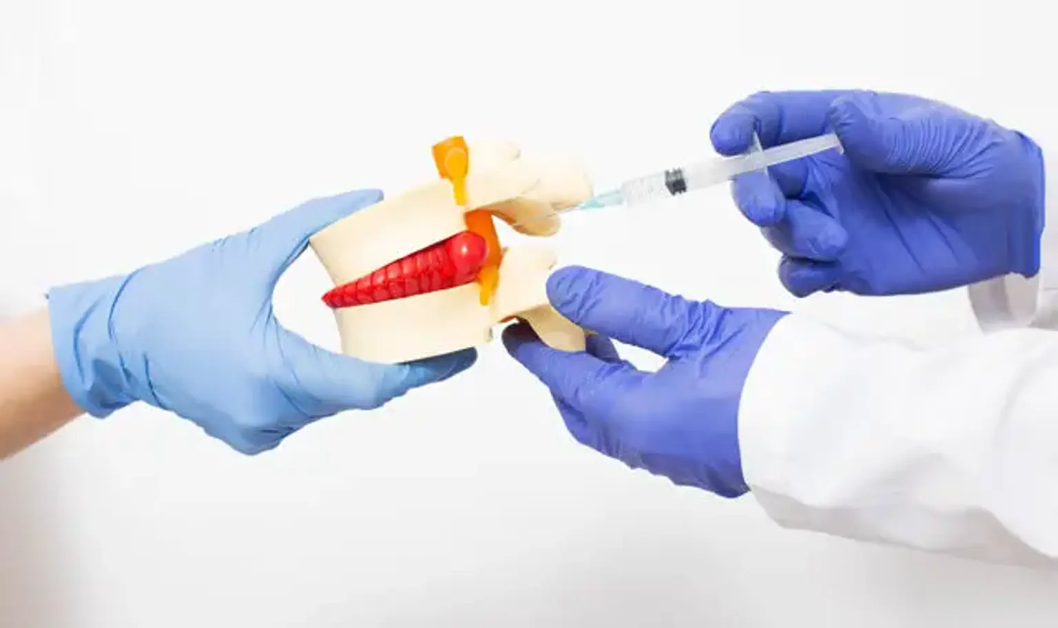
Nucleoplasty (Percutaneous Discectomy) is a minimally invasive, image-guided technique used to relieve back and leg discomfort caused by herniated discs that have not responded to other treatments. Nucleoplasty is the best minimally invasive treatment option for disc bulges. This outpatient surgery employs a needle that produces radio waves to decrease a disc bulge by dissolving extra tissue. This releases pressure inside the disc and on the nerves that cause pain. The operation should take no longer than an hour.
Nucleoplasty is a procedure in which one of the interventional radiologists utilizes imaging guidance to remove a little piece of disc tissue to release pressure on the nerves, therefore reducing discomfort and restoring movement.
People who experience chronic back pain for at least six weeks and debilitation owing to disc herniation after standard therapies have failed are candidates for nucleoplasty. People suffering from osteoporosis may benefit from nucleoplasty as well. The patient may suffer some discomfort or paralysis following nucleoplasty as a result of a disc herniation pressing on a spinal nerve.
Why Nucleoplasty (Percutaneous Discectomy) is indicated?
In cases of cauda equina syndrome with progressive or new motor impairment, an urgent lumbar discectomy is required. Elective lumbar discectomy is suggested in individuals who have failed conservative treatment options and have persisting radicular symptoms that match to radiographic evidence of nerve root compression by a herniated disc. Before undergoing surgery, the patient must have a clear awareness of the expected results. It is also important to emphasize the comparatively higher dependability of improvement in radicular leg pain compared to back pain.
The possibility of recurrence is also a relevant consideration. Should recurrent herniation occur, revision discectomy remains a viable option; however, with the removal of more disc material, and especially in the case of substantial or repetitive annular damage, the option to proceed with spinal fusion at the injured level may be considered. To raise the reasonable possibility of a favourable outcome, a high degree of correlation between a patient's symptoms and pathology on magnetic resonance imaging must be validated prior to surgery.
How herniated disc can be operated?
Open Discectomy:
Following the administration of general anesthesia, the patient is placed prone on a spine frame (Wilson or Allen Bow) or a specialized table. Transverse pads on the iliac crest and chest allow hip flexion to enhance interlaminar space while avoiding abdominal pressure to minimize central venous pressure. The start point and trajectory of the surgical approach may be guided by palpation of bone landmarks, such as the sacrum and iliac crests corresponding to the L4-L5 disc level. Following sterile skin preparation, localisation with a spinal needle and fluoroscopic control verifies the target level. At the midline, a 3 to 4 cm longitudinal incision is made, centered on the radiographic marker.
The skin is slit with a sharp scalpel, and electrocautery subcutaneous dissection reveals the lumbar fascia; this is incised slightly off the midline, as found by palpating the spinous processes ipsilateral to the disc disease treated. At the goal level, this fascia should span the interspinous space. On a lateral fluoroscopic picture, a radiographic marker can be utilized to validate the spinal level and cranially directed trajectory in line with the interspinous space.
Electrocautery is used to complete a subperiosteal elevation of paraspinal muscles from the superior and inferior spinous processes down to the laminar junction. Lateral dissection is continued bluntly with a Cobb elevator as far as the facet joint, taking care not to breach its capsule. Visualization of the interlaminar space is critical, as is the removal of dissected muscle tissue in the operative area, appropriate retraction, and meticulous electrocautery hemostasis. Using a curette, the surgeon separates the ligamentum flavum from its connection on the anterior portion of the superior vertebral lamina.
To protect the dura beneath, an angled Woodson elevator can be placed anterior to the ligamentum and guided caudally. After that, the ligamentum is forcefully cut to allow for retraction using a Penfield elevator and visibility of the departing nerve root and concomitant epidural fat. To allow for appropriate exposure, the medial side of the inferior facet of the superior vertebra may need to be resected. A Penfield or blunt probe is then inserted into the neuroforamen to mobilize the root and allow it to retract medially.
With good visibility of the intervertebral disc area, pituitary rongeurs can be used to remove fractured or herniated tissue. If a part of the herniation persists behind the posterior longitudinal ligament, access may need the use of a scalpel to incise the annulus. It is critical to use a Woodson elevator to probe the epidural area in all directions for any more disc or ligamentous tissue.
Additionally, it is advisable to irrigate the disc space with saline via a bulb syringe to express any loose disc fragments that may have gone unvisualized. Meticulous hemostasis via bipolar electrocautery is achieved, and the wound is liberally irrigated with saline. The fascial and subcutaneous layers are closed with absorbable suture, and the skin closure is by surgeon preference.
Minimally Invasive Procedures:
A lumbar intervertebral disk protrusion is a common cause of leg and back discomfort. Smaller protrusions, if confined, are less likely to resorb spontaneously than larger disk extrusions and have a lower surgical discectomy prognosis. Recently, numerous percutaneous techniques of discectomy or disk decompression have emerged as appealing therapeutic alternatives to open surgical procedures, with potentially comparable clinical success. Furthermore, patients may quickly return to their normal functional performance.
Central decompression is performed by removing or degrading a piece of the nucleus pulposus in the core of the disk without removing the actual ruptured disk material, hence avoiding more significant damage to the annulus.
The benefits of minimally invasive percutaneous techniques include a faster return to work, less pain after surgery, no scar tissue formation around the nerve roots as a result of the procedure, a lower cost, a lower incidence of developing disk space collapse and spinal instability, and fewer complications; success rates for these techniques range from 55% to 90%. Unfortunately, no clinical comparative studies or individual prospective controlled trials exist to support such a claim.
Three techniques that have been introduced recently as minimally invasive treatments for contained herniation of the nucleus pulposus are as follows:
- Nucleoplasty using Coblation technology (RF vaporization of nuclear tissue);
- Catheter disk decompression that uses heat from resistive coil positioned in the area of disk herniation;
- Dekompressor that uses volume reduction to decrease intradiscal pressure.
1. Nucleoplasty using Coblation technology:
Nucleoplasty had positive outcomes in terms of pain relief and functional improvement. Nucleoplasty was discovered to be capable of ablation and coagulation of the nucleus pulposus in order to decompress the disk and thermally change disk tissue. Decompression is clearly minimal or nonexistent in degenerated disks and is more common in nondegenerated ones. Radicular discomfort higher than axial pain for more than 6 months and failure of conservative therapies, including physical therapy, are general signs. A careful examination of patients' MRIs is required, as individuals with less than 50% of the disk height maintained, considerable prolapse above and below the disk level, or extremely high disk protrusion will have less favorable results.
A good candidate for any percutaneous decompression procedure, including nucleoplasty, will have a small (6 mm) contained disk protrusion with proven annular integrity and associated radicular symptoms validated by selective nerve root blocks. In most cases, a selective nerve root block is enough to prove that the discomfort is coming from the target disk.
Provocative discography would be required for a few patients who had either failed selective nerve root block or experienced axial and radicular pain at the same time. Even in individuals with mainly axial back pain and internal disk damage, nucleoplasty appears to be helpful. Another research, however, rejected such a hypothesis.
Contraindications include full disk space collapse, which makes the intervertebral space inaccessible, disk space infection, and medical problems that would preclude safe performance. Furthermore, those with developing neurological impairment, past surgery at the same intervertebral level, substantial canal stenosis, scoliosis, or tumor are not appropriate candidates for nucleoplasty or any other minimally invasive disk decompression operation.
2. Percutaneous decompression (Dekompressor):
The second technique, percutaneous decompression (Dekompressor), was introduced recently and has been used mostly in the United States. Nuclear disk material is extracted by an auger within the cannula whose tip is positioned inside the nucleus. When Dekompressor reduces nuclear volume within closed hydraulic space, significant change in intradiscal pressure follows. Therefore, intact annulus is required in order to retract the protruded part with decrease of the annular wall stress.
This procedure should not be used in patients with disrupted annular wall, and if in doubt, provocative discography should be completed to confirm affected level and rule out annular leak of the contrast. A European study confirmed pain score improvements in the majority of patients treated with Dekompressor, suggesting that somewhat lesser pain scores can be expected in patients with posterolateral foraminal than posteromedian disk protrusion.
3. Electrothermal intervertebral disk decompression:
Finally, another percutaneous method for electrothermal intervertebral disk decompression has just been described. Thermal energy is used by a decompression catheter to achieve targeted decompression of contained herniated discs. It is made up of a short (1.5 cm) resistive coil capable of producing localized heat.
To get the best therapeutic result, the heated portion of the catheter should be placed over the area of contained protrusion. There have been no clinical investigations published, not even case series reports. It might be a good treatment for axial and leg problems. Resistive coils can generate a substantial amount of heat and should not be utilized in individuals with a low disk height (less than 50%).
What happens before surgery?
- You will sign consent and other documents in the doctor's office so that the surgeon is aware of your medical history (allergies, medications/vitamins, bleeding history, anesthetic responses, previous surgeries).
- Discuss with your health care provider all drugs you are taking (prescription, over-the-counter, and herbal supplements).
- Preoperative testing (e.g., blood tests, electrocardiograms, and chest X-rays) may be required many days before surgery.
- Consult your primary care physician about discontinuing certain drugs and ensuring you are surgically cleared.
- Continue to take the medicines prescribed by your surgeon. 7 days before surgery, discontinue all nonsteroidal anti-inflammatory medications (ibuprofen, naproxen, etc.) and blood thinners (Coumadin, aspirin, Plavix, etc.).
- To avoid bleeding and healing issues, stop using nicotine and drinking alcohol one week before and two weeks after surgery.
What happens after Percutaneous Discectomy?
After an uncomplicated discectomy, patients may be discharged on the first postoperative day. Physical therapy rehabilitation or oral pain management may need an extended postoperative stay. Outpatient discectomy has been reported and done in several places. External bracing is not required for spinal stability. Due to concerns about re-herniation, most surgeons suggest restricting substantial bending, lifting, and twisting motions for 3 to 6 weeks after surgery, while more expedient or immediate unrestricted activities may give equal results without higher re-herniation rates.
Your healthcare provider will advise you on how to utilize your back properly. Lifting and bending may need to be restricted. Your physician may require you to wear a back brace for a period of time following the surgery. Most people are able to return to work after a week or so. Following surgery, you may require physical therapy to help strengthen your back.
Some fluid may leak from your little incision. This is quite normal. If there is a lot of discharge from the incision site, notify your physician straight once. Also, if you develop a fever or have a lot of pain in the region, contact your provider.
Sometimes the procedure causes slightly more pain for a while. But you can take pain medicines to ease the pain. Usually this goes away quickly. Your pain should become less than it was before your surgery.
Recovery time & how to prevent unfavourable outcome
Make a follow-up visit with your surgeon two weeks following surgery. Some people may require physical rehabilitation.
The recovery duration ranges from one to four weeks, depending on the underlying condition and your overall health. You may have discomfort at the location of the incision. It is possible that the first discomfort will not be totally alleviated immediately after surgery. Maintain a cheerful attitude and actively execute any prescribed physical therapy exercises.
With professions that are not physically demanding, most people may return to work in 2 to 4 weeks or less. Others may be required to wait at least 8 to 12 weeks before returning to work in positions that demand heavy lifting or operating heavy machinery.
Recurrences of back pain are common. The key to avoiding recurrence is prevention:
- Proper lifting techniques
- Good posture during sitting, standing, moving, and sleeping.
- Appropriate exercise program.
- An ergonomic work area.
- Healthy weight and lean body mass.
- A positive attitude and relaxation techniques (e.g., stress management).
- No smoking.
What are the surgical outcomes?
Good results are achieved in 80 to 90% of patients treated with lumbar discectomy. In a study that compared surgery and nonsurgical treatment for herniated discs, the outcomes were:
- People with leg pain (sciatica) benefit more from surgery than those with back pain.
- People with less severe or improving pain do well with nonsurgical treatment.
- People with moderate to severe pain who had surgery notice a greater improvement than those who did not have surgery.
Similarly, minimally invasive discectomy approaches have been demonstrated to have similar results as open discectomy. While minimally invasive procedures have advantages such as shorter surgical times, less blood loss and muscle stress, and faster recovery, these newer techniques are not acceptable for all patients. Consult your surgeon to see if a minimally invasive microendoscopic discectomy is right for you.
Discectomy may give pain relief faster than nonsurgical therapy. However, it is unknown if surgery affects the type of treatment required later on. A recurrent disc herniation will occur in 5 to 15% of individuals, either on the same or opposite side.
Is Percutaneous Discectomy risky?
A wound or deep infection develops at a rate of 2 to 3%, whereas dehiscence or other wound infections occur at a rate of 1 to 2%. Direct intra-operative nerve root damage has been reported to occur in 1 to 2% of instances. According to the research, the rate of inadvertent durotomy ranges from 0% to 4%. Durotomy may cause CSF fluid leaking, increasing the risk of developing meningitis. Instability following discectomy using the procedures outlined here is thought to be extremely infrequent, while the difficulties in identifying and measuring it makes objective quantification difficult.
The recurrence rate of lumbar disc herniation after discectomy ranges from 1 to 25%, while the risk of intragenic dural rupture reaches 9%. Risk factors include male gender, smoking status, and strenuous labor. According to an 8-year SPORT database follow-up, patients with concomitant retrolisthesis at L5-S1 appear to have the similar long-term clinical results as those without retrolisthesis.
Because recurrence has been studied at widely differing follow-up intervals and using a variety of outcome measures, including recurring symptoms and reoperation, patient-specific characteristics should guide a forthright conversation about the possibility of repeated illness prior to surgery. The patient may have persistent pain following lumbar disc surgery, which may be a part of failed back surgery syndrome.
Conclusion
Nucleoplasty is a therapeutic technique used to treat individuals with mild disc hernia. The central nucleus pulposus within the intervertebral disc is removed in a minimally invasive surgery.
In situations of intervertebral disc hernia, progressive compression of the disc causes the annulus fibrosis to open, allowing the nucleus pulposus to protrude. The protrusion can progress to the point that it compresses surrounding nerve fibers, resulting in pain and other symptoms. Nucleoplasty is the removal of excess nucleus pulposus material that is pinching the nerves and causing discomfort.
The patient may suffer some discomfort or paralysis following nucleoplasty as a result of a disc herniation pressing on a spinal nerve. On discharge, the patients will be informed on how to gradually resume their usual activities over the next few days.