Introduction
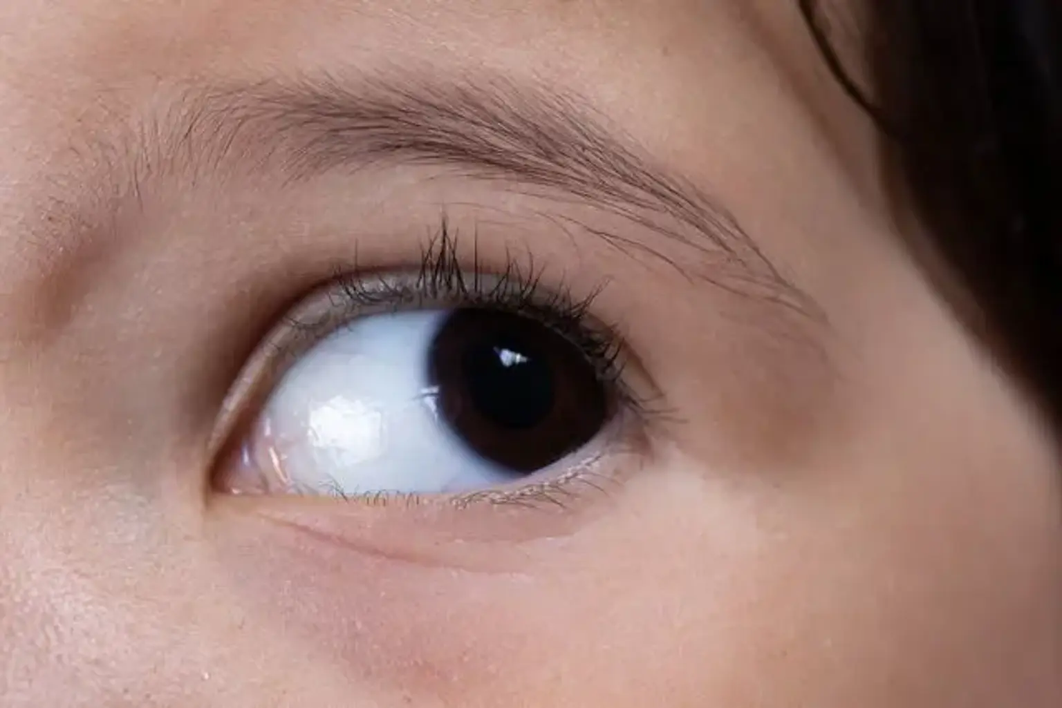
What is Nystagmus? Nystagmus is a condition characterized by involuntary, repetitive eye movements. These movements are typically horizontal, vertical, or circular, and they can affect one or both eyes. While nystagmus may be present at birth (congenital) or develop later in life (acquired), it can significantly impact vision and coordination.
Importance of Early Diagnosis and Treatment Timely diagnosis and treatment are essential in managing nystagmus. Early intervention can help reduce symptoms, improve quality of life, and prevent complications related to vision impairment and balance issues. Treatment options are diverse and can vary depending on the type and cause of the condition.
Understanding Nystagmus
Definition and Classification Nystagmus is defined by involuntary eye movements, which can be jerky or smooth, occurring in different directions. It is generally classified into two types:
Congenital Nystagmus: Present at birth or develops in early childhood.
Acquired Nystagmus: Develops later in life due to various causes like neurological disorders, alcohol intoxication, or vestibular dysfunction.
Types of Nystagmus
Horizontal Nystagmus: The most common type, where eye movements occur side to side.
Vertical Nystagmus: Up-and-down eye movements, often seen in neurological issues.
Pendular Nystagmus: Movements are of equal speed in both directions.
Jerk Nystagmus: The eye moves slowly in one direction and rapidly in the other.
Symptoms of Nystagmus
Involuntary, rhythmic eye movements
Blurry or double vision
Difficulty focusing
Possible issues with balance and coordination
Causes of Nystagmus
Congenital Causes Congenital nystagmus is typically caused by genetic factors or developmental issues during pregnancy. A family history of eye disorders or certain genetic conditions can increase the risk.
Acquired Causes Acquired nystagmus may result from:
Neurological disorders: Stroke, brain injury, or tumors affecting the brain’s visual and motor control centers.
Vestibular disorders: Inner ear problems like Meniere’s disease or vestibular neuritis that impact balance and eye coordination.
Medications: Some drugs, including sedatives and anti-epileptics, can cause nystagmus as a side effect.
Alcohol and drug use: Excessive alcohol or drug consumption can lead to temporary nystagmus.
Other Causes
Multiple sclerosis or Parkinson’s disease: These can affect the nervous system and contribute to eye movement disorders.
Infections: Certain infections, including viral or bacterial ones, can lead to nystagmus if they affect the central nervous system.
How Nystagmus Affects Vision and Daily Life
Visual Disturbances Nystagmus can cause blurry or double vision as the eyes continuously move, making it difficult to focus. The rhythmic eye movements may disrupt clarity, especially when reading or looking at stationary objects.
Impact on Balance and Coordination The condition may affect balance due to its link with the vestibular system (responsible for balance). People with nystagmus may experience dizziness or a sense of unsteadiness, particularly when standing or walking.
Quality of Life Issues Living with nystagmus can be challenging. It can affect everyday activities such as driving, reading, or even recognizing faces. People with nystagmus may also feel self-conscious about their condition, especially if it leads to noticeable eye movements.
Signs and Symptoms: When to See a Doctor
Early Signs of Nystagmus The signs of nystagmus are typically noticeable early on, especially if it is congenital. If a child’s eyes appear to move rapidly and uncontrollably, or if an adult begins to notice unusual eye movements, it may be time to seek medical attention. Common symptoms include:
Uncontrolled eye shaking or jerking
Blurry or double vision
Difficulty focusing on objects or people
How to Recognize the Condition at Home Parents or individuals may notice that eye movements become more pronounced when focusing on distant objects, or that the eyes make jerky movements from side to side or up and down. If these movements persist or worsen, medical evaluation is essential.
Importance of Seeking Timely Medical Advice If nystagmus is suspected, especially if accompanied by other symptoms such as dizziness, balance issues, or sudden vision changes, it’s crucial to seek an eye specialist or neurologist for proper diagnosis. Early intervention can help prevent further complications and guide the patient to the most effective treatment options.
Diagnosis of Nystagmus
Eye Exams and Visual Testing Diagnosing nystagmus begins with a comprehensive eye examination. Doctors assess eye movement patterns and visual acuity to confirm the diagnosis. Specific tests, such as tracking eye movement and checking how the eyes react to different stimuli, help to determine the type of nystagmus.
Tests Used to Diagnose Nystagmus
Frenzel Goggles: These goggles limit visual input to assess eye movements in different conditions.
Electronystagmography (ENG) and Videonystagmography (VNG): These tests use electrodes or cameras to monitor and record eye movements, helping to identify the cause of nystagmus, particularly if it's vestibular in nature.
Magnetic Resonance Imaging (MRI) and CT Scans: These imaging tests are used to rule out underlying neurological causes, such as brain lesions or tumors.
Role of Neuroimaging In cases of acquired nystagmus, neuroimaging (MRI or CT scans) plays a crucial role in diagnosing underlying neurological conditions. If the nystagmus is caused by a stroke, brain injury, or other central nervous system disorders, imaging helps pinpoint the exact problem and guides treatment.
Differential Diagnosis: What Nystagmus May Be Mistaken For
Other Disorders that Mimic Nystagmus Several conditions can produce similar symptoms to nystagmus, making it essential for healthcare providers to distinguish between them. These include:
Vestibular Disorders: Conditions affecting the inner ear, such as benign paroxysmal positional vertigo (BPPV) or Meniere's disease, can cause dizziness and eye movements similar to nystagmus.
Neurological Conditions: Diseases like multiple sclerosis or cerebellar ataxia can also result in abnormal eye movements, which may be mistaken for nystagmus.
Medication Side Effects: Certain medications, especially sedatives, anti-epileptic drugs, or alcohol, can cause eye movements that resemble nystagmus.
How Healthcare Providers Rule Out Other Conditions Doctors often rely on detailed medical history, symptom patterns, and diagnostic tests (such as ENG or MRI) to rule out other possible causes. A thorough examination helps ensure that the proper diagnosis is made and guides the most effective treatment plan.
Treatment Options for Nystagmus
Medical Treatment Options There is no universal cure for nystagmus, but treatments can significantly reduce symptoms and improve quality of life. For many people, the goal is to manage symptoms effectively:
Medications: Drugs such as gabapentin, baclofen, and clonazepam may be prescribed to help reduce the intensity of eye movements and improve stability.
Botulinum Toxin Injections: In some cases, botox injections may help reduce the severity of nystagmus, especially in acquired forms. These injections target the muscles around the eyes to help control involuntary movements.
Surgical Interventions Surgery can sometimes provide relief for congenital or acquired nystagmus. Surgical options include:
Tenotomy or Tendon Surgery: This involves altering the muscles around the eyes to reduce the range of motion and minimize eye movements.
Muscle Repositioning: In some cases, repositioning the muscles controlling the eyes can improve the alignment and control of eye movements.
Vision Therapy and Corrective Lenses While surgery and medications can help control eye movements, vision therapy offers a non-invasive option. Patients may be prescribed corrective lenses, or special glasses, to help improve clarity and focus. Prism glasses can also be used to reduce visual disturbances and enhance depth perception.
Pharmacological Treatments for Nystagmus
Types of Medications Several medications may help reduce nystagmus symptoms, particularly in acquired forms. Commonly prescribed drugs include:
Gabapentin: Often used to treat neurological disorders, gabapentin can help control involuntary eye movements.
Baclofen: This muscle relaxant can help reduce spasms and manage nystagmus symptoms.
Clonazepam: An anti-anxiety medication, it can sometimes be effective in treating the symptoms of nystagmus.
Effectiveness of Medications Medications may not eliminate nystagmus but can reduce the severity of eye movements and improve stability. However, the effectiveness varies from patient to patient, and some may experience side effects such as drowsiness or dizziness.
Side Effects and Risks As with any medication, side effects should be considered. Some patients may experience fatigue, coordination issues, or dizziness, which may affect daily activities. It's important to work closely with a healthcare provider to monitor and adjust treatment as needed.
Non-Surgical Treatments: Therapy and Rehabilitation
Vision Therapy for Nystagmus Vision therapy is a non-invasive treatment that aims to improve eye coordination and reduce visual disturbances. It includes exercises designed to strengthen the eyes’ ability to focus and track moving objects. Regular therapy sessions may help in managing symptoms and improving quality of life.
Occupational Therapy Occupational therapy can help individuals with nystagmus adapt to daily activities. Therapists work on techniques to improve hand-eye coordination, balance, and safety in performing tasks like reading or cooking.
Coordination and Balance Training Since nystagmus can affect balance, rehabilitation programs may focus on improving stability and coordination. This can help reduce dizziness and make it easier for individuals to perform normal daily tasks.
Surgical Treatment for Nystagmus
Surgical Options Available Surgery is typically considered for severe congenital nystagmus or acquired forms that don’t respond to other treatments. Common procedures include:
Tenotomy: This involves cutting and repositioning the eye muscles to help reduce the extent of the involuntary movements.
Tendon Surgery: Muscles around the eyes are surgically adjusted to control eye movements better.
Success Rates of Surgical Procedures Surgical outcomes can vary, but many patients experience significant improvements in the control of their eye movements. While surgery can’t always eliminate nystagmus, it can reduce its severity, allowing patients to focus better and improve their vision.
Post-Surgical Recovery After surgery, patients typically need time for recovery and adjustment. This may involve follow-up visits to monitor the effectiveness of the procedure and assess any changes in eye movements. Most patients can resume daily activities within a few weeks, although full recovery may take months.
Innovative Treatments and Advancements in Nystagmus Care
Ongoing Research in Gene Therapy
Exciting developments are underway in the field of gene therapy for nystagmus. Researchers are exploring potential treatments to address the root causes of congenital nystagmus, particularly those linked to genetic mutations. Gene therapy aims to correct or replace faulty genes responsible for abnormal eye movements, offering hope for a long-term solution.
Botulinum Toxin Injections
Botox injections are sometimes used as a temporary treatment for nystagmus, particularly in cases where other therapies haven’t been effective. The injections target the muscles around the eyes, helping to reduce the severity of involuntary movements. This option is often considered for patients who haven’t had success with medications or surgery.
Emerging Surgical Techniques
Advancements in surgical techniques are making it possible to achieve better outcomes with less invasive procedures. Surgeons are developing more precise methods for adjusting eye muscles, resulting in quicker recovery times and more stable results for patients.
Managing Nystagmus in Children
Impact of Nystagmus on Child Development
Nystagmus in children can affect not only vision but also motor skills and social development. Children with nystagmus may struggle with reading, writing, or participating in physical activities due to impaired focus and coordination. Early intervention is essential to help children adapt and thrive.
Treatment Options for Pediatric Nystagmus
Treatment for children with nystagmus may include vision therapy, corrective lenses, or medication. In some cases, surgical procedures may be considered if the condition significantly affects the child’s ability to engage in daily activities. Pediatric ophthalmologists or neurologists typically manage treatment plans, tailoring them to each child’s specific needs.
Support for Parents and Families
Parents of children with nystagmus may face challenges related to their child’s self-esteem and academic performance. Support groups and resources can help families navigate the emotional and practical aspects of managing nystagmus. Therapy, special accommodations at school, and a supportive home environment can all contribute to positive outcomes.
Coping with the Emotional Impact of Nystagmus
Psychological Effects of Nystagmus
Living with nystagmus can be frustrating and emotionally challenging, particularly for those who experience visible eye movements. Many people with nystagmus may feel self-conscious, anxious, or depressed about their condition, especially if it impacts their social interactions or professional life.
Addressing Self-Esteem and Confidence
Support from loved ones and counseling can help improve self-esteem and reduce anxiety. Coping strategies, such as mindfulness techniques or joining support groups, may also help individuals adjust to living with nystagmus. Open conversations with friends, family, and colleagues about the condition can alleviate some emotional burdens.
Support Networks for Nystagmus
Support groups and online communities can provide valuable emotional support for those living with nystagmus. Connecting with others facing similar challenges can reduce feelings of isolation and provide practical advice on managing the condition.
The Role of Technology in Managing Nystagmus
Assistive Technologies Recent advances in technology have led to the development of tools that can assist people with nystagmus in their daily lives:
Smart Glasses: Specially designed glasses equipped with built-in stabilization can help reduce visual disruptions caused by nystagmus.
Electronic Magnifiers: Devices that magnify text and images can be invaluable for those who struggle to focus or read due to eye movement disorders.
Voice-Activated Technology: Voice-controlled devices, such as smartphones and smart home assistants, can help individuals navigate their environment more easily.
Apps and Software for Visual Assistance There are also various mobile apps available to help those with vision impairment manage their daily tasks. Apps that convert text to speech or provide magnification and contrast adjustment can make reading and navigating environments easier for people with nystagmus.
Telemedicine and Remote Consultations Telemedicine has become an essential tool for people with nystagmus, especially for routine follow-ups or consultations with specialists. Remote consultations save time and offer greater accessibility, particularly for patients in remote areas or those with mobility challenges.
Prognosis and Quality of Life with Nystagmus
Long-Term Outlook for Individuals with Nystagmus
The prognosis for individuals with nystagmus largely depends on the underlying cause and the severity of the condition. Congenital nystagmus typically stabilizes with age, though it may not improve significantly without treatment. In contrast, acquired nystagmus may improve if the underlying condition is treated.
Impact on Daily Life and Independence
With the right treatments and adaptations, many individuals with nystagmus can lead independent and fulfilling lives. Supportive care, such as using assistive devices or making environmental adjustments, can significantly improve quality of life.
Coping Strategies for Better Living
Adaptations in the Home: Simple changes, such as increasing lighting or using magnifying tools, can make it easier to manage daily tasks.
Physical and Occupational Therapy: Ongoing therapy may be recommended to maintain balance and coordination.
Emotional Support: Counseling and support groups play a vital role in helping individuals adapt emotionally to life with nystagmus.
Preventing Nystagmus: Can It Be Avoided?
Preventing Congenital Nystagmus
Unfortunately, congenital nystagmus, which is typically inherited, cannot be prevented. However, genetic counseling can help families understand the risks and prepare for the condition if there is a family history of eye disorders.
Avoiding Acquired Nystagmus
Acquired nystagmus is often a result of other health issues, such as neurological conditions, trauma, or medication side effects. Taking preventive measures such as managing chronic conditions, avoiding head injuries, and following proper medication guidelines may reduce the risk of developing acquired nystagmus.
Importance of Regular Eye Checkups
Routine eye exams can help detect vision problems early, which is essential for catching nystagmus early and managing its progression. Early detection allows for better treatment outcomes and management strategies.
Cost of Nystagmus Treatment
Treatment Expenses
The cost of nystagmus treatment varies depending on the type and severity of the condition. Prescription medications, vision therapy, and corrective lenses can add up, especially if long-term management is required. Surgical options also come with significant costs, including hospital stays, follow-up appointments, and post-operative care.
Insurance Coverage
Many insurance plans cover treatments for nystagmus, especially if the condition affects daily functioning or is a result of an underlying medical condition. However, coverage for experimental treatments, such as gene therapy, may vary. Patients should review their insurance policies and speak with their healthcare provider about the options available to them.
Financial Assistance Programs
There may be financial assistance programs available for patients who are struggling to afford treatments. Non-profit organizations and medical societies often provide grants or help navigate insurance coverage options to ensure people receive necessary care.
Nystagmus in the Global Context
Global Prevalence of Nystagmus
Nystagmus affects people worldwide, with variations in its prevalence based on geographic regions, genetic factors, and access to healthcare. While the condition is more commonly observed in children, it can affect people of all ages.
Awareness and Education
Raising awareness about nystagmus is essential for better diagnosis and treatment. Global initiatives, such as public health campaigns and educational programs, can help reduce the stigma associated with the condition and improve public understanding.
Access to Treatment Around the World
In developed countries, nystagmus treatment is more readily accessible, with advanced medical technologies and specialized healthcare professionals. In contrast, people in low-resource settings may face challenges in obtaining proper care, making access to affordable treatment an ongoing concern.
Conclusion
Embracing Life with Nystagmus While nystagmus presents challenges, many people can lead successful, independent lives with the right support and treatment. Advancements in medical care, technology, and therapy have improved outcomes and provide hope for better management of the condition.
Empowering Those Affected It’s important for individuals with nystagmus to remember that they are not alone. With medical advancements, emotional support, and coping strategies, people with nystagmus can maintain a high quality of life and feel empowered to pursue their goals and dreams.
Hope for the Future Research into gene therapy and other innovative treatments continues to offer hope for a future where nystagmus may be more effectively managed or even cured. Until then, ongoing support and treatment remain key to improving the lives of those living with nystagmus.