Osgood-Schlatter disease (OSD)
Overview
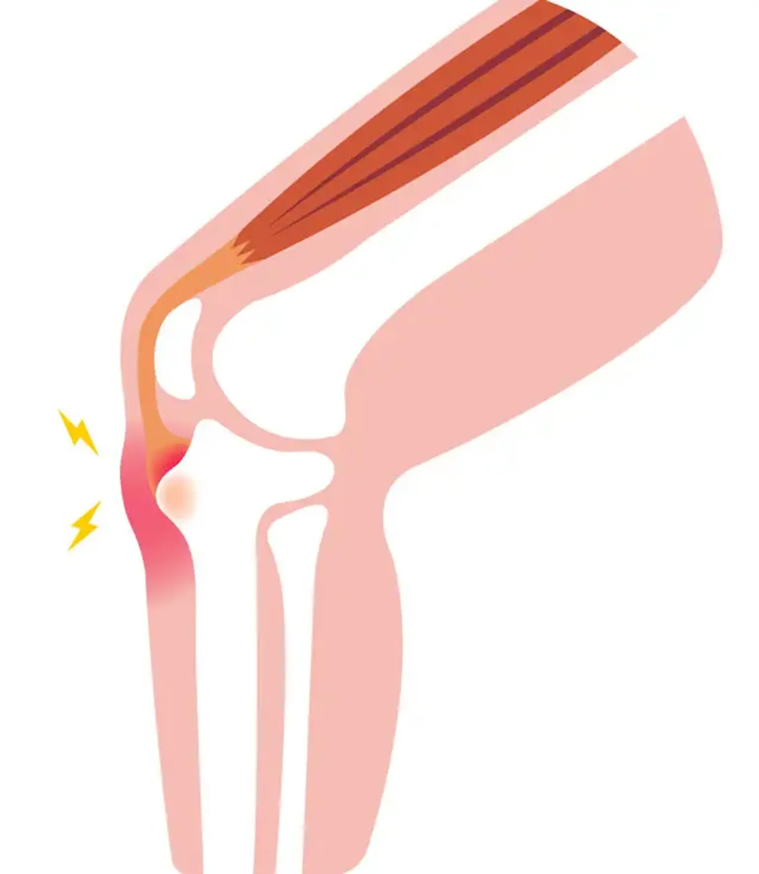
Osgood Schlatter disease, also known as osteochondrosis, tibial tubercle apophysitis, or tibial tubercle traction apophysitis, is a prevalent cause of anterior knee discomfort in the skeletally immature athletic population. Tenderness at the patellar tendon insertion point at the tibial tuberosity is traditionally associated with atraumatic, gradual start of anterior knee discomfort.
The syndrome is self-limiting and develops as a result of recurrent extensor mechanism stress activities like leaping and sprinting. Overall treatment is determined by the level of pain, and management includes symptomatic treatment with ice and NSAIDs, as well as activity modification and relative rest from inciting activities in conjunction with a lower extremities stretching regimen to correct underlying predisposing biomechanical factors.
Osgood schlatter disease in adults
OSD is a traction phenomena caused by repeated quadriceps contractions via the patellar tendon at its insertion on the skeletally immature tibial tubercle. This happens during preadolescence, when the tibial tubercle is vulnerable to tension. The discomfort will be confined to the tibial tubercle and, in rare cases, the patellar tendon itself. Sinding-Larsen-Johansson syndrome is a comparable condition that can arise at the patella-patellar tendon junction.
OSD is a prevalent cause of knee discomfort in children aged 8 to 15 years. This ailment can have a long course and cause time away from athletics. However, it is seldom the source of persistent damage or disability.
Because of the absence of a specific etiology and hence definition, some practitioners may find it difficult to distinguish OSD from tibial tubercle avulsion fractures. An acute avulsion fracture of the tibial tubercle is more likely if the patient is unable to ambulate. OSD sufferers can usually walk, albeit with pain.
OSD generally develops gradually, with patients reporting of discomfort in the tibial tubercle and/or patellar tendon area following repeated activity. Running or leaping exercises that place a considerable strain on the patellar tendon's insertion on the tibial tubercle typically increase the patient's symptoms.
They may also have symptoms that grow and wane in response to changes in their sporting seasons. A quick start of pain in the tibial tubercle region with no antecedent symptoms should alert the doctor to look for an acute tibial tubercle avulsion rather than OSD.
OSD is a self-limiting disorder. In a study by Krause et al, 90 percent of patients treated with conservative therapy were free of all symptoms one year after symptoms began. Patients may continue to experience kneeling issues after reaching skeletal maturity. This is usually caused by discomfort around an unfused tibial tubercle ossicle or a bursa that may need to be resected.
there appears to be little correlation between persistent anterior knee discomfort following OSD and patellar stability. There were no occurrences of recurvatum from early closure of the proximal tibial physis, according to the authors.
Common sports seen in association with the condition include:
- Basketball
- Volleyball
- Sprinters
- Gymnastics
- Football
Epidemiology
One of the most prevalent reasons of knee discomfort in the skeletally immature teenage athlete is Osgood Schlatter disease. Onset occurs between the ages of 10 and 15 years for males and 8 to 13 years for females, during adolescent growth spurts. The disorder is more common in men and occurs more commonly in athletes who participate in running and jumping sports. Osgood Schlatter disease affects 9.8 percent of teenagers aged 12 to 15. (11.4 percent in males, 8.3 percent in females). In 20% to 30% of individuals, symptoms manifest bilaterally.
Etiology
The patellar tendon is made of cartilaginous tissue and inserts at the tibial tubercle. This is followed by tibial tubercle ossification between the ages of 10 and 12 in females and 12-14 in boys. Osgood Schlatter disease develops at this period of bone development. The accepted assumption is that repetitive tension on the tubercle causes microvascular tears, fractures, and inflammation, which manifests as swelling, discomfort, and soreness.
Overuse injuries such as Osgood Schlatter disease occur in active teenage patients. It develops as a result of recurrent strain and microtrauma caused by the strong patellar tendon's insertion into the comparatively soft apophysis of the tibial tubercle. This force causes discomfort as well as partial avulsion of the tibial tubercle apophysis in extreme situations.
Force increases with greater activity, especially following periods of fast development. Trauma may occasionally result in a complete avulsion fracture. Poor flexibility of the quadriceps and hamstrings, as well as other signs of extensor mechanism misalignment, are risk factors.
Risk factors for the disorder include:
- Male gender
- Ages: male 12-15, girls 8-12
- Sudden skeletal growth
- Repetitive activities like jumping and sprinting
Pathophysiology
The tibial tubercle develops as a secondary ossification point, where the patellar tendon is attached. Bone expansion outpaces the muscle-tendon unit's capacity to extend sufficiently to preserve previous flexibility, resulting in increased strain across the apophysis. The physis is the weakest link in the muscle-tendon-bone relationship (as opposed to the tendon in an adult) and, as such, is vulnerable to damage from repetitive stress.
Softening and partial avulsion of the apophyseal ossification center may occur with repetitive contraction of the quadriceps muscle mass, particularly with repeated forceful knee extension as observed in sports involving running and leaping.
The appearance and closure/fusion of the tibial tubercle occurs in the following sequence pattern:
- Tibial tubercle is entirely cartilagenous (age < 11 years)
- Apophysis forms (age 11 to 14 years)
- Apophysis fuses with the proximal tibial epiphysis (age 14 to 18 years)
- The proximal tibial epiphysis and tibial tubercle apophysis fuses with the rest of the proximal tibia (age > 18 years)
In males and females, the primary growth plate of the proximal tibia and the secondary ossification center of the tibial tubercle merge by the end of the next two stages of bony development (eg, epiphyseal and bony stages), and the OSD typically diminishes.
The most widely accepted theory for the onset of OSD is that repeated traction (traction apophysitis) on the anterior portion of the developing ossification center causes multiple subacute microavulsion fractures and/or tendinous inflammation, resulting in a benign, self-limiting disturbance manifested as pain, swelling, and tenderness.
The most prevalent long-term consequences of OSD are adult knee discomfort and the cosmesis of a bony protrusion on the anterior knee. Less common sequelae include a painful ossicle that necessitates surgical excision and a misplaced avulsion of a tibial tubercle.
Osgood Schlatter disease symptoms
An 8 to 15-year-old youngster would often appear with unilateral or bilateral anterior knee discomfort and edema. Pain begins as a dull pain on the tibial tubercle and gradually worsens with movement. The symptoms are indicative of an insidious start without prior trauma.
Pain usually improves with rest and goes away minutes to hours after the triggering activity or sport is discontinued. Running, leaping, direct knee damage, kneeling, and squatting all aggravate the pain. Tenderness over the region of patellar tendon insertion is apparent, as is an increased prominence at the tibial tubercle.
Poor quadriceps and hamstring flexibility may be present as risk factors. Resisted knee extension and active or passive knee flexion can also cause pain.
Physical Examination
The physical examination is very specific, with point tenderness over the tibial tubercle. A firm mass may be palpable.
Other physical examination findings may include the following:
- Proximal tibial swelling and tenderness
- Enlargement or prominence of the tibial tubercle
- Reproducible and aggravated pain by direct pressure and jumping
- Pain with resisted knee extension
- Hamstring tightness
- Quadriceps atrophy
- Erythema of the tibial tuberosity
The following exam findings must be tested and confirmed to verify no concomitant or more severe injury:
- Full range of motion of the knee
- No effusion or meniscal signs
- Negative Lachman test (no knee instability)
- Normal neurovascular examination
- No abnormal findings in the hip and ankle joints
Diagnosis
Osgood Schlatter disease is a clinical diagnosis, and radiographic assessment is rarely required. If the presentation is severe or abnormal, plain radiographs may be utilized to rule out other diseases such as fracture, infection, or bone malignancy. After a traumatic occurrence, a radiographic assessment may be recommended to evaluate for apophysis avulsion damage or other injuries.
Radiographs
Because Osgood-Schlatter disease (OSD) involves a clinical diagnosis, not all patients require radiography. Plain films, on the other hand, should be taken at least once throughout the examination and therapy to rule out various etiologies such as malignancy, acute tibial apophyseal fracture, and infection.
Radiographs may indicate:
- Superficial ossicle in the patellar tendon
- Irregular ossification of the proximal tibial tuberosity
- Calcification within the patellar tendon
- Thickening of the patellar tendon
- Soft-tissue edema proximal to the tibial tuberosity
The Osgood-Schlatter lesion is best viewed from the side, with the knee in a 10-20° internal rotation. The most usual observation is that the knee films are normal, particularly if the youngster is still in the preossification period.
In the acute phase of OSD, the tibial tubercle may be prominent and raised, with anterior soft-tissue edema. Radiographs may indicate radiodense pieces or ossicles detached from the tibial tuberosity in severe situations.
Radiographs may occasionally detect irregularity, fragmentation, or increased density of the tibial tubercle's ossification. In asymptomatic youngsters, this pattern may be a normal variety.
Other Imaging Modalities
Advanced imaging is not required to diagnose Osgood-Schlatter disease (OSD), but it may be required to confirm that another diagnosis is not present.
Ultrasonography may indicate a normal tubercle as well as signal alterations associated with thickening (more echogenic) in the patellar tendon and a hypoechoic region of nearby soft tissue.
Nuclear Medicine If a bone scan is acquired, it may show higher uptake in the region of the tibial tuberosity. Changes near the insertion of the patellar tendon may be seen by computed tomography (CT) scanning or magnetic resonance imaging (MRI)
An MRI may aid in the diagnosis of an unusual presentation. It may potentially be used to stage the illness and predict the clinical course. However, the usefulness of MRI in diagnosis, prognosis, and treatment is still restricted. If MRI is conducted to rule out other diseases, it may reveal increased bone edema of the tibial tuberosity.
Treatment for Osgood schlatter disease
In the end, the condition is self-limiting, however it can last for up to two years until the apophysis merges. Treatment consists of rest and activity modification away from the aggravating activity, as directed by the amount of discomfort. Although there is no evidence that rest accelerates healing, activity limitation is useful in lowering pain.
Patients can engage in sports as long as the discomfort goes away with rest and does not interfere with sports-related activities. For pain relief, ice and NSAIDs can be used locally. To prevent against direct impact, a protective knee pad can be placed over the tibial tubercle. Hamstring stretching, as well as quadriceps stretching and strengthening activities, might be beneficial supplements.
The standard first-line treatment for Osgood-Schlatter disease (OSD) is non-surgical and conservative. The initial therapy consists of applying cold for 20 minutes every 2-4 hours.
Analgesics and nonsteroidal anti-inflammatory medications (NSAIDs) may be used to alleviate pain and reduce local inflammation. NSAIDs, on the other hand, have not been found to shorten the course of OSD.
Historically, patellar tendon insertion injections were not advised. This is mostly related to the potential of subcutaneous tissue atrophy following corticosteroid injections. A recent Japanese research compared lidocaine/dextrose injection to lidocaine alone. There was no difference between the two injection groups, although both groups claimed symptom improvement with no negative consequences.
Long-term immobilization is usually not advised since it might produce greater knee stiffness in mild instances, predisposing the athlete to other sports-related injuries. If a patient is disobedient, the physician may urge immobilization in a knee brace for at least 6 weeks. The brace should be removed solely for stretching and strengthening activities on a regular basis.
Inform the patient to avoid activities that cause discomfort (eg, sports that involve excess amounts of jumping). Infrapatellar straps, pads, or braces can also be utilized for support, however none have been shown to be effective.
After the initial symptoms have subsided, quadriceps-stretching exercises, including hip extension for a full stretch of the extensor mechanism, can be undertaken to relieve strain on the tibial tubercle. Stretching exercises for the hamstrings, which are frequently stiff, are also recommended.
There are no long-term limitations or issues linked with this disorder, other from the presence of an ossicle that produces pain while kneeling.
Physical Therapy
The objective of rehabilitation is for the athlete to be able to return to his or her sport as soon as feasible and in the safest possible way. Because rest, ice, and NSAIDs are the primary treatments, physical therapy has a limited role, if it is utilized at all. The discomfort might last anywhere from 6 to 24 months. If a person returns to exercise too soon, he or she may aggravate the disease. Athletes must concentrate on strengthening the flexibility and strength of the quadriceps and hamstring muscles during the recovery process in order to be ready to return to sports.
Acute phase
The physical therapist may propose a number of strategies to reduce pain and prevent illness recurrence. The severity of the illness influences treatment suggestions.
An infrapatellar strap may be recommended during sports activity but has no proven efficacy.
- Resting when pain arises.
- Ice should be applied to the area for 20 minutes following activity.
- Short-term rest and knee immobilization may be required.
On rare occasions, this author has casted a patient who is in great pain and is refusing conservative treatment. This is frequently with a parent who is determined to alleviate the agony. While a brace can be advised, it is unlikely that it will be utilized on a disobedient patient.
Recovery phase
Straight leg raises can be performed as follows:
- Lie on the floor with your back elevated a few inches by your elbows.
- Bend the unaffected knee to a comfortable posture; using half-pound increments of adjustable ankle weights, establish the weight at which 10 rises on the afflicted leg may be accomplished.
- Tighten the thigh muscles and elevate the afflicted leg 12 inches while maintaining it straight.
- 5 seconds of holding
- Relax as you slowly drop your leg.
- Begin with 10 repetitions of each leg.
- When you've completed 15 reps easily, raise the weight by half a pound and then return to 10 repetitions.
- Increase the weight to a maximum of 7-12 lb after 15 repetitions can be completed comfortably again.
Short-arc quadriceps exercises can be performed as follows:
- Lie back with the unaffected knee bent
- Place a few rolled up towels under the affected knee to raise it 6 inches from the floor
- Tighten the thigh muscles and straighten the leg until it is 12 inches from the floor
- Hold for 5 seconds
- Slowly lower the leg and relax
- Start with 10 repetitions for each leg and increase to 15, using the same ankle weight and repetition progression as for straight leg raises
Differential Diagnosis
The most serious risk is failing to detect another ailment, which might lead to long-term irreparable harm (eg, tumor, osteochondritis dissecans). The clinical examination or history for the majority of other illnesses is more problematic. As a result, always acquire radiographs and examine the likelihood of a hip referred pain condition.
In addition to the conditions listed in the next section, the following should be considered in the differential diagnosis of Osgood-Schlatter disease (OSD):
- Sinding-Larson-Johansson syndrome
- Tumor (bone or soft tissue)
- Perthes disease (often presents with knee pain instead of hip complaints)
- Patellar tendon avulsion/rupture
- Chondromalacia patellae (Patellofemoral syndrome)
- Patellar tendonitis
- Infectious apophysitis
- Accessory ossification centers
- Osteomyelitis of the proximal tibia
- Hoffa's syndrome
- Synovial plica injury
- Tibial tubercle fracture
Prognosis
Osgood-Schlatter disease has a very good prognosis. The illness is self-limiting, although recovery might take months. In roughly 10% of individuals, the symptoms may persist throughout adulthood. Long-term consequences arise when an individual does not seek therapy or does not comply with the suggested treatment. There have been reports of discomfort that lasts for years.
Complications
- Prominence of tibital tubercle
- Ongoing pain
Conclusion
An interprofessional team comprised of a sports physician, physical therapist, primary care provider, orthopedic nurse, and orthopedic surgeon is most suited to handle Osgood Schlatter disease.
Because these injuries frequently occur in very active teenage patients, it may be more useful to reduce sports involvement rather than discontinue exercise entirely. For example, if a patient participates in multiple sports or on multiple teams during the same season, it may be more effective to recommend that the patient discontinue participation in one of these teams or sports to reduce the amount of activity and strain that occurs with frequent and recurrent participation rather than complete rest.
The choice to limit exercise should be made together, taking into account both the patient athlete's and parent's immediate and long-term goals.
The amount of discomfort experienced should ultimately influence the choice. Preventive strategies for Osgood Schlatter include suggesting a moderate increase in workload (less than 10% per week), utilizing proper equipment and procedures, including a stretching program to ensure hamstring and quadriceps flexibility, and considering avoiding early sports specialization.