Osteotomy
Last updated date: 06-Aug-2023
Originally Written in English
Osteotomy
Overview
An osteotomy is a surgical procedure that involves the cutting of one or more bones. The term osteotomy, derived from Greek, is defined in the medical dictionary as a surgical operation in which a bone is divided or a piece of bone is excised (as to correct a deformity).
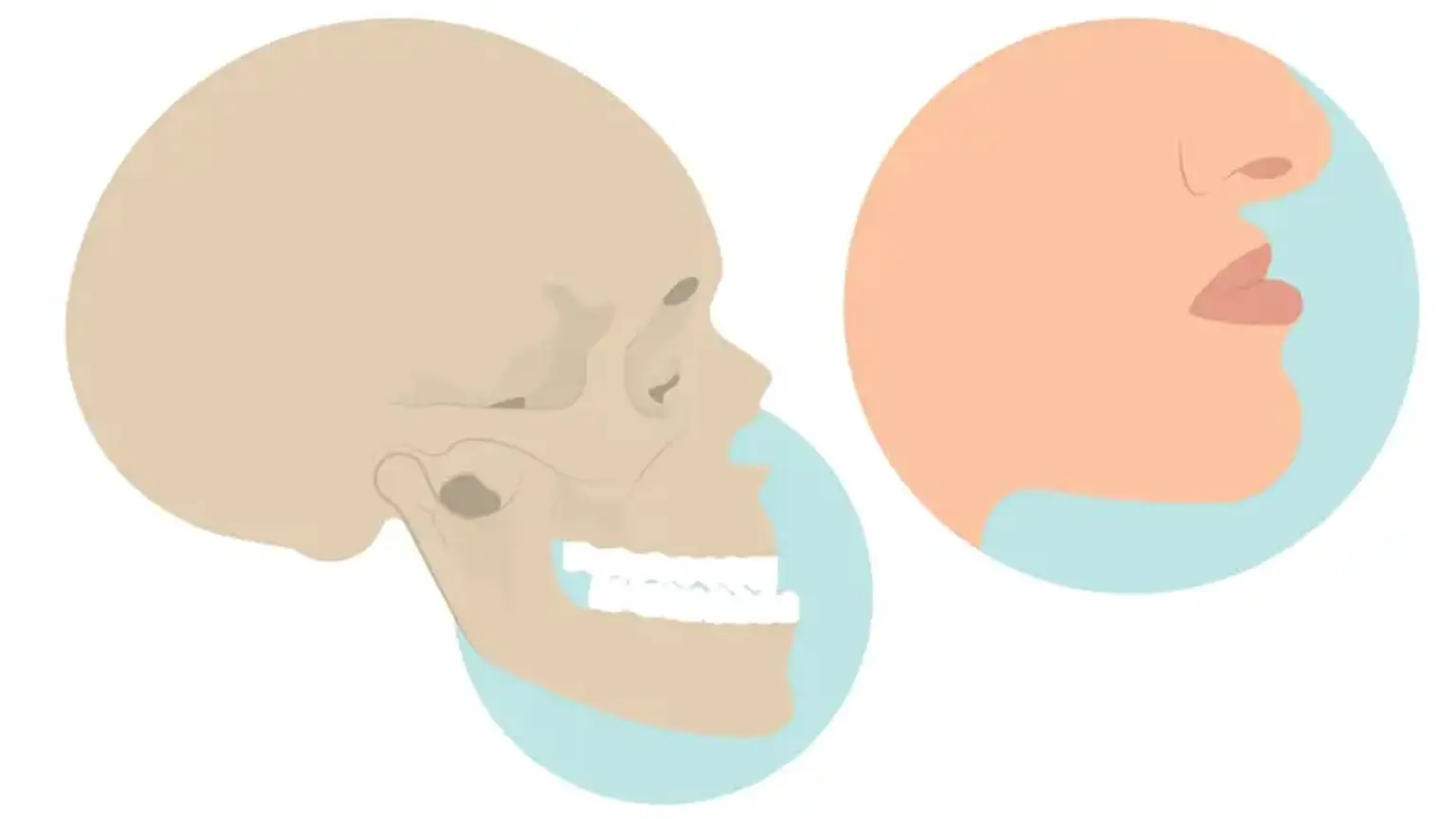
What is Osteotomy?
To repair retreating upper or lower jaws, osteotomy may be done. Osteotomy is frequently used in conjunction with genioplasty (Chin Surgery).
Why would you need an Osteotomy?
You may require an osteotomy for a variety of reasons. Healthcare practitioners refer to one of them as a structural abnormality. A substantial discrepancy in the anatomy of a person's jaw that causes a prominent overbite or underbite is an example of this. An osteotomy may be performed for aesthetic reasons, such as changing the look of your face or jaw, or for medical reasons, such as correcting breathing, eating, or speaking problems caused by the abnormality. Another rationale for an osteotomy is to repair damage caused by an injury or persistent degeneration.
Osteoarthritis is a common cause of leg bone osteotomy, particularly at the hip or knee joints. A surgeon can remove pressure from regions of the joint where you are suffering discomfort and injury by cutting and reshaping bone fragments. To address a disparity in limb length, a surgeon may perform an osteotomy of the leg or arm joints in specific circumstances.
What are the types of Osteotomy?
There are various types of osteotomies, and the one you pick is determined by criteria such as:
- which bone is affected
- why you are having the procedure done
- your overall health
- your surgical goals
Finally, your surgeon will decide which technique to utilize to accomplish the osteotomy. Here are some examples of different types of osteotomies:
- lateral closed-wedge
- medial opening-wedge
- dome osteotomy
- hemicallotasis with external fixator
Some common names for osteotomy types based on the surgical location include:
- Jaw: dentofacial osteotomy, corrective jaw surgery
- Chin: osteotomy of the chin
- Elbow: French osteotomy
- Spine: spinal osteotomy
- Knee: high tibial osteotomy during knee surgery
- Hip: McMurry osteotomy, Pauwel’s osteotomy, Salter’s osteotomy, Chiari’s osteotomy, Pemberton’s osteotomy
Maxillary Osteotomy
A maxillary osteotomy may be performed to correct these issues:
- Significantly receded upper jaw
- Crossbite
- Too much or too little of the teeth showing
- Open bite
The surgeon slices the bone above your teeth (below both eye sockets) from within the mouth so that the complete top jaw — including the roof of your mouth and your upper teeth — may move as one unit. The jaw and upper teeth are advanced until they are appropriately aligned with the lower teeth.
Once the jaw has been straightened, small screws and plates keep the bone in place. These screws, which are smaller than brace brackets, gradually integrate into the bone structure.
Excess bone forms above the molars, causing what is typically a level, even surface to become tilted. When you bite down, your molars make contact but your front teeth do not, making chewing difficult. Your surgeon will shave or remove the extra bone to correct this.
Mandibular Osteotomy
The technique is generally done intraorally. Scars from incisions might be seen on the chin, jaw, or around the mouth. The surgeon makes incisions in the jawbones and slides them into place. When your jaw movement is finished, the miniplate and screws, wires, and rubber bands may be utilized to fix the bones in their new position. Jaw surgery can be performed on the upper or lower jaw, the chin, or any combination of these areas.
1. Upper Mandibular Osteotomy
Upper jaw surgery may perform to correct the following problems:
- Significantly receded or protruding upper jaw
- Crossbite
- Too much or too little of the teeth showing
- Open bite
- Reduced facial growth of the middle of the face
The upper jaw is sliced so that it moves as one unit. The upper jaw and teeth migrate forward or backward until they are properly aligned with the lower teeth. The surgeon next repairs the bone by using plates and screws to keep it in place, after which the extra bone is shaved or removed.
2. Lower Mandibular Osteotomy
Lower jaw surgery may perform to correct the following problems:
- Receding lower jaw
- Protruding lower jaw
The surgeon makes incisions below the teeth and down the jawbone to allow the front of the jaw to move as a single unit. The jaw can be shifted forward or backward to its new location. As the jawbone heals, plates and screws keep it together.
Osteotomy Rhinoplasty
An osteotomy rhinoplasty is a surgical operation in which a patient's nose is "broken" using an osteotome, a bone-cutting instrument. Osteotomy nose jobs require precision and accuracy, and as such, must be performed by a face cosmetic and reconstructive surgeon with extensive experience.
Prior to rhinoplasty, preoperative photographs of the patient is required. Review these images with the patient before to surgery to address the areas of dissatisfaction. Digital photography, in conjunction with computer tools that allow patients to visualize prospective surgical outcomes, has grown in popularity.
Nasal Anatomy
The nasal bones are paired structures that connect superiorly to the frontal bone and laterally to the maxillary nasal process. These bones combine to produce the bony nasal vault. The ethmoid perpendicular plate, a part of the bony septum, connects to the undersurface of the nasal bones in the midline. The nasal bones thin inferiorly and thicken superiorly. Transillumination of the cranium is shown in Figure 10-3.
The varied thickness of the nose's bony bones will impact osteotomy placement. Along its entire length, the nasal septum supports the nose. The septum supports the dorsal profile, and its absence might result in the typical saddle-nose malformation. To maintain support following any nose surgery, sufficient (>1 cm) dorsal and caudal septal struts must be preserved. The anterior nasal spine supports the caudal septum and the medial crura's foot. When the nasal spine is too large, it might actually blunt the nasolabial angle.
Types of osteotomies in rhinoplasty include the following:
- External Osteotomy Rhinoplasty: May help reduce the risk of lateral wall collapse and airway compromise.
- Lateral osteotomies - These are carried out along the frontal portion of the maxilla, sometimes reaching onto the nasal bone.
- Medial osteotomies - These are done to assist guarantee that the surgeon's targeted fracture lines occur; they may be useful if infracturing is problematic with a lateral osteotomy.
- Superior osteotomies - When the nasal bones are abnormally thick and there is a risk of an undesirable fracture line linking the lateral and medial fracture lines, these procedures may be performed.
Workup in osteotomy rhinoplasty
Prior to rhinoplasty/osteotomy, every patient must have a full medical history and physical assessment. The following laboratory tests must be performed:
- Complete blood count (CBC)
- Urinalysis
- Electrolytes
- Electrocardiogram (for men >40 y and women >50 y, unless a history of hypertension, stroke, arrhythmia, diabetes, or smoking, or any other concerning reason, is present)
- Pregnancy test in women of childbearing age
- Human immunodeficiency virus (HIV), hepatitis B, and hepatitis C testing
How Does an Osteotomy Nose Job Differ from a Traditional Rhinoplasty?
An osteotomy nose job differs from typical rhinoplasty surgery in that it involves modifying the position of the nasal bones rather than reshaping or shrinking the nose.
If a patient has a crooked nose, a nasal fracture, or other nose damage, an osteotomy nose job may be advised. The treatment can also be performed to correct a significant nasal hump that causes the nose to seem broader from the front.
What to Expect During Osteotomy Rhinoplasty Recovery
The first healing period following osteotomy nose surgery is one to two weeks. A patient may feel edema and bruising at the treatment site during this time. A patient will also be given a splint and bandage for their nose that will be worn for at least a week. Additionally, around one week following an osteotomy nose job, the dressing on a patient's nose is removed and the nostrils are cleansed.
Following an osteotomy nose surgery, patients are advised to sleep with their heads raised for at least one week. They should rest extensively for the first week following their nose operation and avoid intense exercise for up to six weeks.
Swelling and bruising may continue for several weeks following an osteotomy rhinoplasty, although nose sensitivity and discomfort usually go away within three to six months.
What Are the Risks Associated with an Osteotomy Nose Job?
Osteotomy nose surgery risks include:
- Infection
- Difficulty breathing
- Poor wound healing
- Hole in the nasal septum
- Skin discoloration
How to prepare for an osteotomy?
A healthcare practitioner will undertake diagnostic tests before to an osteotomy to assist determine the exact site and degree of damage that has to be corrected. Your surgeon may use imaging tests such as an X-ray or CT scan to map out the injured bone and devise a treatment plan for reshaping and reconstructing the damaged region.
You will also have routine preoperative testing, such as blood tests to screen for clotting issues and an overall health checkup.
Your particular preoperative instructions will vary depending on the operation and the person performing the surgery. In many cases of leg orthopedic surgery, such as hip or knee osteotomy, your surgeon may advise you to do the following:
- improving your overall health
- losing weight
- taking a preoperative class to review types of therapy
- reviewing current medications and allergies
What to expect during Osteotomy surgery?
On the day of your operation, you will be given instructions on when to cease eating, drinking, or taking medications, as well as when to arrive at the hospital. You will be led to a preoperative area where you will change into a hospital gown and maybe wash in a particular solution to help destroy any bacteria on your skin.
Your medical team will go over the surgery plan, as well as any allergies and concerns you may have. A healthcare expert will insert intravenous catheters, or IVs, to allow you to receive drugs throughout the procedure.
When the operation begins, your surgeon will generally do the following stages, with minor variations depending on the kind of procedure:
- A medical practitioner will accompany you to the operating room and drape you in a sterile drape. The surgical team will validate the procedure and disinfect the spot.
- Sedation, generally in the form of general anesthesia, will be administered to you. This frequently necessitates the use of a breathing tube during surgery, which is afterwards removed in the recovery area.
- To reveal the afflicted bone, a surgeon will make an incision through your skin and surrounding tissues.
- They will cut and sculpt your natural bone using a surgical saw. If necessary, implants or bone transplants may be used to finish the bone remodelling.
- The surgeon may use plates, screws, or other hardware to secure the reconstructed bone.
- They will close the incision and the surgery will be over.
Risks and complication
When done by a qualified oral and maxillofacial surgeon in consultation with an orthodontist, osteotomy is typically safe. However, risks and consequences of the procedure may include:
- Blood loss
- Infection
- Nerve injury
- Jaw fracture
- Relapse of the jaw to the original position
- Problems with bite fit and jaw joint pain
- Need for a second surgery
- Need for root canal therapy on selected teeth
- Loss of a portion of the jaw
Results
Correcting the alignment of your jaws and teeth with jaw surgery can result in:
- The balanced appearance of your lower face
- Improved function of your teeth
- Health benefits from improved sleep, breathing, chewing and swallowing
- Improvement in speech
- Improved appearance
Frequently asked questions about Osteotomies
1. Who is a candidate for an osteotomy?
Although it varies depending on the bone, osteotomy candidates are often active, fit, and under the age of 60. It's also critical that there are still healthy bone sections to preserve and that there are no other underlying joint concerns.
2. Will an osteotomy completely cure my condition?
As in the case of osteoarthritis, an osteotomy is not always curative. Although an osteotomy is frequently performed to treat discomfort and enhance mobility in younger people, a full joint replacement is generally required later in age.
3. Am I limited in how I can use the bone after surgery?
It takes several months to recover from surgery. During this period, your doctor may advise you to reduce the amount of weight you put on the surgery region. Throughout your recuperation, you will gradually increase your weight-bearing capacity, reaching full strength in most cases after 6 weeks or more.
Conclusion
An osteotomy is a surgical technique in which a bone is sliced to reduce or lengthen it or to change its orientation. It is usually used to straighten a crookedly healed bone following a fracture or to correct a hallux valgus. There are several types of osteotomies, the most common of which being a knee osteotomy.
Because this is serious surgery, recuperation may take many months. In the end, you may require further procedures in the future, particularly for knee and hip osteotomies, but this surgery may halt the advancement of problems such as arthritis and lengthen the life of your natural bone.