Pancreatic and biliary tract diseases

Last updated date: 24-Apr-2023

Originally Written in English

Pancreatic and Biliary Tract Diseases

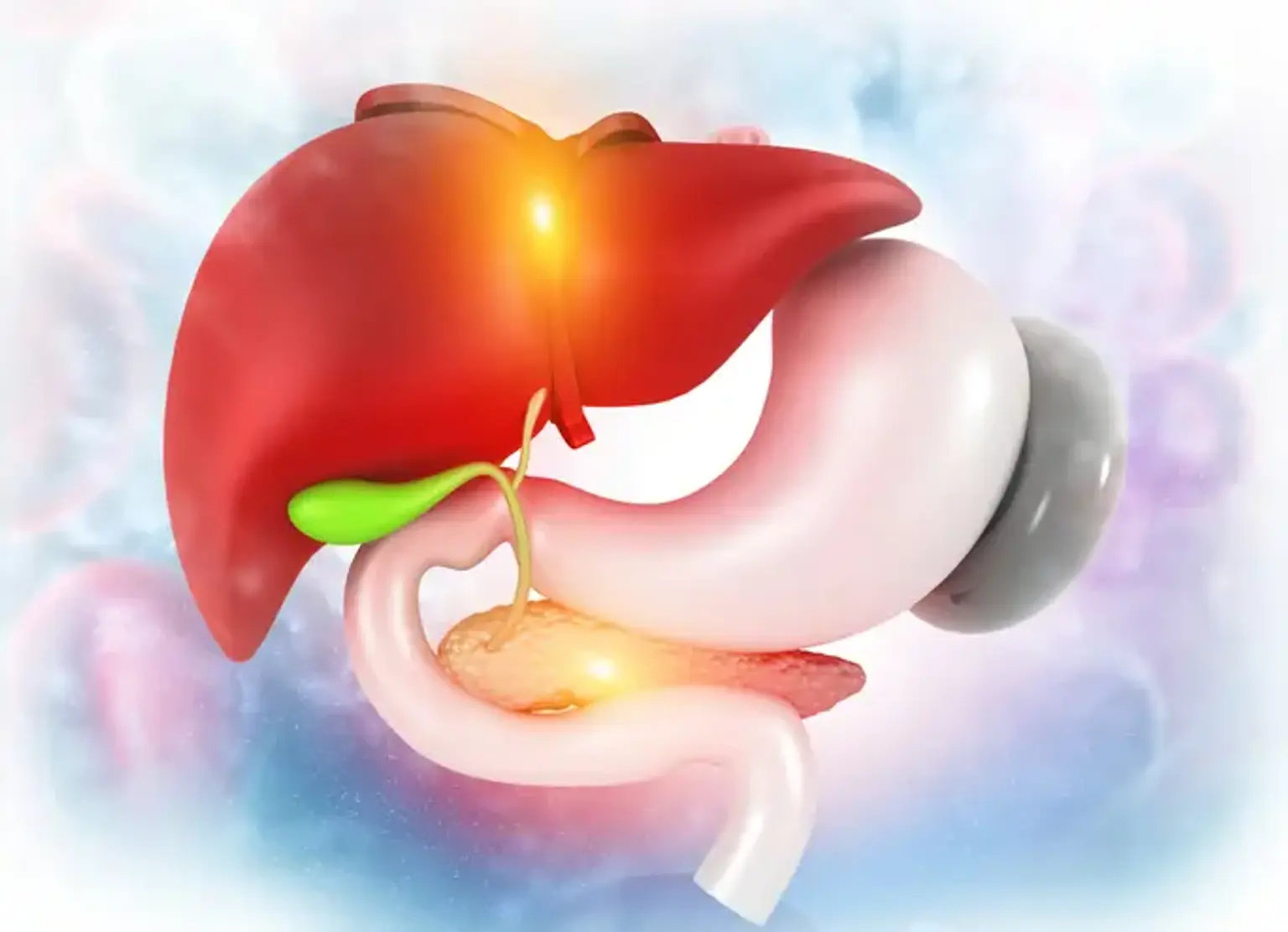
For proper digestion, a healthy pancreas and biliary tract are required. Because pancreatic and biliary tract problems can cause serious symptoms, it's vital to have an appropriate diagnosis and treatment as soon as possible. Through patient-centered care, extensive professional expertise, and the use of the most advanced technologies, nationally known experts are dedicated to evaluating and treating even the most complex pancreatic and biliary conditions, as well as other gastrointestinal diseases.


Pancreatic and Biliary Tract Diseases Risk Factors

While each disease is different, there are a few similar risk factors for pancreatic and biliary disease development:
  • Tobacco consumption
  • Heavy alcohol consumption
  • Pancreatic disease genetics and family history

To provide you with the highest level of clinical treatment, specialists use a multidisciplinary approach to your care, bringing together professionals in gastroenterology, surgery, cancer, pathology, and radiology.


Pancreatic Diseases

Pancreatic Diseases

Acute pancreatitis, chronic pancreatitis, hereditary pancreatitis, and pancreatic cancer are all conditions that affect the pancreas.

Because the pancreas is inaccessible, diagnosing pancreatic disorders can be challenging. There are several ways to assess the pancreas. A physical assessment of the pancreas is the first test, which is challenging because the pancreas is deep in the abdomen near the spine. Blood tests can help determine whether the pancreas is implicated in a certain illness, but they can sometimes be misleading. The CAT (computed tomography) scan, endoscopic ultrasound, and MRI (magnetic resonance imaging) are the commonest radiographic tests for evaluating the anatomy of the pancreas. ERCP (endoscopic retrograde cholangiopancreatography) and MRCP (magnetic resonance cholangiopancreatography) are two tests used to assess pancreatic ducts. The surgical investigation may be the only approach to confirm the diagnosis of pancreatic illness in some cases.


Chronic Pancreatitis

The pancreas becomes damaged as a result of scar tissue and inflammation in chronic pancreatitis. Large stones and calcium deposits can also develop in the pancreatic tissue and ducts.

Chronic drinking and cigarette use are also risk factors for chronic pancreatitis. It's possible that acute pancreatitis will recur.

Chronic pancreatic duct blockage, autoimmune disorders, or genetic composition could all be risk factors.

Symptoms of chronic pancreatitis include abdomen and back pain, weight loss, diarrhea, and diabetes.

Specialists will assist you in eliminating any potential causes of chronic pancreatitis during treatment. Any issues you may have will be managed during your therapy. Medication can be useful in some cases. Other therapy choices could include endoscopic procedures like ERCP or EUS, or surgical techniques to alleviate your discomfort.


Acute Pancreatitis

Acute Pancreatitis

Inflammation of the pancreas causes acute pancreatitis. Severe abdominal discomfort, nausea, and vomiting are common symptoms.

Stones that form and lodge in or travel through the bile duct, strong alcohol consumption, and high triglyceride or calcium levels are all causes of acute pancreatitis. Acute pancreatitis can develop if the pancreatic duct narrows or a tumor forms. It could potentially be a drug side effect. You could also be genetically predisposed to the disease.

A combination of physical exam findings, blood tests, and the results of a computed tomography (CT) scan or magnetic resonance cholangiopancreatography (MRCP), is used to identify acute pancreatitis.

Acute pancreatitis is a medical disease that often necessitates treatment in a hospital, where we can administer intravenous fluids and perform blood tests to keep track of your progress.


Pancreatic Cysts

A pancreatic cyst is an aberrant pancreatic lesion. A cyst looks like a liquid-filled balloon. The pancreas can produce a variety of cysts. After acute pancreatitis, an inflammatory cyst (pseudocyst) might form, and benign neoplastic growths can turn malignant.

Unless the cyst gets very large or cancerous, you may not suffer symptoms such as yellowing of the eyes or skin (jaundice), abdominal discomfort, back pain, weight loss, nausea, vomiting, or diarrhea.

Doctors may remove the pancreatic cyst surgically only if it is symptomatic or poses a health concern. Pancreatic cysts are often followed with frequent imaging examinations to evaluate whether removal or other therapy is required.


Pancreatic Fluid Collections

During acute or chronic pancreatitis, pancreatic fluid can accumulate. Following a serious injury to the pancreas that destroys the pancreatic duct, fluid may accumulate. Leaking of pancreatic enzymes and fluids causes the disease.

biliary duct obstruction, intestinal obstruction, discomfort, and recurring pancreatitis are all major medical disorders that can be caused by pancreatic fluid collections. Endoscopy, surgery, interventional radiology, or a combination of methods may be required to treat infected pancreatic fluid accumulation.


Autoimmune Pancreatitis

When the body's immune system targets the pancreas, autoimmune pancreatitis ensues. Discoloration of the skin or eyes (jaundice), abdominal pain, back pain, weight loss, and diarrhea are all symptoms.

Imaging studies and blood testing are frequently used to identify autoimmune pancreatitis. When blood tests and imaging are unclear, your doctor can use endoscopic ultrasound (EUS) to perform pancreatic biopsies.

Immunosuppressive drugs, such as steroids, are used to treat autoimmune pancreatitis. These treatments reduce inflammation by lowering the immune reaction toward the pancreas.


Pancreatic Cancer

Early detection of pancreatic cancer is critical for effective treatment. The surgeons are specialists in recognizing symptoms and administering the appropriate tests to detect how your disease is progressing. Pancreatic cancer is one of the gastrointestinal cancers we treat.


Biliary Tract Diseases

Biliary Tract Diseases

The gallbladder stores and concentrates bile, which is used by the body during digestion in the small intestines.


Cholecystitis

Cholecystitis is a condition in which the gallbladder becomes inflamed and infected. Gallstones obstructing the gallbladder are the most frequent cause of cholecystitis. Abdominal discomfort on the right side or in the upper central part of the abdomen is common. Nausea, vomiting, and fever are possible side effects. The gallbladder may need to be surgically removed, as well as endoscopic operations to clear the bile duct.


Gallstones and Bile Duct Stones

Gallstones and Bile Duct Stones

In the gallbladder or bile duct system, stones can develop. Blockage, abdominal pain, and infection are all symptoms of these stones. Gallstones are characterized by abrupt and rapidly worsening abdominal discomfort, as well as back pain, nausea, and vomiting.


Choledochal Cysts (Choledochoceles)

Choledochal cysts (choledochoceles) are a relatively uncommon condition. When cysts do appear, it's because you were born with the disease (congenital). The cysts are sac-like enlargements of the bile ducts, a tube-like structure that transports bile from the liver to the small intestine for digestion. Choledochal cysts are divided into five categories based on their location. Cholangitis, a biliary tree infection, can develop if left untreated. Furthermore, several forms of choledochal cysts may increase the risk of bile duct cancer. Surgical treatment usually comprises cyst removal and bile duct reconstruction.


Pancreatic and Biliary Tract Diseases Symptoms

Biliary Tract Diseases Symptoms

If the duct or papilla is obstructed, or if the pancreas is so destroyed by a disease that it cannot produce enough bicarbonate and enzymes, pancreatic juices may not reach the duodenum. Inadequate digestion is caused by a lack of pancreatic enzymes. Large bowel movements, which have a strong smell and are difficult to flush down the toilet due to their high-fat content, are clinically notable. Patients with pancreatic insufficiency may notice an oil slick on the toilet water from time to time. Steatorrhea refers to an excess of fat in the stools. Patients generally lose weight because their food is not well absorbed.

These pancreatic enzymes can be replenished, at least to some extent, by taking a pancreatic enzyme supplement, which is a drug taken by mouth. Replacing the missing bicarbonate output is usually unnecessary.

Bile deficiency can cause steatorrhea and trouble with digestion (especially of fats). The main bile duct, or papilla, is frequently blocked, resulting in a lack of bile in the duodenum. The liver continues to generate bile, which backs up into the bloodstream. This eventually results in body discoloration (jaundice), which is first visible in the whites of the eyes. When bile does not reach the duodenum, bowel movements lose their pigment and resemble pale putty. The retention of bile salts in circulation when the bile ducts are obstructed can cause severe itching (pruritus). Due to overdistention, an obstruction of the bile ducts or pancreatic ducts might produce pain.

Diabetes is caused by a lack of insulin release by the pancreas. When the islets of Langerhans (the source of insulin) overact or become tumorous, it is also common to have too much insulin. As a result, blood sugar levels drop below normal, causing dizziness and finally unconsciousness. Other pancreatic hormones (such as somatostatin, vasoinhibitory peptide, glucagon, and others) can elicit unpleasant symptoms in rare cases.


Biliary Tract Diseases Diagnosis

Diagnostic Laboratory Tests

Diagnostic Laboratory Tests

Patients with suspected biliary pathology are routinely evaluated in the laboratory. It is recommended that a complete blood count and liver function tests be sent. Due to the high rates of concomitance with gallstone pancreatitis, a lipase should be sent whenever gallstone pathology is suspected.

Differentiating cholelithiasis from choledocholithiasis and cholangitis requires laboratory findings. Because cholelithiasis and cholecystitis typically appear with similar labs, lab findings aren't helpful in distinguishing the two.

High bilirubin and alkaline phosphatase levels are common in problems that involve jaundice and block the common bile duct (choledocholithiasis and cholangitis). The white blood cell count can be increased by inflammation and diseases such as cholecystitis and cholangitis.

Laboratory results, like all laboratory evaluations, must be evaluated in context with the patient's symptoms and other findings. The presence or absence of a single lab value should not be used to make management decisions. This is especially true of the white blood cell count. Any of these disorders can occur even if your white blood cell count is normal.


Imaging Studies

Imaging Studies

The cornerstone of biliary tract illness diagnosis is imaging investigations. Endoscopic retrograde cholangiopancreatography (ERCP), magnetic resonance cholangiopancreatography (MRCP), and hepatobiliary iminodiacetic acid (HIDA) scans are used to confirm and, in the case of ERCP, treat these disorders.

  • Ultrasound

For biliary tract illnesses, ultrasound is the preferred first diagnostic test. It's a low-risk, non-invasive procedure that's widely available. When administered by qualified ED practitioners at the bedside, ultrasound can be a reliable tool for detecting cholelithiasis. The presence of possibly mobile echogenic particles within the gallbladder that forms deep shadows is used to identify this condition.

Gallstones outside the gallbladder, on the other hand, may be overlooked by the US, which is substantially less sensitive (80% in some studies) for choledocholithiasis. The presence of a biliary duct stone or other blockage is identified by dilation of the common bile duct, which is defined as > 6mm in adults up to the age of 60 and an additional +1 mm for each consecutive decade, and may be seen in choledocholithiasis and cholangitis.

For acute cholecystitis, ultrasound sensitivity and specificity are predicted to be 88-95 percent and 76-80 percent, respectively. Gallstones, gallbladder wall thickening (more than 3mm), pericholecystic fluid, and a positive sonographic Murphy sign (maximum tenderness with pressure from the US transducer directly over the gallbladder) are all ultrasonography signs of cholecystitis. On Doppler scans, gallbladder distention and increased blood flow to the gallbladder can also be noted. The predictive value of each individual sign on the abdominal US for cholecystitis varies greatly among studies, although most agree that having many abnormalities improves the overall predictive value.

  • CT Scan

CT imaging is one of the most common early modalities utilized in the Emergency Department for the examination of abdominal pain, and it is crucial in the identification of biliary disease.

Because many gallstones are radiolucent, CT imaging is less sensitive (72%) than US imaging for cholelithiasis.

Due to its ability to identify gallbladder distension, wall thickening, and pericholecystic fluid, it is highly sensitive (92%) for the identification of acute cholecystitis. In the diagnosis of rare consequences of acute cholecystitis, such as emphysematous cholecystitis or GB perforation, CT may outperform ultrasonography. It detects biliary blockage in choledocholithiasis and cholangitis with high sensitivity (>96%).

Because of its reduced risk and higher sensitivity for discovering stones, ultrasonography is usually favored over CT as the first examination. A CT scan in the Emergency Department is feasible if the US findings are unclear, a HIDA scan is not easily available, or a broad differential to the patient's abdominal pain is still being explored.

  • HIDA Scan (Hepatobiliary iminodiacetic acid scan)

The HIDA scan is 90-94 percent sensitive for acute cholecystitis and is recommended if the US is inconclusive or negative for cholecystitis despite a high clinical suspicion.

A HIDA scan is a nuclear imaging method that evaluates gallbladder function using a radioactive tracer (technetium-99m). The tracer is administered intravenously, circulated to the liver, and then eliminated through the biliary system. The gallbladder is seen within 1 hour of tracer injection in a normal (or negative) HIDA. A positive study suggests the existence of cholecystitis or cystic duct obstruction if the gallbladder is not visible within 4 hours of the injection.

The scan functions poorly if the patient has eaten within the last 24 hours, and it takes a considerable period of monitoring after tracer injection (up to 4 hours), which are two common downsides of using a HIDA scan in the ED.

  • Magnetic Resonance Cholangiopancreatography (MRCP) and Endoscopic Retrograde Cholangiopancreatography (ERCP)

The gold standard for diagnosing obstructive duct pathology such as choledocholithiasis has traditionally been ERCP. The ERCP procedure is used to identify and treat biliary and pancreatic duct disorders. An endoscope is inserted through the GI tract and into the biliary system in this procedure. Fluoroscopy allows for the detection of dye introduced into the biliary tree, as well as the detection of blockages. Many blockages will be removed during the dilatation of the Sphincter of Oddi and dye injection. Pancreatitis (5% risk), perforation of the GI tract or ductal system, allergic reactions, and bleeding are all concerns related to this invasive procedure. As less invasive diagnostic procedures such as MRCP and endoscopic ultrasonography become more frequently available, its application strictly for diagnosis is declining due to these concerns.

When compared to ERCP, MRCP is more accurate (both in terms of sensitivity and specificity) and does not expose patients to an invasive technique, contrast material, or documented complications, morbidity, and mortality rates associated with ERCP. MRCP can now be utilized to ensure that ERCP is only performed on patients who require it for therapeutic reasons.


Pancreatic Diseases Diagnosis

Pancreatic Diseases Diagnosis

Due to the inaccessibility of the pancreas, diagnosing pancreatic illnesses can be difficult. Well-trained and experienced professionals are required to make an accurate diagnosis that leads to effective therapies. Medical history is taken, a physical examination is performed, blood tests are performed, and additional imaging procedures are performed to assess the pancreas. The following diagnostic procedures are commonly used to detect pancreatic abnormalities:

  • Pancreatic enzymes, such as amylase and lipase, are measured in the blood. These enzymes normally rise quickly when acute pancreatitis occurs. However, because other factors might affect the levels of these pancreatic enzymes, laboratory findings must be carefully analyzed by a specialist.
  • CA19-9, a unique protein or tumor biomarker produced by pancreatic cancer cells, is measured via a blood test. CA 19-9 levels that are too high are often a marker of pancreatic cancer. However, because the sensitivity and specificity of CA19-9 in detecting pancreatic cancer are pretty limited, the concentration of CA19-9 should not be used as a confirmatory indicator to make a definite diagnosis of pancreatic cancer. Instead, the rise of CA19-9 can indicate other types of cancer or certain non-cancerous diseases. However, because some pancreatic cancer patients do not have increased CA19-9 levels, the test is not used alone to screen for or diagnose pancreatic cancer.
  • Imaging procedures that produce detailed images of the inside organs. These procedures, such as computerized tomography (CT) scan of the pancreas and magnetic resonance imaging (MRI) or magnetic resonance cholangiopancreatography (MRCP), support the specialist in identifying any abnormalities in the pancreas and surrounding areas of the abdomen. These imaging procedures can provide high-resolution images that can tell whether the observed mass is malignant. If the suspected tumor is cancerous, the risk of metastasis, or cancer cells spreading to new parts of the body, must be considered.
  • An endoscopic ultrasonography (EUS) is used. The pancreas is evaluated using an ultrasound probe from inside the abdomen. To obtain the images, the instrument is introduced down an esophagus and into the stomach through a thin, flexible tube (endoscope). During a EUS, tissue is usually collected by passing specialized tools via the endoscope. The pathologic evaluation is then used to confirm the diagnosis of pancreatic cancer and provide treatment options.


Pancreatic and Biliary Tract Diseases Treatment

Treatment of Biliary Tract Diseases

Treatment of Biliary Tract Diseases

Biliary colic caused by cholelithiasis is usually self-limiting and should be managed with antiemetics, fluids, and painkillers (NSAIDs or opioids). Medical treatments like ursodeoxycholic acid and chenodeoxycholic acid (which both reduce the hepatic release of biliary cholesterol and increase gallstone breakdown) are available but ineffective. Extracorporeal shock wave lithotripsy (similar to what is used to break up stones in the kidneys) is a rarely used procedure to break up stones in the gallbladder, usually in conjunction with ERCP. These are indicated for patients who are not fit for surgery. Surgical excision (cholecystectomy) of the gallbladder is the only way to cure symptomatic cholelithiasis. Dietary modifications (low-fat foods) may be beneficial till the surgery. Choledocholithiasis is managed by removing the stone surgically or endoscopically (ERCP).

Symptom control with fluids, antiemetics, and painkillers is the first line of treatment for cholecystitis and cholangitis, just as it is for biliary colic. Additionally, broad-spectrum intravenous antibiotics should be started as soon as possible. In both cases, the antibiotic choice should account for enteric gram negatives and anaerobes. In moderate cases, a cephalosporin (such as ceftriaxone) plus metronidazole (for anaerobic coverage). Piperacillin-tazobactam (tazocyn) or meropenem as single agents are appropriate in moderate to severe cases. Vancomycin is usually reserved for infections that arise after surgery.

A timely surgical evaluation is essential. Cholecystectomy is recommended for cholecystitis, but it is sometimes postponed, especially in extremely unwell patients. Cholangitis caused by an obstructing stone in the common bile duct may be treated with an ERCP +/- sphincterotomy. Both surgical and percutaneous decompression (cholecystostomy) with biliary drainage may be considered in critically unwell patients.

Patients with cholelithiasis who are pregnant should be treated symptomatically till after delivery. With the exception of utilizing pregnancy-safe antibiotics (avoid fluoroquinolones), pregnant patients with cholangitis should be treated the same as non-pregnant patients (parenteral antibiotics and early biliary drainage). Cholecystectomy will most probably be performed on pregnant women with acute cholecystitis in the first or second trimester. Due to higher postoperative complications, pregnant patients in their third trimester with acute cholecystitis are often trialed with nonoperative therapy to postpone cholecystectomy to the postpartum period.

Morphine is an acceptable narcotic choice for pain treatment in patients with acute pain caused by these diseases that are not managed by NSAIDs. All narcotics are highly sensitive to the peristaltic movement and pressure of the Sphincter of Oddi, but no other narcotic (including meperidine) has been demonstrated to be better than morphine in any outcome-based studies in biliary illness for pain control. Morphine has the advantage of being widely available, well-known, and lasting longer than most other opioids. Other opioids, in comparison to meperidine, have a lower risk of seizures.


Treatment of Pancreatic Diseases

Treatment of Pancreatic Diseases

Treatment for pancreatic illnesses is determined by the severity of the disease. Pancreatitis is frequently treated using non-surgical methods. If a pancreatic tumor or cancer is suspected, surgery is the most common treatment option. The stage and location of cancer, as well as the patient's condition and personal preferences, decide the type of surgery and following therapies. Pancreatic surgery must be done by a surgeon who specializes in the hepatopancreatobiliary system due to the pancreas' complex nature. Minimally invasive surgical procedures with higher precision and safety optimize surgical outcomes, thanks to cutting-edge technology and medical developments in surgery. If pancreatic tumors are present together with vascular compression or invasion, vascular removal and reconstruction can be performed. Furthermore, minimally invasive pancreatic procedures are becoming more popular around the world due to their greater benefits, which include less pain, fewer postoperative problems, and a faster recovery period.

To reduce your chances of acquiring pancreatic illnesses, you should:

  • Stop or reduce your alcohol consumption.
  • Choose a low-fat diet that includes fresh fruits and vegetables as well as lean proteins.
  • Exercise daily.
  • Have an annual medical assessment.

Do not wait if any warning sign or symptom appears. It is important to get medical seeking as soon as possible.


Conclusion

The pancreas, gallbladder, and bile ducts are all affected by pancreatic and biliary tract diseases. These diseases can be complicated, necessitating a specialist's assessment, care, and treatment.

To provide a collaborative approach for patients, the Pancreatic and biliary center with gastroenterologists, surgeons, anesthesiologists, radiologists, and pathologists is required. The team approach allows for improved treatment and service coordination, resulting in better patient outcomes.