Partial Mastectomy

Partial Mastectomy

Overview

As the surgical management of breast cancer evolves, the medical community is providing the most up-to-date techniques, such as breast-conserving strategies such as partial mastectomy, as well as skin- and nipple-sparing approaches. A partial mastectomy is another name for a lumpectomy, which is a breast-conserving surgery that removes only the cancerous portion of the breast and a small rim around it to help prevent recurrence.
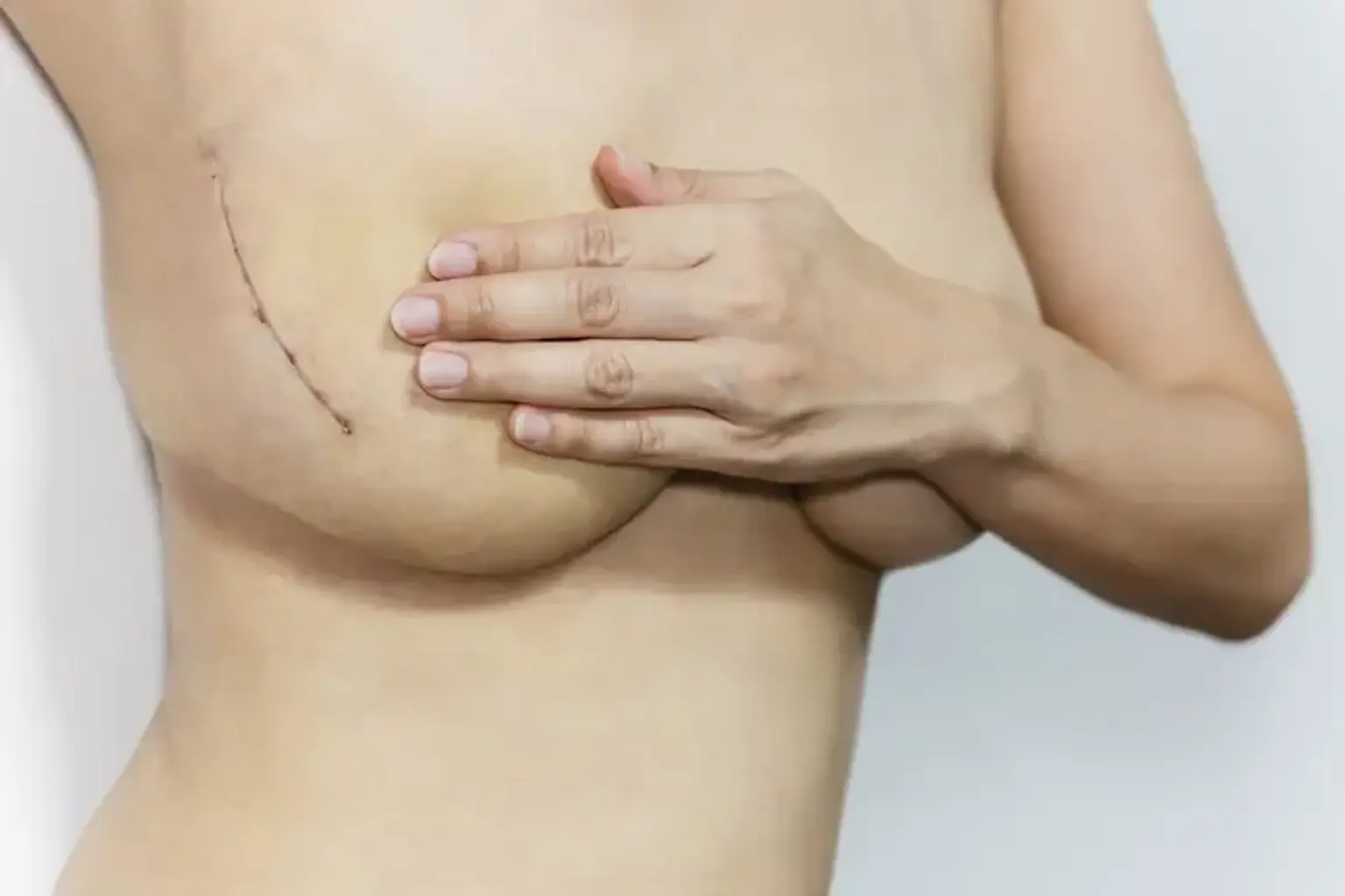
 

Types of Mastectomy 

Mastectomy 

There are several different types of mastectomy. You and your doctors will work together to decide which is best for your individual situation.

    1. Unilateral vs. bilateral mastectomy 

You can have a mastectomy to remove one or both breasts (called unilateral or single mastectomy). If you have cancer in both breasts or are at high risk of developing a second breast cancer due to a strong family history of breast cancer or an inherited genetic mutation linked to breast cancer, your doctors will most likely recommend a bilateral mastectomy (such as a BRCA1, BRCA2, mutation)

Preventive (prophylactic) mastectomies are also done bilaterally in people who are at high risk but have not been diagnosed with breast cancer. Many women who have early-stage cancer in one breast have the option of having a unilateral or bilateral mastectomy. It's not always an easy choice.

Some women choose a bilateral mastectomy even though unilateral mastectomy is an option for them because of:  

  • Fear that a new,
  • Second breast cancer may develop in the unaffected breast, raising concerns about the ongoing need for surveillance (via breast imaging tests such as mammograms) and
  •  The possibility of future biopsies in the unaffected breast, as well as concerns about the cosmetic appearance of the chest if only one breast is removed or reconstructed.

Over the last two decades, an increasing number of women in the United States who have been diagnosed with early-stage breast cancer in one breast have opted for a bilateral mastectomy. Contralateral prophylactic mastectomy refers to the removal of the other breast that does not have cancer. Some doctors are concerned that too many women are undergoing contralateral prophylactic mastectomy because they believe their risk of developing breast cancer in the other breast is higher than it is.

According to research, removing the other healthy breast does not improve survival in women at average risk. Their chances of getting cancer in the other breast are about 1% or less per year. Consult your doctors about what is best for your specific situation. To make an informed treatment decision, you must first understand your actual risk of the breast cancer returning (recurrence) or developing a new cancer.


    2. Simple or Total mastectomy 

For a simple mastectomy (also called a total mastectomy), the surgeon removes:

  • All of the breast tissue
  • The skin of the breast
  • The nipple and the areola (the dark area around the nipple) 

Typically, the surgeon will also perform a sentinel lymph node dissection, which involves the removal of 1-3 lymph nodes under the arm on the side of the tumor to determine whether the cancer has spread there. Sentinel lymph nodes are the first lymph nodes to which cancer can spread.

Lymphatic mapping is performed the day before, the morning of, or during surgery to assist the surgeon in locating the sentinel nodes. In this procedure, a radioactive liquid, a blue dye, or both are injected beneath the nipple or near the tumor site.


    3. Modified radical mastectomy

The surgeon removes all of the breast tissue, skin, nipple, and areola during a modified radical mastectomy. The surgeon will also perform an axillary lymph node dissection, which means that the surgeon will remove several lymph nodes (usually about 10) under the arm on the side of the tumor to see if cancer has spread there.


    4. Nipple-sparing mastectomy 

A nipple-sparing mastectomy removes all of the breast tissue but leaves the nipple, areola, and skin of the breast intact. Lymph nodes are typically removed as well in order to determine whether the cancer has spread beyond the breast (unless the mastectomy is prophylactic). Depending on your specific diagnosis, a sentinel lymph node dissection or an axillary lymph node dissection will be performed. In addition, tissue from beneath the nipple and areola is removed to look for cancer cells. If cancer is discovered in that area, the nipple and, in most cases, the areola must be removed.

This is usually done in a separate procedure later on. Another reason the nipple may require removal in a separate procedure is if it lacks adequate blood supply and develops necrosis (tissue breakdown). During a nipple-sparing mastectomy, the breasts are usually immediately reconstructed with tissue expanders, breast implants, or tissue flaps. If the nipples must be removed, they can be rebuilt later using nipple reconstruction surgery, nipple tattoos, or both.

Because it produces good, natural-looking cosmetic results with minimal scarring, nipple-sparing mastectomy with immediate reconstruction has grown in popularity. However, if you have a nipple-sparing mastectomy, you should not expect much (or any) sensation in your nipples after the surgery. Also, after this surgery, your nipples may appear to be erect all of the time. 

If you have a tumor near or involving the nipple or areola, you may not be a candidate for nipple-sparing mastectomy. Furthermore, the procedure is not advised for people with inflammatory breast cancer. Being a smoker, having scarring around the nipples from previous surgeries, or having had radiation to the breast in the past can reduce your chances of getting a good result from a nipple-sparing mastectomy.


What is Partial Mastectomy? 

Partial Mastectomy

A partial mastectomy is a surgical procedure used to treat breast cancer. Breast-conserving surgery is so named because it removes only the cancerous tissue and a margin of healthy tissue around it. It is a less invasive alternative to a full mastectomy, which involves the removal of the entire breast. In general, breast-conserving surgery is the preferred treatment method for early-stage breast cancer.

In contrast to a Total mastectomy, which removes the entire breast, a partial mastectomy attempts to preserve as much of the breast as possible while not altering its appearance. The amount of breast removed during a partial mastectomy is determined by several factors, including the size and location of the tumor.

When a biopsy reveals that the cancer is small and in its early stages, doctors recommend a partial mastectomy. If the patient only has one area of cancer or multiple areas close enough to be removed without significantly altering the appearance of the breast, surgery may be a good option. In addition, the patient must be willing to undergo radiation therapy after surgery and cannot have certain genetic predispositions or complicating disorders.


Anatomy and Physiology

Anatomy and Physiology of Partial mastectomy

The breast is located on the anterior thoracic wall and is located above the pectoralis major muscle. The mature female breast's superior border approaches the level of the second or third rib and then extends inferiorly to the inframammary crease or fold. The sternal border is the breast's medial boundary. The breast extends laterally to the mid-axillary line. Approximately two-thirds of the breast overlies the pectoralis major muscle posteriorly, and the remaining portion overlies the serratus anterior and upper portion of the oblique abdominal muscles.

The axillary tail of Spence refers to the portion of the upper breast that extends superior-laterally toward the axilla. The breast is divided into four quadrants, allowing for consistency in the documentation of physical examination or breast imaging findings. Upper inner, upper outer, lower inner, and lower outer are the four quadrants. The upper outer quadrant of the breast contains the majority of the breast tissue, including the axillary tail of Spence. As a result, it has the highest incidence of breast cancer.

The breast is made up of mammary tissue and is surrounded by subcutaneous fat and skin, as well as superficial and deep fascial layers. The superficial layer of fascia extends deep into the dermis and covers the anterior breast before extending over the medial and lateral breast. The deep layer of superficial fascia covers the breast's posterior surface and is located anterior to the pectoralis major fascia. 

Suspensory ligaments of Cooper are fibrous bands of connective tissue that run through the breast parenchyma and insert perpendicular to the dermis from the deep layer of superficial fascia. The weakening of these ligaments is what causes breast ptosis.


Indications for Mastectomy

Indications for Mastectomy

Breast cancer is the most common reason for having a mastectomy. Most breast cancer patients require localized surgical treatment (either mastectomy or breast-conserving surgery), which can be combined with neoadjuvant or adjuvant therapy, such as radiation, chemotherapy, or hormone antagonist medications, or a combination of these.

Tumor characteristics such as size and location, as well as patient preference, play an important role in the decision-making process, given that survival rates for patients undergoing mastectomy or lumpectomy with adjuvant radiation therapy are often comparable. 

In brief, breast cancers can have both invasive and non-invasive histologies. Invasive ductal carcinoma is the most common type of breast cancer, accounting for approximately 85% of all invasive breast cancers. In contrast, invasive lobular carcinoma and other uncommon histologies, such as breast sarcoma or lymphoma, are much less common. Breast non-invasive carcinomas include ductal carcinoma in situ and lobular carcinoma in situ. The latter is frequently regarded as a risk factor for future breast cancer and may be best classified as a benign precursor lesion.

Patients with Paget's disease of the breast may also be candidates for mastectomy. Paget's disease is a rare form of breast cancer in which neoplastic cells are found in the nipple-areolar complex epidermis. While the disease may be confined to this area, 80 to 90% of cases will have an associated cancer elsewhere in the involved breast.

The traditional approach to surgical management of Paget's disease has been total mastectomy with axillary sentinel node biopsy. When combined with whole breast radiation therapy, central lumpectomy with complete removal of the nipple-areolar complex has been shown to be effective for local control in patients with no other cancer in the breast.

Due to the volume and distribution of disease, mastectomy may be indicated in patients whose disease is multifocal or multicentric within the breast. Patients with advanced locoregional disease, such as large primary tumors (T2 lesions larger than 5 cm) and skin or chest wall involvement, may also benefit from mastectomy in many cases. Due to tumor burden within the dermal lymphatic channels and more diffuse involvement of the underlying breast parenchyma, patients with inflammatory breast cancer are treated with mastectomy in addition to systemic chemotherapy and radiation treatment.

Patients who have had breast-conserving surgery (lumpectomy or partial mastectomy) and have margin involvement with tumor cells may be candidates for mastectomy if margin re-excision fails or is not technically or cosmetically feasible. Clear or negative margins after resection of a primary tumor are an important factor in lowering the risk of recurrence. Mastectomy is also recommended for patients with recurrent breast cancer who have previously undergone lumpectomy and radiation treatment.


Contraindications to Breast Conservation

Contraindications to Breast Conservation

If medically indicated, mastectomy can usually be performed safely and easily. There are a few critical factors to consider as surgical contraindications. These are frequently divided into two categories: systemic and locoregional. In patients with proven distant metastatic disease, mastectomy may be contraindicated. Furthermore, due to the burden of their overall health and poor performance status, frail or elderly patients with significant medical co-morbidities or systemic organ dysfunction may not be candidates for surgery.

Patients who are at a high risk of death from surgery or anesthesia are not candidates for surgery. Mastectomy may be relatively contraindicated in patients with advanced locoregional disease at the time of diagnosis if there is skin or chest wall involvement and concerns about the ability to close the surgical wound or obtain a negative surgical margin. In these cases, neoadjuvant chemotherapy, radiation, or endocrine therapy may be beneficial in reducing the volume or extent of local disease and allowing for surgery.


Mastectomy: How to Prepare?

Mastectomy preparation

Mastectomy is typically an elective procedure, and patients are expected to report to the hospital or surgical center on the day of their procedure. Patients undergoing mastectomy, with or without axillary surgery or reconstruction, should receive pre-operative antibiotics to reduce the risk of surgical site infection. Unless the patient is allergic or has a history of methicillin-resistant Staphylococcus aureus infection, a first-generation cephalosporin is the antibiotic of choice for prophylaxis.

After anesthesia is administered, the patient is placed in a supine position in the operating room, and the breast, chest wall, axilla, and upper arm are exposed. Many surgeons will include the contralateral breast in the surgical field. The surgical field is sterilely prepared with an agent that reduces the presence of skin flora and the risk of surgical site infection. For surgical antisepsis, alcohol-based skin preps such as chlorhexidine gluconate are commonly used.

Surgeons should prepare patients for their procedure and discuss the expected postoperative course and care with them perioperatively. Many surgeons choose to insert a drain during mastectomy to evacuate any fluids that may accumulate in the wound bed and to promote flap adherence to the chest wall. Patients benefit from education on drain care and keeping an accurate output log. Patients should also be counseled on postoperative restrictions such as lifting, driving, and any other limitations in the immediate recovery period.


How is the Procedure Performed?

Partial mastectomy procedure

The procedure entails locating the abnormality in the breast, removing it with a band of surrounding tissue, and sending the removed area to a pathologist for analysis. If the tumor is too small to be seen or felt on a patient's exam, the surgeon can use a pre-operative wire to guide their partial mastectomy.

In 1972, John Madden described the current standard for a mastectomy. This method entails making an elliptical incision around the breast, including the nipple areolar complex, and keeping the tumor site as a central landmark. The mammary tissue is separated from the skin flaps and resected alongside the pectoralis major fascia, while both pectoral muscles are preserved. As a result, less disruption of surrounding neurovascular and lymphatic structures is required.

Some patients who choose to have breast reconstruction may be candidates for skin-sparing and nipple-sparing mastectomy as reconstructive techniques have evolved and become more popular. Whatever approach is used, oncologic principles take precedence over cosmetic concerns. Skin-sparing mastectomy, as the name implies, is intended to preserve a healthy skin envelope for immediate breast reconstruction, assuming adequate margins can be achieved.

Nipple-sparing mastectomy removes the mammary tissue and pectoral fascia while leaving the nipple-areolar complex and the entire breast skin envelope intact. Only patients undergoing immediate breast reconstruction are given this surgical approach. It is critical to maintain blood supply to the nipple-areolar complex with this approach to avoid flap ischemia or breakdown.

All of the described mastectomy techniques create uniform flaps by dissecting just above the superficial layer of the breast's superficial fascia. The ideal flap thickness has been much debated, with the ultimate goal of removing all possible breast tissue while preserving skin viability. As previously stated, regardless of the type or location of skin incision used, mastectomy dissection should continue to the anatomic boundaries of the breast.

While the surgeon retracts the breast tissue away from the overlying tissue, the flaps are elevated and retracted at a right angle to the chest wall. The skin flap is elevated superiorly to the clavicle, laterally to the anterior border of the latissimus dorsi, medially to the sternal border, and inferiorly to just below the inframammary crease.

Once the overlying flaps have been dissected, the breast is removed from the superomedial to the inferolateral borders. In most cases, a closed suction drain is placed beneath the skin flaps in the wound bed, and the incision is closed in two layers with absorbable suture.

After the surgery is finished, a pathologist will examine the tissue that was removed under a microscope. If the pathologist finds no cancer cells at any of the removed tissue's edges, the margins are said to be negative or clear. Breast cancer cells can sometimes spread beyond what preoperative imaging studies can detect. Positive margins are defined as the presence of cancer cells at the edges of the removed tissue.

Positive margins indicate that some cancer cells may still be present in the breast after surgery, so the surgeon may need to return to the operating room and remove additional tissue. This procedure is known as a re-excision. If cancer cells are discovered at the edges of the removed tissue after the second surgery, a full mastectomy may be required.


Possible problems after mastectomy

Partial mastectomy complications

In most cases, patients tolerate mastectomy well, with low morbidity and mortality. However, several complications are possible. Seroma or hematoma formation wound infection, skin flap breakdown or necrosis, and lymphedema are examples of these. A seroma is a collection of fluid in a surgically created cavity caused by vessel and lymphatic transection. To reduce the rate of seroma formation, most surgeons used closed suction drains beneath the skin flaps. Approximately 8% of patients undergoing breast surgery develop surgical site infections.

Staphylococcus aureus and Streptococcus epidermis are the most common organisms involved, and infections should be treated with an appropriate antibiotic, with or without wound opening. Similarly, flap necrosis affects approximately 8% of patients and is associated with insufficient blood supply to the flap, wound closure under tension, obesity, and type of incision (vertical versus transverse). Necrosis is treated with debridement and, if necessary, skin graft coverage.

Lymphedema has become less common since the introduction of modified mastectomy techniques. With a reported incidence of more than 20%, axillary lymph node dissection is the most significant risk factor for the development of lymphedema. Lymphedema affects 3.5 to 11% of patients who have sentinel lymph node biopsy. Early intervention with physical therapy and decompressive massage techniques in patients who develop lymphedema can help prevent progression and, in some cases, reduce lymphedema.


Conclusion 

Breast removal

Partial mastectomy is a less invasive surgery than total breast removal, and it has been shown to be just as effective in removing cancer and preventing recurrence in early-stage breast cancer as mastectomy. The procedure entails locating the abnormality in the breast, removing it with a band of surrounding tissue, and sending the removed area to a pathologist for analysis.

As with any surgery, there are risks of side effects such as bleeding, infection, temporary swelling, tenderness, pain, the formation of hard scar tissue, and possible breast distortion if a large portion is removed.