Pectus Excavatum Surgery
Overview
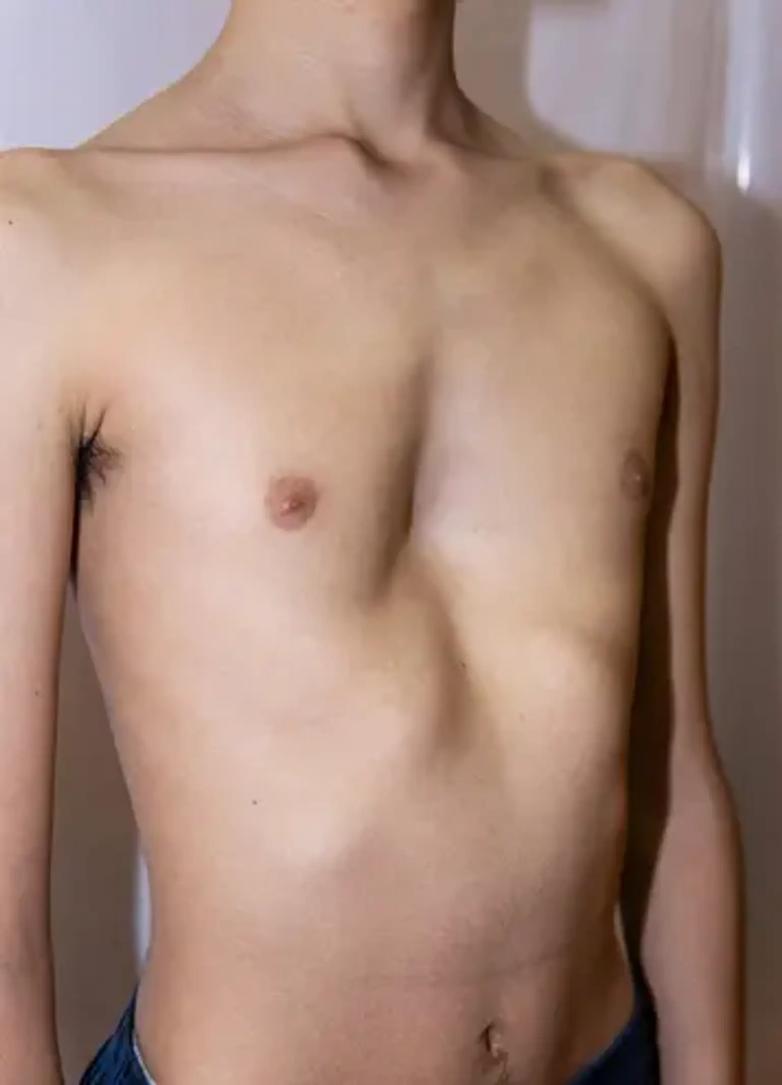
Pectus abnormalities are responsible for around 95 percent of congenital chest wall defects, with pectus excavatum being the most frequent. Pectus excavatum is distinguished by an anterior chest wall depression that results in a "funnel chest."
While the abnormality affects the third to seventh costocartilages or ribs, the xiphisternum is the most severely affected location. Although the malformation can be symmetrical, it is more often asymmetrical and can affect other parts of the thorax. A pectus deformity might be detected in a newborn or develop later in infancy.
What is Pectus Excavatum?
Pectus excavatum is a congenital chest wall deformity caused by improper development of the cartilage connecting the ribs to the breastbone (sternum). This results in a sternum depression and a "sunken in" or "funnel chest" look. Boys are more affected than females by this condition.
What is the prevalence of Pectus Excavatum?
The reported frequency ranges from 1/300 to 1/1000 live births, with a male to female ratio of 5:1. Pectus excavatum accounts for 90% of all chest wall abnormalities. The majority of birth abnormalities are discovered within the first year of life, with severe malformations visible at birth. During the pubertal development surge, the funnel-shaped chest becomes more prominent. Pectus excavatum can appear as a single defect or as part of a group of congenital illnesses. Connective tissue abnormalities are seldom (less than 1%) related with pectus excavatum among various clinical conditions.
Pectus Excavatum Causes
There are several possibilities about the underlying genesis of pectus excavatum. The funnel-shaped chest has been linked to sternum weakness and aberrant flexibility, rib overgrowth, and developmental failure of the bony thorax. Regardless of the method, the consequence is different degrees of depression and dorsal deviation of the sternum and neighboring ribs or costal cartilages. Although no specific genetic abnormality has been found, the existence of a favorable family history supports a genetic susceptibility in more than 40% of instances.
Symptoms of Pectus Excavatum
The typical presenting patient is a skinny, tall guy who looks to be slouching, and thoracic scoliosis may be seen. Sternal depression and rotation can depress the heart, resulting in a variety of cardiovascular symptoms, including exercise intolerance. An audible murmur can be caused by a faulty mitral valve (i.e., prolapse, regurgitation). Though the actual reason is unknown, exercise intolerance is one of the most prevalent presenting ailments. Furthermore, the psychological impact on patients, particularly throughout adolescence, can be severe.
How Pectus Excavatum is Diagnosed?
The sternal defect is plainly visible in the lateral image of the chest radiograph. Further imaging investigations may detect misplaced vertebral bodies as well as varying degrees of scoliosis. Patients with moderate pectus excavatum may have few or no symptoms; nonetheless, every one to two years, a cardiopulmonary examination is recommended to establish a baseline and monitor for progression.
To assess for secondary or related abnormalities of a clinical condition, a complete evaluation comprises a chest radiograph, pulmonary function tests (spirometry, plethysmography, and respiratory muscle strength assessment), EKG, and echocardiography. In elderly individuals, pulmonary function tests may indicate obstructive or restrictive lung disease, as well as air trapping with an increase in residual volume (RV). A mechanical disadvantage limiting respiratory muscle function might be causing the air trapping. Axis deviation on an electrocardiogram (EKG) indicates a leftward heart deviation.
Arrhythmias such as first-degree heart block, right bundle branch block, and Wolff-Parkinson-White occur in 16% of this patient population. Cardiopulmonary exercise testing may show cardiopulmonary limitations that are not apparent at rest. Echocardiography is recommended to check for cardiac compression, valvular abnormalities, and myocardial function. A patient with pectus excavatum may have left heart deviation and conduction abnormalities.
The Haller index (HI) is the gold standard for determining the severity of a pectus excavatum deformity. It is the ratio of the transverse diameter to the anteroposterior diameter. The readings are from a computed tomography (CT) scan; the typical value is 2.5 or less. Measurements greater than 3.2 are deemed serious. A restrictive pattern on pulmonary function tests is four times more prevalent in patients with a Haller Index greater than 7.
MRI with breath-holding MRI has been utilized to determine the morphology of thoracic abnormalities prior to surgery for pectus excavatum. Recently introduced techniques such as oculo-electronic plethysmography (OEP) can show that the depressed portion of the sternum and adjacent chest wall does not move with respiration and there is a reduction in lung volume, providing a better understanding of the functioning of the lungs of patients with pectus excavatum deformity.
Management of pectus excavatum
Surgery can be used to treat pectus excavatum, although it is normally reserved for those who have moderate to severe indications and symptoms. Physical therapy may benefit those who have modest indications and symptoms. Certain exercises can help to improve posture and enhance chest expansion.
Tests Needed Before Surgery
Your kid must have the following outpatient tests before being admitted to the hospital for surgery:
- An echocardiogram (or Echo) of the heart is performed to search for valve issues related to pectus excavatum.
- A CT scan of the chest to determine the effectiveness of the repair procedure.
- Pulmonary function study, is a breathing test that may be required to determine how much air your child can get into and out of his lungs.
Home Preparation before surgery
When general anesthesia is required, there are strict food and drinking guidelines that must be observed in the hours preceding the operation. Based on your child's age, the nurse will offer you particular feeding and drinking guidelines. The following are the standard eating and drinking instructions. Regardless of your child's age, you must follow the particular directions provided to you over the phone by the nurse.
For children older than 12 months:
Do not provide any solid meals or non-clear drinks after midnight the night before the procedure. This includes milk, formula, pulp juices, coffee, and chewing gum or candies.
For infants under 12 months:
- Formula-fed newborns may be given formula up to 6 hours before their planned arrival time.
- Breastfed newborns may breastfeed up to 4 hours before their planned arrival time.
For all children:
- Give only clear beverages up to 2 hours before the anticipated arrival time. Clear drinks include water, Pedialyte®, Kool-Aid®, and see-through juices like apple or white grape juice.
- Give nothing to eat or drink for 2 hours before the anticipated arrival time.
How is Pectus Excavatum surgically repaired?
The extensive excision and chest wall rebuilding conducted for asphyxiating thoracic dystrophy (Jeune's syndrome ) served as the foundation for early surgical repair of the pectus excavatum. The current approach is to postpone the repair until pubertal development has occurred, and the procedure has been refined to allow for limited cartilage resection. The most prevalent reason for surgical correction is impaired cardiopulmonary function, not cosmesis. It is critical that surgical correction takes place following the child's pubertal growth spurt.
Ravitch conducted the first reported surgical correction of pectus excavatum in 1949. Ravitch conducted a subperichondrial excision of all malformed costal cartilages and the xiphoid process, combined with a transverse sternotomy, based on the aggressive chest wall resection used to treat asphyxiating thoracic dystrophy (Jeune's syndrome).
Six years later, Rehbein and Wernicke changed the surgery, implanting a variety of metallic bars longitudinally and/or transversely to support the sternum. This repair can be addressed either with a median longitudinal incision along the sternum or, in female patients, using a submammary skin incision. Patients return one year later to have the implanted bars removed. The Ravitch treatment has been modernized by the use of orthopedic metal plates and screws. These devices may be customized to the patient's defect and do not require a second surgical operation for hardware removal.
Nuss and Kelly's minimally invasive procedure entails inserting a metal bar beneath the sternum at the most depressed zone and most everted costal line on both sides of the chest. The retrosternal bar is inserted thoracoscopically through 3cm incisions along the mid-axillary lines. Skin flaps are raised and tennelled up to the most-everted intercostal gaps previously found. Under thoracoscopic guidance, a metal introducer is used to slice a retrosternal plane between the anterior pericardium and the posterior sternal table.
The guide is exteriorized through the incision on the left side in order to be linked to a titanium rod and then removed. The retrosternal curve var is turned to drive the concave side rearward, pressing the sternum ventrally. To keep the bar from moving, metallic stabilizers and/or subperiostal cable wires are utilized. Although the initial stabilization time was two years, it is currently more typical to leave the ban in place for three years.
After mobilizing the pectoralis muscles, the Leonard modification to Ravitch's operation involves excision of the lower costal cartilage while leaving the perichondrium intact. Robicsek discusses posterior sternum stabilization following transverse osteotomy with Marlex mesh fixation to costal cartilage remains. Erlangen's modification of the Nuss method is defined by minimal cartilage resection, with transternal implantation of an elastic metal bar through stitch incisions.
Intraoperative tensiometry is used to reduce cartilage excision. The magnetic mini-procedure pulls the sternum anteriorly by using magnetic forces between a sternal magnet and another on a recommended brace worn by the patient. Lacquet's modification is the only one that avoids the use of prosthetic material by repairing the deformity with a sternochondroplasty. For asymmetric flaws, this technique appears to outperform the Nuss approach.
Negative pressure applied to the thorax provides a non-surgical option for pectus excavatum. A vacuum bell is placed on the chest wall defect, and the patient applies negative pressure using a handpump. While long-term results are unclear, this may be a feasible alternative in the future for the management of less severe abnormalities. Furthermore, this treatment might be appropriate for younger symptomatic kids for whom prepubertal surgical correction is not required.
Hospital Stay After Surgery
The Nuss technique is uncomfortable in the days following the surgery. While in the hospital, your kid will be given an epidural catheter. During surgery, an epidural catheter will be implanted to provide for a continuous infusion of pain medicine for a few days after surgery. Your youngster will be given a modest pain reliever by oral for roughly 2 weeks following the procedure.
Following an open operation, your kid will be given a powerful pain medicine that your surgeon has recommended. The drug must be taken for roughly 5 to 7 days following surgery. Your child may also require an epidural catheter for a few days following surgery.
Small tubes will be put into the incision to drain fluid if your kid has the open operation. To remove air from the chest cavity following the open surgery, a chest tube is usually always required. It is also used to drain blood from the chest cavity during an open operation. The chest tube is used until no more air or blood can be expelled, which normally takes 2 to 4 days.
If your kid underwent the Nuss operation, these tubes are unlikely to be required, although they may be utilized in specific situations.
- If an epidural catheter is used, your child's activities will be slightly restricted following either treatment. The length of time your child will have an epidural catheter will be determined by his or her surgeon. It is normally removed four days after surgery.
- If an epidural catheter is not utilized during an open operation, exercise will be encouraged as soon as feasible. Deep breathing, walking, and sitting up in a chair will all be encouraged for your youngster.
- Your child's food will be resumed when the surgical team believes it is safe. Typically, liquids are given the evening before or the day following surgery, followed by a regular diet during the next several days.
Recovery at Home
Before you leave the hospital, you will be given a prescription for your child's pain medicine as well as instructions on how to use it. Most routine activities can be resumed for your youngster.
- In 5 to 7 days, your youngster will be able to bathe on a daily basis.
- When your kid is no longer using pain medication, he or she may return to school.
- For a few months, he or she will be excused from gym class.
- You should discuss with the surgeon how long your youngster should avoid contact sports.
- For around 6 months, your kid should avoid twisting at the waist or any action that might cause the bar in the chest to shift.
- Your child should not wear a backpack until after the initial consultation with your surgeon. Please leave a set of your child's school books at home if feasible, or purchase a wheeled bag.
- If an MRI of the chest or abdomen is ordered while either type of bar is in the chest, you should notify your child's doctor.
What are the Complications of pectus excavatum?
Minor pectus excavatum deformities are typically asymptomatic and so do not cause issues. More severe abnormalities, on the other hand, might have a deleterious influence on cardiopulmonary function, resulting in a number of symptoms:
- Chest pain
- Fatigue
- Exertional dyspnea
- Recurrent respiratory tract infections
- Asthma
- Palpitations
- Heart murmur (mitral regurgitation)
- Arrhythmia
- Syncope
Complications are more commonly appreciated in patients who undergo surgical correction of the defect:
- Migration of the sternal bar
- Infection
- Pneumothorax
- Bleeding
- Cardiac laceration
- Chronic pain
- Recurrence
According to a recent meta-analysis, the overall morbidity and length of stay for Ravitch and Nuss operations are comparable. However, the Nuss surgery has a greater rate of postoperative hemothorax, pneumothorax, and return to OR.
What is the outcome for a child with pectus excavatum?
Historically, pectus excavatum was mistakenly thought to be just a cosmetic problem. Recent research, however, has shown that children with major abnormalities also have cardiac and respiratory problems. The sunken chest limits the capacity of the chest and prevents the lung from fully extending. Lung capacity may be diminished, making it harder for youngsters to tolerate exercise or severe activities.
The sunken chest can also constrain the heart, resulting in decreased blood flow and cardiac function. These effects on the heart and lungs may be monitored and reversed by raising the chest. It is crucial to highlight that these effects on heart and lung function are not life-threatening, and that people with pectus excavatum can live a full and normal life.
As a result of the condition, some individuals experience psychological and emotional distress. Although many patients live happily and easily with their abnormality, many others battle with poor body image, low self-esteem, and social discomfort. This is especially true for teens since the pectus deformity frequently develops throughout the adolescent years when the youngster is looking for peer approval.
Conclusion
Pectus excavatum is a congenital chest wall malformation that causes numerous ribs and the breastbone to expand inward. Children with minor pectus excavatum who are unconcerned about their looks and do not have respiratory issues usually do not require therapy. If there are additional health issues, such as difficulty breathing, surgery is usually recommended. Surgery to enhance the look of the chest is also an option.