Pediatric Blood Disorders

Last updated date: 03-Mar-2023

Originally Written in English

Pediatric Blood Disorders

Overview

Pediatric blood disorders are a type of noncancerous illness that mostly affects newborns, children, and teenagers. Such disorders include bone marrow failure, anemia, and hemophilia. The severity of the problems varies and might have a severe impact on functioning.
 

What are Pediatric Blood Disorders?

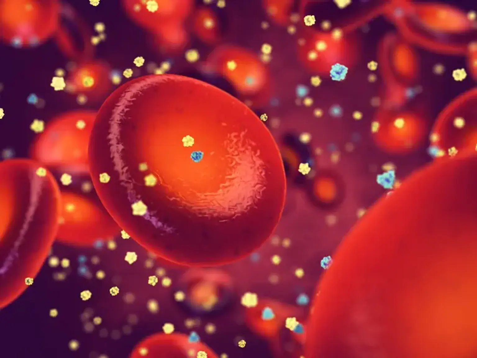
Blood disorders and illnesses impair the capacity of the blood to operate correctly. Blood diseases might entail abnormalities with red blood cells, white blood cells, platelets, blood arteries, bone marrow, lymph nodes, or proteins involved in bleeding and clotting.

Noncancerous blood disorders can be related to issues with:

  • Red blood cells, which carry oxygen throughout the blood
  • White blood cells, which defend the body against disease or infection
  • Platelets, which causes blood to clot after injury
  • Bone marrow, the part of a bone where blood cells and platelets are produced
  • Lymph nodes, which are immune system glands that act as filters for the body
  • Proteins responsible for bleeding and clotting


Blood cell disorders Symptoms 

Blood cell disorders symptoms

The symptoms of blood cell diseases differ according to the type of condition your kid has.

The symptoms of the different kinds of anemia can include:

Some of the more serious symptoms of a blood disorder include:

  • Damage to any organ, including eyes, kidneys, liver, and/or gall bladder
  • Sudden loss of vision
  • Stroke
  • Frequent, serious infections

Anemia in your child may be an indication of another disease. Your child's doctor can do blood tests to determine what is causing the anemia.


Sickle cell disease

Sickle cell disease

Sickle cell disease (SCD) is a hereditary category of red blood cell diseases. SCD is discovered in children born in the United States via a newborn screening program. SCD treatment is complicated and needs the expertise of a pediatric hematologist.


Medicines and vaccines

The spleen does not function properly in children with SCD, making it easier for them to contract serious infections. As a result, children are given daily penicillin to reduce the risk of infection from the pneumococcus bacteria.

Most children with SCD should take hydroxyurea, a daily medication that increases hemoglobin F levels, which has been demonstrated to decrease or avoid some SCD consequences.

Children with SCD should receive all recommended childhood vaccines including:

  • Pneumococcus
  • Meningococcus
  • Influenza


Screenings and tests

Children with SCD require periodic blood tests to establish a "baseline" for problems including anemia.

Transcranial Doppler (TCD) ultrasonography screening should be done yearly on children aged 2 to 16 to determine if they are at a higher risk of stroke. If the test results are abnormal, frequent blood transfusions can reduce the risk of suffering a stroke.


Transfusions

To treat and avoid specific SCD problems, some children will benefit from a red blood cell transfusion program. Transfused red blood cells have normal hemoglobin and can help reduce the likelihood of blood vessel blockage and increase oxygen delivery to tissues and organs.

We also provide erythrocytapheresis, a type of continuous transfusion treatment in which patients have their blood filtered to replace sickle red blood cells with normal hemoglobin.


The cure for children with sickle cell disease

At the moment, the only treatment for SCD is hematopoietic stem cell transplantation (HSCT). The following is how it works:

  • Donor stem cells are extracted from the bone marrow or blood of someone who does not have SCD but is genetically related to the kid.
  • After the donor's stem cells are extracted, the kid with SCD is given medicines that kill or diminish his or her own bone marrow stem cells.
  • The donor stem cells are then injected into the infant and settle into the bone marrow, where they begin to replace the recipient's cells.
  • The new stem cells will produce red blood cells with normal hemoglobin that will no longer sickle.


Childhood anemia

Childhood anemia

A hemoglobin or hematocrit level lower below the age-adjusted reference range for healthy children is referred to as pediatric anemia. Anemia is a physiological condition in which low hematocrit or hemoglobin levels result in decreased oxygen-carrying capacity, which does not satisfy the body's metabolic demands efficiently.

Anemia is a disorder caused by a variety of underlying pathologic processes rather than a unique disease entity. It can be either acute or chronic. This page presents an overview of anemia in general, with a focus on the acute type. Furthermore, cases in which anemia is the only hematologic abnormality are highlighted.

The primary physiologic function of red blood cells (RBCs) is to transport oxygen to tissues. To compensate for the shortage of oxygen supply, an individual with anemia may undergo several physiologic changes. These are some examples:

  1. Increased cardiac output; 
  2. Shunting of blood to vital organs; 
  3. Increased 2,3-diphosphoglycerate (DPG) in the RBCs, which causes reduced oxygen affinity, shifting the oxygen dissociation curve to the right and thereby enhancing oxygen release to the tissues; and 
  4. Increased erythropoietin to stimulate RBC production.

The clinical consequences of anemia are determined by its length and severity. Because the body does not have enough time to make the required physiologic adaptations when anemia is acute, the symptoms are more likely to be strong and dramatic. When anemia develops gradually, the body is able to respond, employing all four processes outlined above, alleviating symptoms according to the degree of anemia.


Complications in pediatric acute anemia

Acute and severe anemia can impair the cardiovascular system. Furthermore, if those suffering from acute anemia are not treated promptly and effectively, the resultant hypoxemia and hypovolemia can cause brain injury, multiorgan failure, and death. Long-term anemia can cause children to fail to flourish.

Many studies have demonstrated that iron deficiency anemia or iron deficiency without anemia has a negative impact on children's neurocognitive and behavioral development. Congestive heart failure, hypoxia, hypovolemia, shock, seizure, and an acute silent cerebral ischemia event are all possible consequences.


Workup in pediatric acute anemia

pediatric acute anemia

To assess anemia, acquire preliminary laboratory tests such as a complete blood count (CBC), reticulocyte count, and a peripheral smear review. Chest radiography is used to screen out mediastinal mass in individuals who may have congestive heart failure (CHF).


Management of pediatric acute anemia

Management of pediatric acute anemia

The most common therapy for severe acute anemia is transfusion with packed red blood cells (PRBCs). The decision to transfuse should not be based exclusively on hemoglobin or hematocrit levels; rather, the clinical impact or signs and symptoms of the individual with anemia must be considered.


Thalassemia

Thalassemia

Thalassemias are a diverse set of hereditary illnesses caused by a reduction in the production of hemoglobin's alpha or beta chains (Hb). Hemoglobin is the component of red blood cells that transports oxygen. It is made up of two proteins, an alpha and a beta. If the body does not produce enough of one or both of these proteins, red blood cells may not form correctly and cannot carry enough oxygen, resulting in anemia that begins in childhood and lasts throughout life. Thalassemia is a hereditary condition, which means that at least one of the parents must be a carrier. It is caused by a genetic mutation or the loss of critical gene sequences.

  • Alpha thalassemia is caused by the loss of the alpha-globin gene, which leads in diminished or nonexistent synthesis of alpha-globin chains. The alpha globin gene has four alleles, and illness severity varies based on the number of allele deletions. The most severe type is four allele deletion, in which no alpha globins are formed and the extra gamma chains (existing throughout the prenatal stage) form tetramers. It is harmful to life and causes hydrops fetalis. The mildest variant is one allele loss, which is typically clinically asymptomatic.


  • Beta thalassemia is caused by point mutations in the beta-globin gene. It is classified into three groups according on the zygosity of the beta-gene mutation. A heterozygous mutation (beta-plus thalassemia) causes beta-thalassemia minor, in which beta chains are underproduced. It is often asymptomatic and mild. A homozygous mutation (beta-zero thalassemia) of the beta-globin gene results in the complete lack of beta chains in beta thalassemia major. It causes jaundice, growth retardation, hepatosplenomegaly, endocrine problems, and severe anemia that need life-long blood transfusions. 


Because thalassemia causes your body to produce fewer red blood cells, you may have symptoms of a low blood count, often known as anemia. You may feel fatigued or weak if you have anemia. You may also encounter:

  • Dizziness
  • Shortness of breath
  • A fast heart beat
  • Headache
  • Leg cramps
  • Difficulty concentrating
  • Pale skin

Your body will work hard to produce more red blood cells. The bone marrow, the black spongy component in the center of bones, is the primary site of blood cell production. Because your bone marrow is working harder than usual, it may expand. This causes your bones to expand, causing them to become thinner and more easily shattered.

The spleen is another organ where blood is produced. It is located on the left side of your abdomen, slightly behind the lower ribs. The spleen performs several functions. Two of the most important are blood filtration and monitoring for infections. When it discovers these infections, it may begin combating them.

When you have thalassemia, your spleen might balloon as it attempts to produce blood cells. Because it is working so hard on its duty, it cannot filter blood or monitor for and combat infections as well. As a result, persons with thalassemia are considered "immunocompromised," which implies that part of the body's defenses against infection are ineffective. When your immune system is impaired, you are more susceptible to illnesses, and you may require additional protection, such as flu shots and other immunizations.


How is Thalassemia treated?

The sort of treatment a person receives is determined on the severity of their thalassemia. The more severe the thalassemia, the less hemoglobin the body contains, and the greater the risk of anemia.

One method of treating anemia is to provide the body extra red blood cells to transport oxygen. This can be accomplished with a blood transfusion, which is a safe and popular surgery in which blood is administered through a small plastic tube put into one of your blood arteries. Because their bodies produce so little hemoglobin, some persons with thalassemia, mainly thalassemia major, require regular blood transfusions. People with thalassemia intermedia (not as severe as major, but not as mild as trait) may require blood transfusions from time to time, such as when they are sick or have an infection. People with thalassemia minor or trait do not frequently require blood transfusions since they either do not have anemia or have a moderate anemia.

To treat anemia, persons with thalassemia are frequently offered folic acid, an additional B vitamin. Folic acid can aid in the development of red blood cells. Folic acid treatment is typically used in conjunction with other medications.


G6PD deficiency

Glucose-6-phosphate dehydrogenase (G6PD) deficiency is a hereditary metabolic disorder caused by a G6PD enzyme deficiency. This enzyme is essential for red blood cell activity; if the amount of this enzyme is too low, red blood cells may break down early (hemolysis). Anemia occurs when the body is unable to compensate for rapid loss. However, a lack of this enzyme is insufficient to produce hemolysis on its own; other variables must be present to "trigger" the beginning of symptoms.

Certain viral infections, certain medicines, and eating fava beans can all trigger hemolysis in G6PD-deficient people, resulting in a potentially fatal acute hemolytic anemia known as favism. Fatigue, pale complexion, jaundice or yellow skin tone, shortness of breath, fast heartbeat, black urine, and an enlarged spleen are some of the symptoms.

Most importantly, in the absence of triggering conditions, the majority of persons with G6PD deficiency are normal, and they go about their lives with no knowledge of the illness or any obvious symptoms. G6PD insufficiency is caused by changes (mutations) in the G6PD gene, which is located on the X chromosome.


G6PD Diagnosis

G6PD Diagnosis

A diagnosis is made based on the recognition of specific physical features and symptoms, a complete clinical assessment, a full patient history, and/or specialized testing. If a person has a symptom, such as blood in the urine, and spontaneously remembers eating fava beans, and comes from a region or community where G6PD deficiency is frequent, the illness should be suspected.

If a doctor suspects a patient is G6PD-deficient, he or she will do a battery of blood tests to confirm the diagnosis and rule out other illnesses that produce similar symptoms. The diagnosis is based on establishing reduced G6PD enzyme activity using a quantitative assay or a screening test such as a fluorescent spot test.

Molecular genetic testing can discover mutations in the gene known to cause G6PD, but it is only offered as a diagnostic service in specialist laboratories.


G6PD Treatment

The majority of those impacted do not require therapy. G6PD insufficiency is frequently best handled by prophylactic interventions. Before receiving certain treatments, such as antibiotics, antimalarials, and other medications known to cause hemolysis in G6PD-deficient persons, individuals should be tested for the G6PD deficiency. Hemolytic anemia from fava beans or recognized medicines should not arise in G6PD-deficient patients since exposure may be avoided.

If an episode of hemolytic anemia is caused by the use of a certain medicine, the causative drug should be stopped under the guidance of a physician. If such an incident is caused by an underlying infection, the infection should be treated appropriately.

Some adults may require short-term fluid therapy to prevent hemodynamic shock (inadequate blood flow to the organs) or, in extreme situations when the rate of hemolysis is very high, blood transfusions. Blood transfusions are more likely to be required in children than in adults, and they can be life-saving in children with favism.

Neonatal jaundice is treated by exposing the newborn to specific lights that help to reduce the condition. In more serious situations, an exchange transfusion may be required. The blood of an afflicted newborn is removed and replaced with fresh donor blood or plasma.


Hemophilia

Hemophilia

Hemophilia is a genetic bleeding condition in which the blood fails to clot normally. This can result in both spontaneous and post-injury or surgical bleeding. Blood includes several proteins known as clotting factors that can aid in the prevention of bleeding. Hemophilia patients have low levels of either factor VIII (8) or factor IX (9). The amount of factor in a person's blood determines the severity of their hemophilia. The less of the component there is, the more probable bleeding will occur, which can lead to major health concerns.

Hemophilia can occur later in life in rare situations. The majority of instances involve persons in their forties or fifties, or young women who have recently given birth or are in the last stages of pregnancy. With the right therapy, this illness may typically be resolved.


Hemophilia Causes

A mutation or alteration in one of the genes that provides instructions for generating the clotting factor proteins required to create a blood clot causes hemophilia. This alteration or mutation can cause the clotting protein to malfunction or be absent entirely. These genes are found on chromosome X. Males have one X chromosome and one Y chromosome (XY), whereas females have two X chromosomes (XX). The X chromosome is passed down from mothers to sons, and the Y chromosome is passed down from dads to sons. Females receive one X chromosome from each of their parents.


Hemophilia Types

Hemophilia is classified into numerous categories. The two most prevalent are as follows:

  • Hemophilia A (Classic Hemophilia)
    This type is caused by a lack or decrease of clotting factor VIII.
  • Hemophilia B (Christmas Disease)
    This type is caused by a lack or decrease of clotting factor IX.


Hemophilia Diagnosis

Many people who have or have had hemophilia in their family would request that their baby boys be tested immediately after delivery. About one-third of newborns with hemophilia have a novel mutation that is not found in other family members. If a newborn exhibits specific hemophilia symptom, a doctor may perform a hemophilia test.

Doctors would run blood tests to see if the blood was clotting properly in order to make a diagnosis. If it does not, they will do clotting factor testing, commonly known as factor assays, to determine the etiology of the bleeding issue. These blood tests would reveal the type and degree of hemophilia.


Hemophilia Treatment

Hemophilia Treatment

The most effective treatment for hemophilia is to restore the missing blood clotting factor, allowing the blood to clot normally. This is accomplished by infusing (administering) commercially produced factor concentrations through a vein. People with hemophilia can learn how to make these infusions on their own, allowing them to stop bleeding episodes and, if done on a regular basis (called prophylaxis), even avoid most bleeding episodes.


Fanconi anemia

Fanconi anemia (FA) is a rare genetic condition that affects all three blood cell lines. It is the most prevalent cause of hereditary bone marrow failure with pancytopenia. Furthermore, it affects nearly all of the body's organs. Fanconi anemia is primarily caused by a deficient homologous recombination DNA repair pathway, as well as abnormalities in proteins and other enzymes involved in the repair of damaged DNA after exposure to alkylating chemicals, irradiation, and cytotoxic drugs.

It's also known as the hereditary type of aplastic anemia. The discovery of Fanconi anemia had far-reaching consequences beyond the disease itself. An comprehensive research of various bone marrow failure syndromes and chromosomal fragility illnesses has improved scientific understanding of Fanconi anemia bone marrow failure. It is frequently accompanied with other congenital abnormalities and is predisposed to hematological and solid malignancies. It is more frequent in children, with an average age of diagnosis of 7 years. The progress of molecular genetic research has aided in the thorough investigation of Fanconi anemia.


Diamond-Blackfan anemia

Diamond Blackfan anemia (DBA) is a form of anemia that occurs at birth and is characterized by pure red cell aplasia as well as congenital bone abnormalities. It is a kind of macrocytic-normocytic anemia that is persistent. DBA is a hereditary disorder that is inherited as an autosomal dominant trait in 40 to 45 % of cases. The remaining, around 55 to 60% of cases, often appear sporadically. Less often, examples of autosomal recessive inheritance have been reported. Hydrops can occur on rare occasions.


Prevention

blood diseases

The prevention measures for blood diseases vary depending on the type of blood illness your child has. Not all blood cell diseases are avoidable. Some ways of prevention include:

  • Making sure your child gets enough iron from their food.
  • Avoiding injuries or infections if possible, by washing their hands often and keeping them away from sick people.
  • Making sure your child drinks enough water, sleeps enough, and has a good diet.
  • Knowing if you carry any blood diseases so that when you make family planning decisions, you’re aware of the risk of passing on a blood disease.

Blood abnormalities, sometimes known as blood illnesses, affect many components of your child's blood. White blood cells, red blood cells, platelets, plasma, and hemoglobin are all affected by several blood illnesses. Children with blood problems may struggle to move oxygen throughout their bodies, replace lost blood, or fight off other diseases. Blood illnesses can be treated with medication, bone marrow transplants, a better diet, or by addressing the underlying ailment.


Conclusion 

Pediatric blood diseases

Pediatric blood diseases are not malignant, but they have an effect on a child's quality of life and, in certain situations, can be fatal. Many blood diseases need clinical treatment from a physician or other health care expert.