Pediatric flexible bronchoscopy
Overview
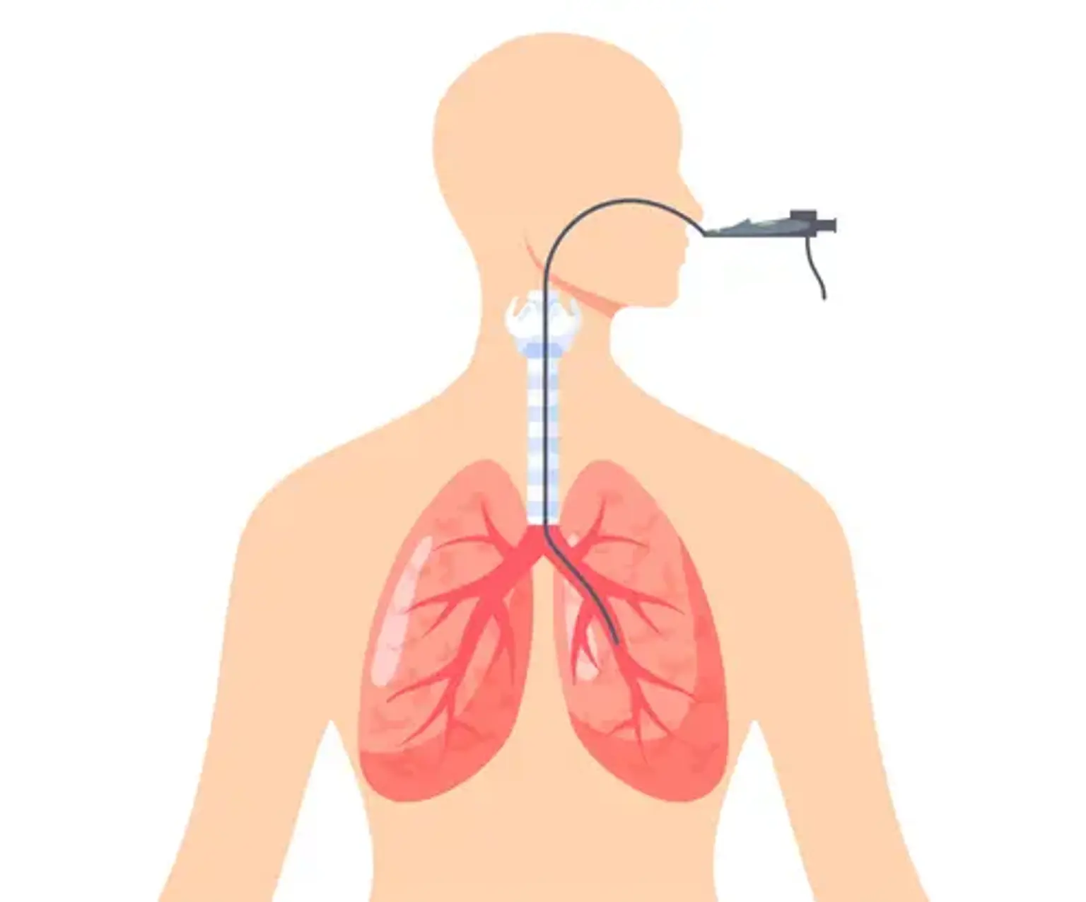
Flexible bronchoscopy (FB) is a critical procedure for assessing the pediatric airway, voice cords, trachea, bronchi, and bronchioles. A flexible bronchoscope is introduced via the nose or mouth into the lungs while your kid is sedated. The bronchoscope is a specialized equipment equipped with a small camera that allows the pediatric pulmonologist to observe the anatomy through the scope or on a video display.
Other procedures, such as Bronchoalveolar lavage (cleaning), transbronchial biopsy (removing tiny pieces of tissue), endobronchial ultrasonography, electrocautery, or laser therapies, are possible using the bronchoscope.
Bronchoscopy, in general, assists in the visualization of airway anatomy (e.g., agenesis), the assessment of airway dynamics (e.g., malacia), the localization and treatment of obstructions (e.g., mucus plugs), the collection of fluid samples (i.e., bronchoalveolar lavage (BAL), and brushing/biopsy for microbiology and histopathology. Suctioning, re-inflation, therapeutic wash out, medicine delivery, and counseling for difficult intubation are other therapeutic reasons.
In children, bronchoscopy is an invasive treatment that necessitates anaesthesia. Complications such as desaturation, airway damage, and laryngeal spasm are possible. Several knowledge gaps persist, particularly in terms of approaches to improve diagnostic capacities and reduce problems. As a result, further research to achieve these goals is required.
What is a Flexible Bronchoscopy Used for?
Exploration of the Pediatric Airways:
- Persistent stridor:
Its primary cause is laryngomalacia, which normally resolves within the first year and hence does not require endoscopic revision, except in cases of parental worry. A complete exploration is recommended in cases of atypical presentation, biphasic character, prolonged persistence, suffocation crisis, eating difficulties, association with syndromes or malformations, history of intubation, or diagnosis of severe laryngitis in children under the age of 6 months, as there may be anatomical, congenital, or acquired anomalies or suspicion of a Foreign Body.
The presence of undetected foreign bodies can be related with respiratory symptoms and/or repeated or persistent radiological findings. In these conditions, RB has a significant risk of false negatives. FB, on the other hand, has a diagnostic exactitude of 100 percent. As a result, if a diagnosis is incorrect, it is suggested that FB be performed first. Once extracted, FB can be quite valuable for validating the absence of residues or fragmented material, as is the case with nuts.
- Difficult-to-Control, Persistent Wheezing:
FB is suggested in cases with difficult-to-control wheezing bronchitis, particularly in small children, with asymmetries on auscultation and/or radiological alterations, which might be caused by a foreign body or structural abnormalities of the tracheobronchial wall.
- Hemoptysis:
Haemoptysis is a rare and worrisome sign. FB is necessary in the absence of a recognized cause (otorhinolaryngological infections or bronchiectasis). Problems with artificial airways, tracheostomies, primary alveolar bleeding, congenital vascular malformations, lung cancer,ILDs, and viral or inflammatory endobronchial diseases are the most common causes, with tumors being the exception.
- Persistent/Recurring Atelectasis:
Persistence of more than 6 weeks, together with unexplained symptoms, makes FB recommendable. The most frequent findings are mucus plugging, foreign bodies, extrinsic compression in cases of congenital cardiopathy, Hypersensitivity pneumonitis, inflammatory granulation tissue or endobronchial tuberculosis.
- Persistent/Recurring Pneumonia:
Middle lobe syndrome is a relatively frequent entity in pediatrics due to the anatomical characteristics of this bronchus. The observation of bacterial growth, a predominance of neutrophils in the bronchoalveolar lavage, and evolution towards bronchiectasis in more than 50% of children with non-atopic wheezing has renewed interest in this syndrome. It has been proposed that the syndrome if it cannot be resolved with treatment, indicates a study with FB.
- Localized pulmonary hyperlucency:
When it is not due to congenital or post-infectious causes, localized hyperlucency necessitates FB because there may be air trapping due to a valvular mechanism, which is usually secondary to an intrinsic obstruction (foreign body, bronchomalacia, granulation tissue) or extrinsic compression due to adenopathies, vessels that are aberrant, anomalous, and/or increased in size, or congenital or acquired mediast.
- Problems Related With Artificial Airways
FB is required for the identification of difficulties that emerge during intubation and extubation, as well as for monitoring and following tracheostomy patients. The most common findings are laryngeal edema, subglottic stenosis, and tracheobronchial granulation tissue as a result of endotracheal tube and cannula damage or recurrent aspirations.
Therapeutic Applications in Pediatrics:
- Aspiration of secretions:
FB can be used to treat atelectasis caused by secretion retention or mucus plugging. In pulmonary alveolar proteinosis and acquired diseases such as pneumonia, repeated high volume BAL is the preferred therapy.
- Difficult and selective intubations:
In situations of craniofacial deformities and numerous malformation syndromes, bronchoscopy can serve as a guideline for intubation. It has also been utilized for selective bronchial intubation and intubation in unusual circumstances such as suspected or proved cervical spine injury.
- Removal of foreign body:
In children, removing a foreign body with FB is a difficult surgery. Some papers support the effectiveness of FB for foreign body removal. Though FB is the method of choice for foreign body diagnosis, rigid bronchoscopy remains the gold standard for retrieval in both children and adults. Rigid bronchoscopy is favored in children because it provides the benefits of general anesthetic, assisted breathing, bigger tools, and a wider range of accessories. The optimum method would be, to begin with, FFB to allow for a broader reach in the exploration and identification of the foreign body, followed by extraction using a rigid bronchoscope and, if necessary, the last revision with FB to rule out any residual foreign bodies.
- Transbronchial biopsy:
Transbronchial biopsy (TBB) is the removal of a biopsy sample from the lymph nodes around the carina or the pulmonary parenchyma for microscopic examination. Lymph node samples aid in the diagnosis of conditions such as extrapulmonary TB and lymphoma. For the diagnosis of rejection in children having lung transplants, samples from the pulmonary parenchyma are recommended. The yield of TBB is determined by the size of the TBB sample collected. Multiple tiny lymph node samples are preferable to fewer bigger samples for reliably diagnosing cancer and ILDs.
- Bronchial washing:
Bronchial washing can be used to study the mucosa of the bronchi and to culture bronchial secretions. It is an effective tool for diagnosing ciliary dyskinesia, TB, and pneumonia in mechanically ventilated patients, as well as lung infections in immunocompromised individuals.
- Balloon dilatation:
Rigid bronchoscopy and FFB can be used to dilate stenosed or restricted airways. Web-like stenosis, benign strictures, endotracheal intubation complications, and granulomatous disorders are ideal candidates for balloon dilatation. It is a process that is minimally invasive, safe, and quick.
Preparing Your Child for a Bronchoscopy
- Documentation:
A bronchoscopy should be documented utilizing image recording devices/software. Video capture is better for documenting dynamic airway changes, allowing for an excellent assessment of follow-up tests. Uncertain results or prospective indications for surgery can be examined with the assistance of a consulting professional without the need for a fresh examination. Furthermore, video documentation enables for post-processing of the clip, such as slow motion. There are signs that simultaneous recording of respiratory sounds might help in diagnosis.
- Personnel:
A bronchoscopist and a bronchoscopy helper should make up the bronchoscopy team. If the bronchoscopy is conducted under general anesthesia, anesthetists and assistants with experience in child care of various ages should be used. Another physician and assistant may provide sedation if both have competence in procedural sedation of children of all ages, including the capacity to handle respiratory and cardiovascular problems.
- Consent and Information:
This Guidelines Group believes that bronchoscopy education and consent are critical in the preparation stage. Because the entire procedure risks are a function of the intervention and anesthesia/sedation hazards, all parties involved must be well briefed. In the educational debate, the influence on the respiratory tract should be given specific emphasis. It is advised to utilize standardized information and permission forms.
Informed consent documentation should include:
- Fasting, a risk of aspiration of stomach contents and saliva with the danger of lung failure and permanent lung damage, especially with incompliance with the necessary fasting period.
- Reaction to applied medication (allergic reactions, cardiovascular reactions, nausea/vomiting, etc..)
- Laryngospasm/bronchospasm.
- Possible respiratory depression including intubation and ventilation.
The anesthesiologic information should include:
- Fasting, a risk of aspiration of stomach contents and saliva with the danger of lung failure and permanent lung damage, especially if the fasting period is not observed.
- Laryngospasm/ bronchospasm.
- Swallowing difficulties and hoarseness.
- Damage to teeth, throat, jaw and larynx, vocal cords, and injuries to the trachea.
- Shortness of breath and permanently impaired tongue sensation.
- Life-threatening metabolic disorders (e.g., malignant hyperthermia).
- Unwanted alertness during anesthesia.
- Risks of intravenous cannulas and injections.
- Allergic reaction to the applied medication.
Contraindications
Bronchoscopy using flexible scopes is often well tolerated. To reduce risk, appropriate actions to optimize the patient's condition should be adopted. The indication for FB should be tailored to the person. The treatment should be carried out only when the advantages outweigh the hazards. Severe refractory hypoxemia, hemodynamic instability, uncorrected hemorrhagic diathesis, and a lack of parental or guardian authorisation for the surgery are absolute contraindications to doing bronchoscopy.
The relative contraindications are determined by the team's experience and the degree of critical care at the hospital. A risk-benefit analysis must be performed in extremely preterm neonates and children with congenital cyanotic cardiomyopathies who have an increase in bronchial collateral circulation, severe pulmonary hypertension, or coagulation abnormalities.
Sedation and Monitoring
The goal of pediatric sedation is to keep the kid safe, pleasant, and relatively calm during the procedure while ensuring appropriate oxygenation and breathing. Bronchoscopy can be conducted under general anesthesia or sedation with benzodiazepines or opioids. 'Conscious sedation,' in which the patient can follow verbal instructions and retain reflexes, is not encouraged.
During the procedure, an anesthesiologist or intensivist must be present to give medicines and continually monitor the patient. During diagnostic procedures, spontaneous breathing is preferable, hence the dose of sedation should be adequate. Diagnosis of dynamic airway abnormalities in a deeply sedated kid with no spontaneous breathing may be challenging.
Positive pressure ventilation is used during diagnostic procedures to help maintain appropriate breathing. Local anesthetic medicines of choice include 2% lidocaine jelly for the nose and 1% lidocaine spray for the pharynx and larynx. Midazolam, fentanyl, or ketamine are utilized for analgesia and sedation.
Continuous heart rate, respiratory rate, color, head position, and gas exchange measurement using continuous pulse oximetry are all part of the patient's monitoring. Continuous ECG and invasive monitoring are preferred in critically ill patients, such as kids with heart disorders. A sedated patient should never be left alone or unattended. The juvenile patient must be closely observed until he or she awakens. After the procedure, supplemental oxygen should be administered until the patient has recovered from anesthesia and sufficient oxygenation on room air is recorded. When the operation is done as an outpatient, the kid should be able to tolerate oral intake before being discharged
Pediatric Bronchoscopy Procedure
Flexible bronchoscopy is commonly done trans-nasally, however, it can also be done orally or through an artificial airway. While utilizing the nasal or oral route, an oxygen mask or nasal prongs can be utilized simultaneously. While the patient is receiving high flow nasal cannula ventilation, the nasal route can be used. Special noninvasive ventilation facemasks (interfaces) are available for FFB while the patient is still receiving positive pressure ventilation. When the nasal route is not possible due to choanal atresia, nasal hemorrhage, or trauma, the oral route is employed. A laryngeal mask airway can be used for oral ventilation. Endotracheal and tracheostomy tubes with a minimum diameter of 4 mm are utilized in severely sick patients with a suitable bronchoscope.
To prevent the equipment from being bent, the operator stands or sits at the patient's head with the gurney in a low position. Forced bends in the scope's fibers might damage them and make it difficult to handle. The size of the bronchoscope is determined by the child's age. If the access is nasal, the morphology and functions of the pharyngeal and laryngeal structures (sublingual glands, tonsils, arytenoids, epiglottis, and voice cords) are examined.
Further advancement via the larynx is accomplished by positioning the bronchoscope end in the angle of the anterior corner of the vocal cords and introducing it by posterior flexion when the patient inhales. A topical dosage of 1% lidocaine can be sprayed via the working channel to ease the passage and avoid the emergence of laryngeal spasm. To control coughing while navigating the trachea and bronchi, the lidocaine spray and continue approach is employed after reaching the subglottic region. The bronchoscope is advanced until it is jammed in the desired sub-segmental bronchus at the desired place.
Pediatric Bronchoscopy Complications
In general, pediatric FB is a safe procedure. Complications are likely depending on the patient's risk factors, the kind of surgery performed, insufficient sedation/anesthesia, the use of inappropriately sized equipment, and the bronchoscopist's inexperience. The following are the primary complications.
- Epistaxis and Nasal Trauma:
These are the most common. It can be controlled by compression and/or topical adrenalin.
- Hypoxia and desaturation:
These might be caused by factors inherent in the surgery and patient, or by an excess or deficit of sedation. FB raises airway resistance, which, when combined with sedation, can cause hypoxemia and hypercapnia, which can last for 15–20 minutes after the investigation. BAL and prolonged aspiration movements, particularly in neonates and infants, promote and increase the risk of alveolar collapse. Bronchospasm may occur as a result of vagal irritation or in children with bronchial hyperreactivity. These alterations can be minimized by keeping a stable hemodynamic state, using appropriate-sized equipment, providing bronchodilators and supplementary oxygen beforehand, optimizing the sedation anesthesia, and conducting the treatment fast.
- Bronchospasm and/or Cough:
These might be attributed to insufficient sedation or the application of topical anesthetic.
- Trauma and Airway Obstruction:
Also associated with the use of an incorrectly sized bronchoscope or pushing the way through a stenotic region, resulting in edema, hypersecretion, and increased blockage.
- Hemorrhage:
Small transient hemorrhages are frequently caused by aspiration, brushing, or biopsies, which are assisted by the presence of granulation tissue, tumors, bronchiectasis, or hemorrhagic diathesis. Severe bleeding is unusual in children and can be related with highly vascularized endobronchial lesions, BAL in patients with coagulation abnormalities, and especially transbronchial biopsies.
In most situations, hemorrhaging will stop on its own within a few minutes, but in severe cases, lavage with cold physiological saline solution (5ml aliquots) or adrenalin at 1/20 000–1ml of adrenalin at 1/1000, diluted in 20ml of physiological saline solution can be used. If the hemorrhage persists, the patient should be placed in lateral decubitus, with the bleeding lung in a decreasing posture, and the bronchoscope inserted into a segmental or subsegmental bronchus, aspirating continuously for 3–5 minutes to cause a collapse of the distal bronchial walls.
On rare situations, compression with a Fogarty catheter or selective guided intubation is required. In certain circumstances, RB may be required to investigate the source of the bleeding and aspirate big clots. Endobronchial instillation of pro-coagulation drugs, such as tranexamic acid, has been indicated in one research, albeit the evidence for this advice is currently lacking.
- Pneumothorax:
Although pneumothorax is an uncommon consequence of transbronchial biopsy, it can occur. In other circumstances, they might be caused by bronchoscope usage (uncontrolled pressure with the tip, rapid movements, especially if coughing, or employing either O2 or pressurized air via the working channel).
- Lung Infections and Fever:
Fever is common, occurring in up to 15% of surgeries, especially if BAL was used. It is associated with cytokine release or temporary bacteriemia, particularly in immunocompromised individuals. More infrequently, FB can cause infectious issues for both the patient (dissemination to healthy lung regions or transmission of contaminated material from a prior examination) and the healthcare practitioner (transmission of M. tuberculosis).
Conclusion
To summarize, the flexible bronchoscope is a significant tool in the diagnosis and treatment of pediatric airway problems. It has a favorable safety profile, with only a few cases of life-threatening or long-term problems documented. Furthermore, the ability to do BAL using this process can play a crucial role in the early distinction of infections vs non-infection diseases, and assists in determining the exact pathology and optimum therapy.
Early intervention and use of bronchoscopy should be recommended after careful examination of which patients will benefit from this technique and a thorough assessment of its advantages and pitfalls.
Pediatric bronchoscopy has matured and is now utilized for diagnostic and therapeutic reasons in a growing range of clinical settings. In the future, prospective studies will be required to assess its efficacy and patient benefit while also enhancing its technical capabilities. It has grown in importance as a research tool for investigating pathophysiologic ideas, monitoring, and even implementing medicines locally.
The use of electronic media as a training tool, as well as the presentation of scientifically accurate information on illnesses identified or treated by bronchoscopy, will necessitate additional effort, as well as scientific and clinical advice. It does, however, offer improved treatment and information for kids and parents, as well as extra services for a well-educated pediatric pulmonologist.