Pediatric Solid Malignancies

Last updated date: 14-Jun-2023

Originally Written in English

Pediatric Solid Malignancies

Children's solid brain tumors are different from adults' solid tumors. Neuroblastoma, Wilms' tumor, rhabdomyosarcoma, osteosarcoma, Ewing's sarcoma, and retinoblastoma are examples of solid tumors that originate in children but never occur in adults. The most prevalent types of solid tumors in children include brain tumors, neuroblastoma, rhabdomyosarcoma, Wilms' tumor, and osteosarcoma.


Brain Tumors

Brain Tumors incidence and Etiology
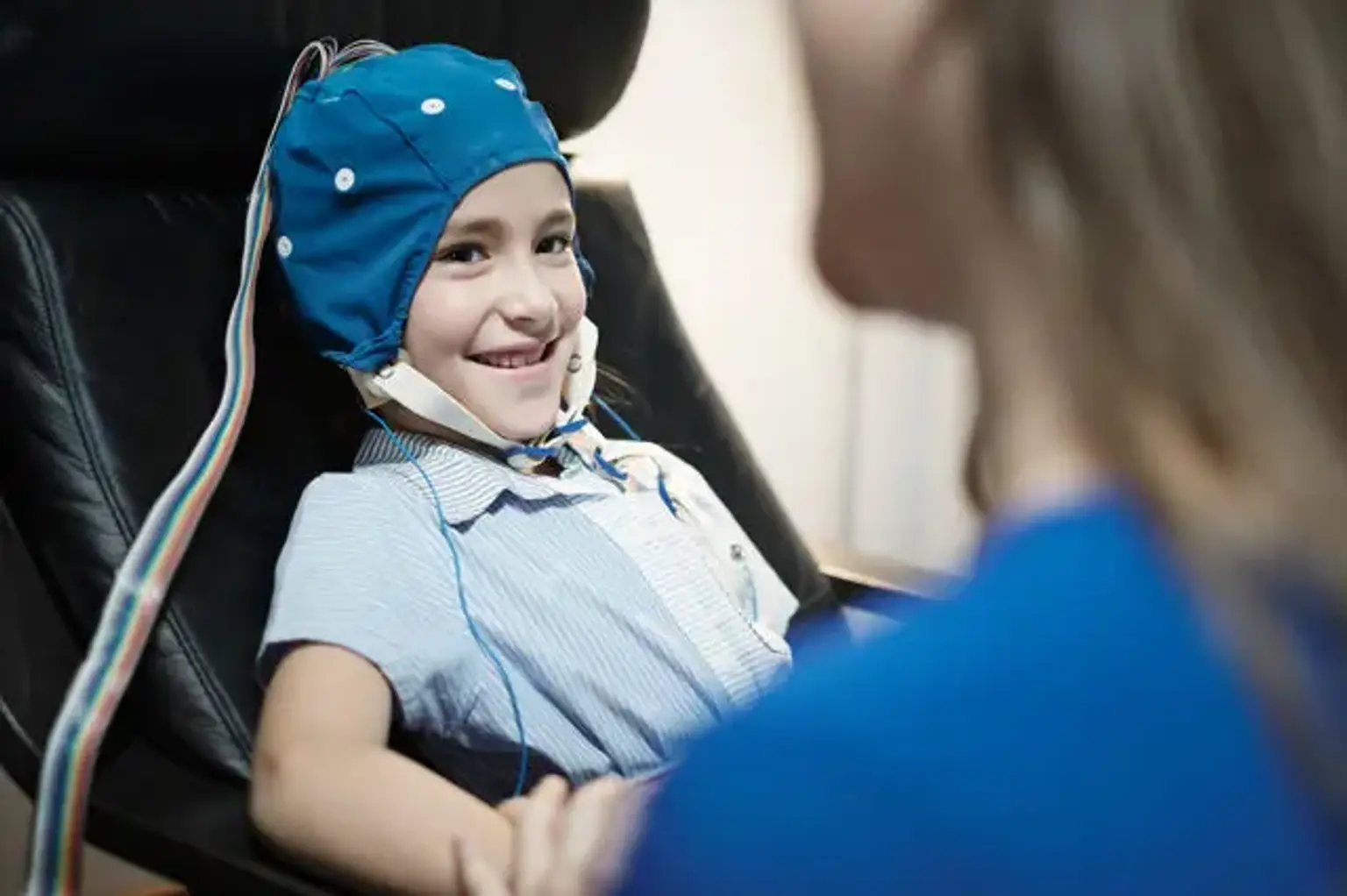
In 1999, approximately 1,700 children in the United States were diagnosed with brain cancer. The most prevalent type of solid tumor in children is central nervous system tumors, which are also the second most common pediatric malignancy.

Infratentorial tumors account for about 56 percent of these cancers, while supratentorial tumors account for 44 percent. Although children with neurofibromatosis type I are more likely to develop optic gliomas, and children with tuberous sclerosis are more likely to develop subependymal giant cell astrocytomas, the origin of brain tumors in children remains mainly unclear.


Brain Tumors Clinical Findings

Brain Tumors Clinical Findings

Increased intracranial pressure is typically present in the signs and symptoms of brain tumors. The tumor mass squeezing essential structures, restriction of cerebrospinal fluid flow, or tumor-associated edema all contribute to increased intracranial pressure. The indications and symptoms that the kid displays are also linked to the tumors' location. Infants and children under the age of two years may have vague, nonspecific symptoms and, as a result, may be detected late. Headaches, gait problems, instability, loss of coordination, head tilting, visual problems (e.g., blurred vision, diplopia), cranial nerve impairments, forceful vomiting in the morning, papilledema, and hydrocephalus are common in children with posterior fossa tumors. Symptoms in children with brain stem tumors vary depending on which cranial nerves are impacted.

Headaches, nausea, vomiting, papilledema, involuntary movements, visual field abnormalities, and difficulties with smell are all symptoms of midline tumors (cranial nerves 1-4). Tumors in the pons (cranial nerves 5-8) impair facial movement and sensation, induce ocular movement issues, and make hearing difficult. The perception of taste is affected by tumors that grow from the medulla oblongata (cranial nerves 9-12), and the gag reflex might be inhibited. The patient may experience tongue fasciculations, heart rate fluctuations, and decreased spinal accessory muscle strength. The signs and symptoms that children with supratentorial tumors experience vary depending on which part of the brain is affected. Convulsions, hemiplegia, memory problems, poor judgment, behavior problems, visual field abnormalities, hormone inadequacy, handwriting difficulties, and hydrocephalus are all possible outcomes for these youngsters.

Macrocephaly, instability, hyperreflexia, cranial nerve palsies, and a protruding fontanelle can all be signs of a brain tumor in children under the age of two. Convulsions and weakness are less common in younger children than they are in older children. Vague findings are also possible, and they could be the sole signs and symptoms that appear. Vomiting, lack of coordination, tiredness, and irritation are some of the symptoms. These kinds of misleading signs can last for weeks before causing neurologic symptoms.


Brain Tumors Diagnosis

Brain Tumors Diagnosis

For detecting brain malignancies, magnetic resonance imaging (MRI) has become the recommended diagnostic method. It displays the tumor in axial, coronal, and sagittal planes, and contrast indicates tumor-associated edema and tumor expansion into surrounding brain tissue, providing better resolution than computed tomography scan. To minimize normal postoperative alterations being misconstrued for residual disease, postoperative MRI scans should be obtained as soon as feasible following surgery, preferably within 24 hours. The degree of tumor removal is determined by the postoperative neurosurgeon's assessment and the postoperative MRI scan.


Brain Tumors Treatment

Brain Tumors Treatment

Tumor tissue is required for the pathologist to make a histologic diagnosis and identify treatment options. In many cases, the extent of surgical excision of the tumor corresponds with the prognosis. Radical resections are especially critical in children under the age of two because cranial radiotherapy can be delayed as long as feasible in these patients. If a child's hydrocephalus continues, a ventriculoperitoneal shunt may be required. A patient's best chance of long-term survival is with combined therapy (surgery, radiation, and/or chemotherapy). Radiation therapy is effective for most brain tumors, and it may be used as part of the post-surgery treatment plan. Because of the related morbidity, radiotherapy is indicated for patients above the age of two years.

Five days a week, conventional external beam radiotherapy is administered. Cranial radiation takes about 4 weeks to complete. Spinal radiotherapy will be given to children with posterior fossa disease, leptomeningeal or spinal metastases, tumor cells in the cerebrospinal fluid, or malignant tumors of the pineal or suprasellar areas for about 2 weeks. Cranial radiotherapy has a wide range of side effects that might arise during treatment or after it is finished. Chemotherapy is one of the most effective treatments for brain tumors. Combination chemotherapy, like other types of children’s cancer, is more effective than single-agent therapy. Some of the chemotherapy medications used to treat brain tumors include vincristine, cyclophosphamide, cisplatin, carboplatin, and etoposide. Chemotherapy-related side effects are most common during treatment, but there are some late effects as well. The nurse's position in multimodal treatment techniques for what is typically an aggressive disease process makes nursing care for a child with a brain tumor challenging.

The prognosis for children with brain tumors varies significantly depending on the histology, surgical excision, and metastases. Tumor recurrence, like other types of pediatric cancers, indicates a bad prognosis.


Neuroblastoma

Neuroblastoma Incidence and Etiology

In the United States, around 600 new instances of neuroblastoma are identified each year. It is the most frequent malignancy in newborns and the fourth most prevalent kind of cancer in children over the age of one year. Because 51 percent to 63 percent of children will have metastases and have had few signs of the disease until that time, it is not uncommon for extensive metastatic disease to be discovered upon diagnosis. Neuroblastoma is more common among boys and non-Hispanic white children. At the time of diagnosis, the average age is two years. The majority of cases of neuroblastoma are unknown. Neuroblastoma may be linked to maternal drug consumption or paternal occupational exposure to magnetic waves, according to certain theories.


Neuroblastoma Clinical Findings

Neuroblastoma Clinical Findings

Neuroblastoma can develop at any point in the sympathetic nervous system. Symptoms at the time of diagnosis are specific to the location where the tumor grows. More than half of the cases will arise from the adrenal gland in the retroperitoneal area. These children have a palpable, hard, non-tender abdominal mass that may cross the midline, as well as abdominal pain. The head, neck, mediastinum, and pelvis are all common sites of disease. Proptosis and periorbital ecchymosis are symptoms of the orbital disorder, which can lead to vision loss if not treated quickly. Primary malignancies in the neck can obstruct or impede breathing by compressing the airway. Tumors that develop along the spine can grow into the intervertebral foramina, compressing the spinal cord.

Cord involvement can spread quickly, resulting in irreversible paralysis. Numbness, tingling, or incontinence are significant symptoms that signify an oncologic emergency that necessitates prompt treatment. The bone, bone marrow, liver, lungs, brain, and soft tissue are all potential sites for metastasis. Pain is caused by bone metastasis, and if the legs are affected, the children may refuse to bear weight. Other non-tender, permanent, and bluish-colored bone tumors may form on the skull. Anemia, thrombocytopenia, and/or neutropenia can result from tumor cells infiltrating the bone marrow, which crowd out the normal hematopoietic cells. The presence of metastatic tumors in the brain might result in localized neurologic symptoms or seizures.


Neuroblastoma Diagnosis

Neuroblastoma Diagnosis

The diagnostic examination is used to determine the severity of the condition. The degree of surgical removal and the sites of metastases are factors in disease staging. CT scans of the primary site, as well as secondary sites, can help determine tumor size and location. A skeletal assessment and a bone scan revealed the presence of bone disease. To assess whether the bone marrow is implicated, aspirates and biopsy examinations are required. Bilateral specimens improve the chances of discovering tumor cells that might otherwise go undetected if just one site was collected. Catecholamines (homovanillic acid and vanillylmandelic acid) are secreted by 90% to 94% of neuroblastoma tumors and discharged in the urine.

Before the initial surgical treatment, urine samples for homovanillic and vanillylmandelic acid will reveal whether the tumor is a catecholamine secreting tumor and will aid in the diagnosis. Urine homovanillic and vanillylmandelic acid can be used to track disease progression both on and off therapy. Ferritin and lactate dehydrogenase levels are high in certain children with advanced-stage neuroblastoma. As a result, including these investigations when collecting blood samples is beneficial.


Neuroblastoma Treatment

Neuroblastoma Treatment

The stage of the disease and the child's age influence the treatment for neuroblastoma. Multimodal therapy is done if treatment is required. Complete surgical resection is generally not possible at the time of the initial operation in advanced-stage disease. After a biopsy is taken to confirm the diagnosis, induction chemotherapy is administered. Vincristine, cyclophosphamide, doxorubicin, cisplatin, carboplatin, and etoposide are some of the most regularly utilized chemotherapy medicines. The effectiveness of therapy is determined by disease restaging after multiple sessions have been administered, and it may be possible to remove the residual tumor at that time. Following the second surgical resection, additional chemotherapy, particularly high-dose chemotherapy with bone marrow rescue, may be necessary.

Infection, hemorrhage, mucositis, and malnutrition are all frequent chemotherapy side effects that the nurse must be aware of. Because neuroblastoma is particularly radiosensitive and responds quickly to this treatment, it is frequently delivered at the primary tumor site as part of the initial treatment. Palliative radiotherapy can help manage pain in patients who are approaching the end of their lives.

Children with limited disease, as well as those who respond completely to treatment after the first treatment, have a substantially higher chance of achieving disease-free status and long-term survival. Those aged 1 year or above with broadly disseminated disease or unfavorable histologic indicators upon diagnosis, or those who have quick relapse after finishing treatment, have a worse prognosis.


Rhabdomyosarcoma

Rhabdomyosarcoma Incidence and Etiology

Rhabdomyosarcoma is that the commonest soft-tissue sarcoma in pediatric age. Rhabdomyosarcoma is most commonly caused by mesenchymal cells that are generally dedicated to the creation of skeletal muscle, although it can also be caused by smooth muscle cells (i.e., urinary bladder). Sarcomas that are undifferentiated can't be traced to a distinct mesenchymal lineage.  Rhabdomyosarcoma has the same incidence as all other types of soft-tissue sarcomas combined. Each year, about 250 new cases of rhabdomyosarcoma are diagnosed in the United States, with over two-thirds of cases occurring in children aged 6 and less. Primary head and neck tumors are most common in children under the age of eight, while extremities tumors are more common in adolescents. Although the formation of rhabdomyosarcoma has been linked to certain familial disorders, the great majority of cases occur spontaneously (e.g., neurofibromatosis, Li-Fraumeni syndrome).


Rhabdomyosarcoma Clinical Finding

Rhabdomyosarcoma Clinical Finding

Clinical outcomes are influenced by the initial tumors' location as well as distant metastasis. The metastatic disease affects less than a quarter of newly diagnosed individuals, with lung metastasis being the most prevalent.

1. Head and neck

Proptosis is caused by malignancies of the orbit or eyelids, which can affect eyesight. Tumors of the nasopharynx or sinuses can restrict the nasal or sinus cavities, resulting in serosanguinous discharge. Increased intracranial pressure may result if intracranial tumor development occurs as the tumor erodes through bone (e.g., headache, vomiting, lethargy).

2. Genitourinary tract

Genitourinary rhabdomyosarcoma most commonly affects the bladder and prostate. Hematuria and urinary blockage are symptoms of bladder tumors. Urinary problems are sometimes caused by prostate tumors, but not usually. Constipation is one of the early symptoms of primary prostatic rhabdomyosarcoma in children. At the time of diagnosis, large pelvic masses are prevalent. A mucosanguinous discharge, identical to that seen with a foreign body, is produced by vaginal tumors. Cervical and uterine tumors are frequently associated with a pelvic mass, which may or may not be accompanied by vaginal discharge. Para-testicular tumors cause painless, unilateral scrotal or testicular enlargement and are frequently associated with lymph node involvement in the region.

3. Extremities

When the affected area starts to swell, sarcoma of the extremities is diagnosed. Tenderness and erythema are also possible side effects, however, many of these tumors are asymptomatic. The presence of a non-tender mass, particularly in the absence of trauma, raises the possibility of cancer. These tumors usually spread through the fascia and involve the lymph nodes in the region.

4. Other sites

Rhabdomyosarcoma can develop in any skeletal or smooth muscle tissue. A primary rhabdomyosarcoma can develop in the intrathoracic, retroperitoneal, perineal, and perianal regions and be untreated for a long time. Before they are discovered, these tumors can grow to be quite large or spread to other parts of the body. Other organ structures, such as the liver, brain, heart, breast, or ovary, can develop primary tumors, albeit they are uncommon. Symptoms that are now present are linked to specific organ involvement.


Rhabdomyosarcoma Diagnosis

Rhabdomyosarcoma Diagnosis

The surrounding tissue and lymph nodes should be examined closely during the physical examination. A plain film of the damaged body part and a skeletal survey should be included in the radiographic examination. An MRI is required if a primary tumor is discovered on a radiograph. CT scans of the chest, abdomen, and pelvis should be ordered for patients with worrisome abdominal or pelvic tumor findings. All patients should have nuclear medicine scans to look for bone metastasis, and a chest x-ray or CT scan to look for pulmonary metastasis.


Rhabdomyosarcoma Treatment

Rhabdomyosarcoma Treatment

Staging is important for establishing treatment and prognosis since it determines the degree of the disease. Surgery, chemotherapy, and radiotherapy are the three current therapeutic options. Complete surgical removal is preferable, however due to tumor invasion of neighboring structures, this is often not achievable.

At best, the initial surgery will be a biopsy or debulking treatment. If the residual disease persists after the first treatment, a second surgical procedure may be needed. All patients with rhabdomyosarcoma receive chemotherapy. The most often utilized first-line drugs include vincristine, dactinomycin, doxorubicin, cyclophosphamide, ifosfamide, and etoposide. Topotecan, irinotecan, and paclitaxel are among the agents that could be used in the future.

Rhabdomyosarcoma is vulnerable to high doses of radiotherapy. If a second surgical procedure is not sought after the first 9 weeks of chemotherapy, radiation therapy may be employed. Due to the high doses of chemotherapy and radiation therapy used in treatment, nursing care for patients with rhabdomyosarcoma necessitates constant supportive care.


Wilms’ Tumor

Wilms’ Tumor Incidence and Etiology

Wilms' tumor is the most frequent kind of pediatric kidney tumor. In the United States, about 465 new cases are diagnosed each year. Aniridia, hemihypertrophy, cryptorchidism, and hypospadias are some of the malformations associated with Wilms' tumor in children. It has been found in children with overgrowth syndromes such as Beckwith-Wiedemann, Perlmann, and Soto. Wilms' tumor is linked to chromosomal abnormalities, such as gene deletions on chromosomes 11p13 and 11p15.


Wilms’ Tumor Clinical Findings

Wilms’ Tumor Clinical Findings

Wilms' tumor causes an abdominal mass in children that is usually painless. Hematuria, hypertension, and discomfort are uncommon occurrences. Often, the tumor is detected during a routine medical assessment, or a family member feels it during daily activities. Focal neurologic symptoms can emerge as a result of metastatic disease to the brain.


Wilms’ Tumor Diagnosis

Wilms’ Tumor Diagnosis

Hemihypertrophy, aniridia, swollen abdominal veins, and an abdominal mass should all be checked during a physical examination. To reduce the risk of rupturing the tumor capsule before surgery, palpation of the abdominal mass should be limited to a minimum. To evaluate the extent of the underlying tumor and metastatic cancer, radiographic investigations such as abdominal ultrasonography, abdomen CT, and chest radiograph or CT should be conducted. Hematuria will be detected by urinalysis, and kidney function will be determined by blood urea nitrogen and creatinine levels.


Wilms’ Tumor Treatment

Wilms’ Tumor Treatment

Wilms' tumor is treated primarily by nephrectomy. Chemotherapy and radiation treatment are prescribed according to the stage of the disease. The duration of chemotherapy, as well as the specific medicines used, are all influenced by the stage of the disease. Vincristine, dactinomycin, doxorubicin, and cyclophosphamide are among the first-line chemotherapy medicines used to treat Wilms' tumor. If both kidneys are affected, a biopsy and staging of both kidneys are performed first, followed by 5 weeks of chemotherapy. Following this initial treatment, a disease restaging will determine which kidney has the most significant disease and requires full resection.


Osteosarcoma

Osteosarcoma Incidence and Etiology

The most prevalent type of bone cancer in children is osteosarcoma, which affects about 450 children in the United States each year. Osteosarcoma is most common in the second decade of life when teenagers are quickly growing vertically. At the time of diagnosis, about 20% of patients will have metastasis, the majority of which will be pulmonary in type.


Osteosarcoma Clinical Findings

Osteosarcoma Clinical Findings

The long bones are the most commonly affected by osteosarcoma. Before being diagnosed, the youngster may have been experiencing dull, excruciating discomfort for several months. As the pain develops, the child's walk may vary as he or she becomes hesitant to bear weight. Point pain, swelling, warmth, and erythema are common palpable symptoms at the site of disease. There may or may not be a soft-tissue mass present.


Osteosarcoma Diagnosis

Osteosarcoma Diagnosis

A unique sunburst pattern of the diseased bone, lytic lesions, sclerosis, and pathological fractures at the site of disease are all possible radiographic findings. To rule out pulmonary metastases, a chest radiograph and chest CT are required. A bone scan might help you find other regions of bone disease or areas that are mending. Elevated alkaline phosphatase levels can occur, but they can also occur in a healthy adolescent going through normal bone growth.


Osteosarcoma Treatment

Osteosarcoma Treatment

To confirm the diagnosis, a surgical biopsy procedure is required. To treat the microscopic disease, all children with osteosarcoma require chemotherapy. The initial surgical treatment will be conducted after many courses of induction chemotherapy. The surgery's purpose is to remove the diseased bone while leaving a large margin of healthy bone around it. Amputation or limb-sparing procedures are frequently performed, albeit this is depending on the patient's age, preoperative treatment response, and tumor location. The patient, his or her family, and the physician must all agree on the optimum procedure for the child. The entire limb, or affected expendable bone, is amputated (e.g., ulna, fibula, rib, toe, finger).

The diseased bone is removed and replaced with an endoprosthetic device or cadaver bone in a limb-sparing technique. Following the original surgical surgery, the patient is given further chemotherapy and is continuously evaluated for recurring disease and physical function. In children with recurrent disease, radiation therapy is not offered as part of the core treatment plan, but it may be utilized as palliative therapy. Patients with osteosarcoma require nursing care that focuses on therapy, side effects, recovery, and body image.

Adjuvant chemotherapy has markedly increased survival rates in children with osteosarcoma. After chemotherapy and before the initial operation, children who achieve a 97 percent necrosis of the main tumor have the best prognosis. Recurrent disease is frequent within three years of diagnosis, with pulmonary recurrence accounting for 86 percent of all recurrences.


Conclusion

Solid tumors account for 65% of all malignant tumors in children, with roughly 3750 new cases diagnosed in the United States every year. The spectrum of tumor types seen in children differs significantly from those seen in adults. Tumors of the central nervous system; neuroblastoma; soft tissue sarcoma, such as rhabdomyosarcoma; Wilms tumor; bone tumors, such as osteosarcoma and Ewing sarcoma; retinoblastoma; and various tumors, such as hepatoblastoma, germ cell tumors, and melanoma. Since the first demonstration of Wilms tumor chemosensitivity to actinomycin D in 1966, significant progress has been achieved in the detection and management of these malignancies. Since the 1970s, cure rates for most children’s neoplasms have improved by as much as 50%. A better understanding of prognostically essential biological and clinical characteristics, improved accuracy of clinical staging, consistent use of supportive therapy, and development of better treatment, which often combines chemotherapy, surgery, and radiotherapy, are all contributing to the rise.