Pediatric Tumor
Last updated date: 30-Mar-2023
Originally Written in English
Pediatric Tumor
Overview
Cancer is the most common cause of mortality among children and adolescents. Childhood cancer is uncommon in general. Many malignancies that develop in children also occur in adults. Leukemia is by far the most frequent, accounting for around 28% of all children's malignancies; brain tumors account for approximately 27%; lymphomas account for approximately 12%; and some bone cancers account for approximately 12%.
Childhood cancer treatment is determined by the kind of malignancy, stage, and/or risk categorization. Chemotherapy, surgery, radiation therapy, and stem cell transplantation are all common therapies. Immunotherapy is a newer kind of treatment that uses the patient's own immune system to target the tumor and may be beneficial in the treatment of some children malignancies.
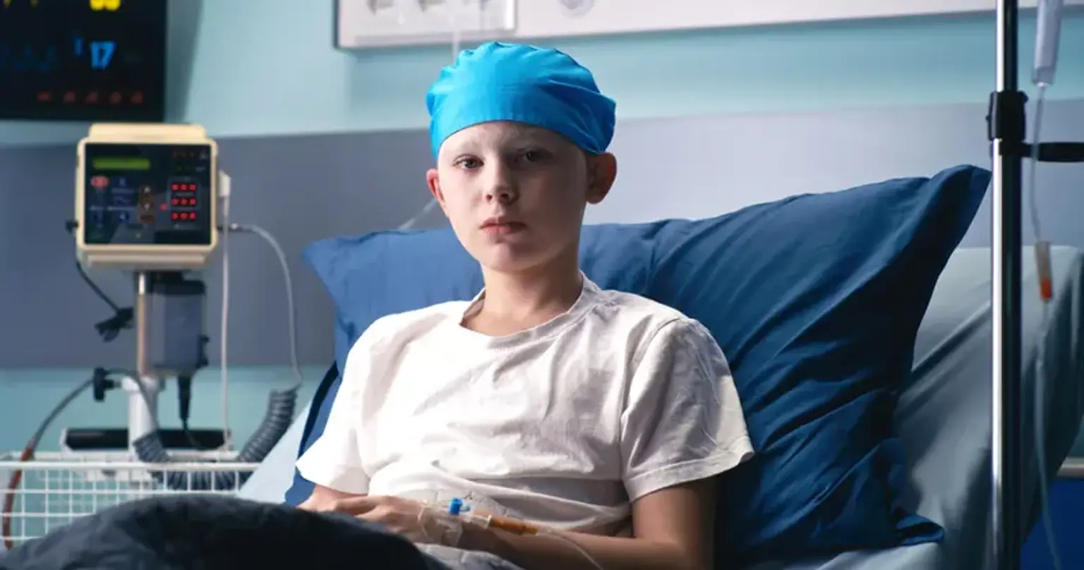
What is Pediatric Tumor?
A word used to characterize malignancies that develop between the ages of birth and 14 years. Pediatric malignancies are extremely rare and may differ from adult cancers in their growth and spread, therapy, and response to treatment. Leukemia, brain and spinal cord tumors, lymphoma, neuroblastoma, Wilms tumor (a kind of kidney cancer), retinoblastoma, and bone and soft tissue malignancies are the most prevalent types of pediatric cancer.
What causes cancer in children?
Cancer affects people of all ages and can arise in any region of the body. It starts with a genetic alteration in single cells, which then expand into a mass that invades other regions of the body and causes injury and death if not treated. Unlike adult malignancies, the great majority of children's cancers have no recognized etiology. Many types of research have been conducted to determine the causes of pediatric cancer, however, relatively few are caused by environmental or lifestyle factors. Children's cancer prevention initiatives should concentrate on habits that will keep the kid from acquiring avoidable cancer as an adult.
Childhood cancer is influenced by chronic illnesses such as HIV, Epstein-Barr virus, and malaria. Other infections can raise a child's risk of developing cancer as an adult, so it's critical to get vaccinated (against hepatitis B to help prevent liver cancer and against human papillomavirus to help prevent cervical cancer) and to pursue other methods, such as early detection and treatment of chronic infections that can lead to cancer.
According to current research, around 10% of all children with cancer have a hereditary susceptibility. More study is needed to understand the variables that influence cancer development in youngsters.
Neuroblastoma
Neuroblastoma is a disorder in which malignant (cancer) cells grow in the adrenal glands, neck, chest, or spinal cord.
Neuroblastoma is frequently seen in the nerve tissue of the adrenal glands. In the rear of the upper abdomen, there are two adrenal glands, one on top of each kidney. The adrenal glands produce vital chemicals that assist regulate heart rate, blood pressure, blood sugar levels, and how the body responds to stress. Neuroblastoma can start in the nerve tissue of the neck, chest, abdomen, or pelvis.
Neuroblastoma is most commonly diagnosed in childhood. It is often identified between the first month of life and the age of five. It is discovered when the tumor begins to develop and create symptoms. It can develop before birth and be discovered during a fetal ultrasound.
Cancer has frequently spread by the time it is diagnosed (spread). In babies and children, neuroblastoma most commonly spreads to the lymph nodes, bones, bone marrow, liver, and skin. Adolescents may potentially develop lung and brain metastases.
Neuroblastoma-related gene mutations can occasionally be inherited (passed from the parent to the child). Neuroblastoma frequently develops at a younger age in children with a gene mutation, and several tumors may arise in the adrenal glands or nerve tissue in the neck, chest, abdomen, or pelvis.
Symptoms of neuroblastoma
Signs and symptoms of neuroblastoma include a lump in the abdomen, neck, or chest or bone pain.
The most frequent neuroblastoma signs and symptoms are produced by the tumor pushing on neighboring tissues as it develops or by the malignancy spreading to the bone. These and other signs and symptoms might be the result of neuroblastoma or another ailment.
Check with your child’s doctor if your child has any of the following:
- Lump in the abdomen, neck, or chest.
- Bone pain.
- Swollen stomach and trouble breathing (in infants).
- Bulging eyes.
- Dark circles around the eyes ("black eyes").
- Painless, bluish lumps under the skin (in infants).
- Weakness or paralysis (loss of ability to move a body part).
Neuroblastoma diagnosis
Tests that examine many different body tissues and fluids are used to diagnose neuroblastoma.
The following tests and procedures may be used to diagnose neuroblastoma:
- Physical exam and health history: An examination of the body to look for general symptoms of health, such as tumors or anything else that appears peculiar. A history of the patient's health habits, as well as previous diseases and treatments, will be collected.
- Neurological exam: A sequence of questions and tests to assess the function of the brain, spinal cord, and nerves. The test assesses a person's mental state, coordination, ability to walk normally, and the efficiency of the muscles, senses, and reflexes. This is also known as a neuro exam or a neurologic exam.
- Urine catecholamine studies: A urine test that measures the number of particular chemicals, vanillylmandelic acid (VMA) and homovanillic acid (HVA), produced when catecholamines degrade and are discharged into the urine. A higher-than-normal level of VMA or HVA might indicate neuroblastoma.
- Blood chemistry studies: A blood test that measures the quantities of specific chemicals produced into the blood by organs and tissues in the body. A chemical with an abnormally high or low concentration might be a symptom of the disease.
- MIBG scan: A technique for detecting neuroendocrine cancers such as neuroblastoma. A very small quantity of radioactive MIBG is injected into a vein and circulates through the circulation. A scanner detects neuroendocrine tumor cells that take up radioactive MIBG. Scans can be taken over a period of 1-3 days. To prevent the thyroid gland from absorbing too much MIBG, an iodine solution may be administered before or during the test. This test is also used to determine how effectively a tumor responds to therapy. MIBG is used to treat neuroblastoma at high dosages.
- CT scan (CAT scan): A process that produces a series of detailed images of parts of the body taken from various angles. A computer coupled with an x-ray machine creates the images. To make the organs or tissues more visible, a dye may be injected into a vein or ingested. This is also known as computed tomography, computerized tomography, or computerized axial tomography.
- PET scan (positron emission tomography scan): A method of locating malignant tumor cells in the body. In a vein, a little quantity of radioactive glucose (sugar) is injected. The PET scanner revolves around the body, capturing images of where glucose is utilized. Because malignant tumor cells are more active and take up more glucose than normal cells, they appear brighter in the image.
- X-ray of the chest or bone: An x-ray is a form of energy beam that may go through the body and onto film to create an image of places within the body.
- Ultrasound exam: High-energy sound waves (ultrasound) are rebounded off inside tissues or organs, creating echoes. The echoes combine to generate an image of bodily tissues known as a sonogram. The image can be printed and viewed later. If a CT/MRI has been performed, an ultrasound exam is not performed.
Treatment of Neuroblastoma
Neuroblastoma treatment options include:
- Surgery: Surgeons may remove the tumor at the time of diagnosis or after shrinking it with chemotherapy or radiation.
- Chemotherapy: Several chemotherapeutic drugs have been shown to be successful in the destruction of neuroblastoma cells. Chemotherapy is used to shrink the tumor, remove the malignancy, and prevent further tumor spread.
- Radiation therapy: Radiation treatment is used to remove neuroblastoma cells that remain after chemotherapy or surgery. It is also occasionally utilized for individuals who require quick tumor shrinking to avoid or treat a problem (such as the tumor pressing on an important nerve or the spinal cord).
- Stem cell transplant: A treatment in which part of a child's healthy blood cells are temporarily removed and very high doses of chemotherapy and, in certain situations, radiation therapy are administered to overwhelm and eradicate the neuroblastoma. To support regular body and organ function, we reinfuse the child's stored healthy blood cells back into their body.
- Biologic therapy: These medicines include antibody therapy and isotretinoin, a medication that takes use of what we've learned about the biologic properties of neuroblastoma cells. The goal of biologic treatment is to destroy any residual tiny tumor cells.
- Immunotherapy: Immunotherapy is exclusively used to treat high-risk neuroblastoma. Unituxin (dinutuximab), an antibody that attaches to the surface of neuroblastoma cells, is usually given to patients in conjunction with chemotherapy.
What is Wilms Tumor?
Wilms tumor (also known as Nephroblastoma) is a kind of pediatric cancer that begins in the kidneys. It is the most frequent kind of pediatric kidney cancer. Wilms tumors account for about 9 out of 10 kidney malignancies in children.
The majority of Wilms tumors are unilateral, affecting only one kidney. The majority of the time, there is only one tumor, however, a tiny percentage of children with Wilms tumors have several tumors in the same kidney. The bilateral disease affects around 5% to 10% of children with Wilms tumors (tumors in both kidneys).
Wilms tumors often grow to be quite big before being detected. The average freshly discovered Wilms tumor is several times larger than the kidney where it began. The majority of Wilms tumors are discovered before they have spread (metastasized) to other organs.
Signs and symptoms of Wilms tumor
Wilms tumor is characterized by:
- Abdominal swelling, often toward one side
- Fever
- Abdominal pain
- Constipation
- High blood pressure (hypertension)
- Large swollen veins across the abdomen
- Blood in the urine (hematuria)
- Fatigue
- Loss of appetite
- Weight loss
- Frequent urinary tract infections
Diagnosis of Wilms tumor
Following a thorough medical history and physical examination of your kid, the doctor may request the following tests to establish whether the cancer has spread (metastasized):
- Abdominal ultrasound: This imaging test creates images of inside organs, blood arteries, and tissues using high-frequency sound waves and a computer.
- Bone scan: This bone cancer imaging test employs a particular radioactive substance that is injected into a vein. The material accumulates in damaged bone regions and is detected by specific cameras that detect radioactivity.
- Abdominal computerized tomography scan (CT or CAT scan): This imaging test use X-rays and computer technologies to provide comprehensive cross-sectional pictures (slices) of the body's bones, muscles, fat, and organs. It can reveal the presence of a tumor in the kidneys as well as if the cancer has migrated to other organs such as the lungs.
- Chest X-ray. This imaging test produces images of the heart, lungs and bones.
- Magnetic resonance imaging (MRI): An MRI scan employs radio waves, powerful magnets, and computer technologies. MRI provides more comprehensive pictures than CT and ultrasound and can help physicians determine whether the cancer has spread to one of the major blood veins surrounding the kidney.
- Blood and urine tests. These laboratory tests help evaluate kidney and liver function.
- Biopsy. A sample of tissue is extracted and studied under a microscope, which aids in the diagnosis and treatment plan.
Treatment for Wilms tumor
Your kid will be treated by a multidisciplinary team of surgeons, oncologists, urologists, radiologists, radiation oncologists, pathologists, and nurse practitioners who will collaborate to develop a treatment plan tailored to your child's individual needs. You and your kid will meet with the treatment team so that your family can develop a trusting connection with them.
Proton therapy, a more focused kind of radiation therapy, may be used to treat Wilms tumor.
Wilms tumor has an overall cure rate of 85%. However, like with any disease, prognosis and long-term survival might differ substantially from kid to child. For the best prognosis, prompt medical treatment and intensive therapy are essential. Continuous follow-up treatment is needed for a kid diagnosed with Wilms tumor since radiation and chemotherapy side effects, as well as second malignancy, may emerge.
Surgery for Wilms Tumor
The majority of Wilms tumor patients will require surgery as part of their treatment regimen. You and your kid will meet with the treatment team so that your family can develop a trusting connection with them. Prior to surgery, scans are performed to identify the size of the tumor in the kidney and whether it has migrated to other regions of the body, such as the lungs.
The damaged kidney may be surgically removed, either whole or partially. A nephrectomy is when the entire kidney is removed. Surgeons can conduct nephron-sparing surgery (NSS), also known as partial nephrectomy, which removes only a portion of the kidney. In situations of bilateral Wilms tumor, when the only option is bilateral nephrectomy, multifocal unilateral disease, or Wilms tumor predisposition syndromes, NSS has been advised. NSS has a low complication rate and no increased risk of tumor recurrence, and it should be regarded as the standard current method for the care of selected instances of Wilms tumor.
Several assessments and surgical procedures have been used to optimize effective surgical results. We use various parameters to determine the chance of problems following partial nephrectomy and arrive at a complexity score. We discovered that the majority of Wilms tumors with high-intermediate complexity (a high score) can still be adequately treated with NSS rather than complete nephrectomy, as is commonly suggested. This accomplishment is aided by 3D models of each patient's kidneys. To better comprehend anatomical linkages, the surgeon can use a 3D model to visualize the organ and tumor location from multiple perspectives. In some cases, a tumor will be checked further utilizing intra-operative ultrasonography to determine the extent of the tumor.
A three- to five-day hospital stay is normal following surgical recuperation. We collaborate with our anesthetic experts to maximize pain control following surgery. We also advocate early walking after surgery and anticipate a one- to two-week return to typical activity.
Retinoblastoma
Retinoblastoma is a kind of cancer that begins in the retina, the very back of the eye. It is the most frequent kind of childhood eye cancer. Other types of eye cancer in children include medulloepithelioma and ocular (eye) melanoma.
How does retinoblastoma develop?
The eyes begin to grow long before birth. The eyes have cells called retinoblasts that proliferate to generate new cells that fill the retina during the early stages of development. These cells eventually cease proliferating and develop into adult retinal cells.
Occasionally, something goes wrong with this procedure. Instead of maturing, some retinoblasts continue to grow uncontrollably, resulting in retinoblastoma, a kind of cancer.
How does retinoblastoma grow and spread?
Retinoblastoma tumors can develop and fill most of the eyeball if not treated. Cells may break away from the original tumor on the retina and go to other areas of the eye, where they might develop other tumors. These tumors may obstruct the channels that allow fluid to move within the eye, increasing intraocular pressure. This can result in glaucoma, which causes discomfort and visual loss in the afflicted eye.
Most retinoblastomas are discovered and treated before they extend beyond the eyeball. Retinoblastoma cells, if not treated, can spread to other regions of the body. Cells can develop along the optic nerve and eventually reach the brain. Retinoblastoma cells can also develop through the eyeball's covering layers and into the eye socket, eyelids, and surrounding tissues. Once cancer has gone beyond the eyeball, it can spread to lymph nodes (small bean-sized collections of immune system cells) and other organs such as the liver, bones, and bone marrow.
Rhabdomyosarcoma
Rhabdomyosarcoma develops from cells that ordinarily form skeletal muscles. These are the muscles that we use to move various portions of our bodies. This cancer can originate almost anywhere in the body, including the head and neck, groin, belly (abdomen), pelvis, or arm or leg. It might result in discomfort, swelling (a lump), or both. In children, this is the most prevalent kind of soft tissue sarcoma. It accounts for roughly 3% of all pediatric malignancies.
Brain tumors
Brain cancer accounts for 15% of all childhood cancers. The symptoms are determined by the tumor's location. Gliomas and medulloblastomas are the two most common kinds of brain tumors in children. Gliomas arise from the brain's supporting cells, which hold nerve cells in place, and brainstem gliomas are almost exclusively found in youngsters. The typical developmental age is around 6 years old. Medulloblastoma develops in the cerebellum or posterior fossa and can spread to other areas of the brain or the spinal cord. The majority of medulloblastomas develop before the age of ten.
Bone cancers
Cancers that begin in the bones (primary bone cancers) are more common in older children and teenagers, although they can occur at any age. They account for around 3% of all pediatric malignancies.
Two main types of primary bone cancers occur in children:
- Osteosarcoma is most frequent in teenagers and typically occurs in regions where the bone is rapidly expanding, such as towards the ends of the leg or arm bones. It frequently causes bone discomfort that worsens at night or with activity. It can also cause edema in the bone's surrounding region.
- Ewing sarcoma is a rare kind of bone cancer. It is particularly common in early adolescents. The pelvic (hip) bones, the chest wall (such as the ribs or shoulder blades), or the center of the leg bones are the most likely areas for it to begin. Bone discomfort and edema are possible symptoms.
Improving outcomes of childhood cancer
Because cancer cannot be prevented in children, the most successful method for reducing the burden of cancer in children and improving outcomes is to focus on early, accurate diagnosis, followed by effective, evidence-based therapy and customized supportive care.
- Early diagnosis
Cancer is more likely to react to appropriate therapy when detected early, resulting in a higher chance of survival, less suffering, and typically less expensive and rigorous treatment. Significant gains in the lives of children with cancer can be realized by finding cancer early and minimizing care delays. A precise diagnosis is critical for treating children with cancer since each malignancy has a unique treatment plan that may involve surgery, radiation, and chemotherapy.
Early diagnosis consists of 3 components:
- Awareness of symptoms by families and primary care providers;
- Accurate and timely clinical evaluation, diagnosis, and staging (determining the extent to which cancer has spread); and
- Access to prompt treatment.
Early detection is important in all contexts and increases survival rates for many malignancies. Programs to encourage early and proper diagnosis have been implemented effectively in nations of all economic levels, frequently via the coordinated efforts of governments, civil society, and voluntary organizations, with parent groups playing critical roles. Childhood cancer is associated with a variety of warning signs, including fever, severe and persistent headaches, bone pain, and weight loss, all of which can be spotted by families and competent primary care professionals.
Screening is not typically beneficial for pediatric malignancies. It can be explored in some limited circumstances in high-risk groups. For example, certain childhood eye tumors can be caused by an inherited mutation, therefore if that mutation or illness is detected in the family of a kid with retinoblastoma, genetic counseling can be provided, and siblings can be followed with regular eye examinations early in life. Only a tiny percentage of children with cancer have genetic origins of their disease.
Conclusion
Although pediatric cancers are less prevalent than adult tumors, their impact on children and their families is devastating. Leukemias, neuroblastomas, Wilms tumors, brain malignancies, rhabdomyosarcomas, lymphomas, retinoblastomas, osteosarcomas, and Ewing's sarcomas are the most common forms of juvenile cancers.
Children's malignancies are not usually treated in the same manner as adult tumors are. Pediatric oncology is a medical specialty that focuses on the treatment of cancer in children. It is critical to understand that this knowledge exists and that there are successful therapies for many children malignancies.
The shock of a cancer diagnosis, as well as the intensity of therapy, is overwhelming for the kid and family. It is challenging for the youngster to maintain a feeling of normalcy, especially given the need for frequent hospitalizations, outpatient visits, and possibly painful treatments. Overwhelming stress is common as parents strive to continue working, be attentive to siblings, and care to the myriad demands of the cancer kid. When the child is being treated at a specialist center far from home, the situation becomes much more tough.