Percutaneous nephrolithotomy (PCNL)
Overview
This percutaneous endoscopic method is widely used to treat upper urinary tract stones that are not susceptible to conventional treatment modalities (e.g., shockwave lithotripsy, retrograde ureteroscopic therapy). Percutaneous treatment of contaminated stone loads is commonly used because it allows for not only stone fragmentation but also quick evacuation of the diseased material.
PCNL procedure definition
The primary therapy for renal calculi is percutaneous nephrolithotomy (PCNL). Since it was initially defined in the 1980s, PCNL has progressed dramatically.
Kidney stones occur in the urinary system as a result of the crystallization of chemical components in urine. PCNL is a technique for removing stones in the kidney or upper ureter (the tube that drains urine from the kidney to the bladder) that are too big for conventional stone removal methods such as shock wave lithotripsy or ureteroscopy.
Relevant Anatomy
The kidneys are retroperitoneal organs that are positioned obliquely on the anterior aspect of the psoas muscles. Because of the presence of the liver on the right side, the right kidney is frequently 2-8 cm lower than the left. It is located near the 12th rib, the liver, the duodenum, and the hepatic flexure of the colon. The 11th and 12th ribs, pancreas, spleen, and splenic flexure of the colon are all located next to the left kidney.
With variations in patient posture and breathing, the kidney's location may shift. When a patient stands from a supine posture, the kidneys should not move more than 8-9 cm. The kidneys normally migrate vertically during the breathing cycle. Because the kidney is so near to the pleural cavity, there is a danger of pleurotomy with a percutaneous puncture, especially with upper-pole renal access. Percutaneous renal access above the 12th rib should be conducted at the end of the rib to assist avoid pneumothorax.
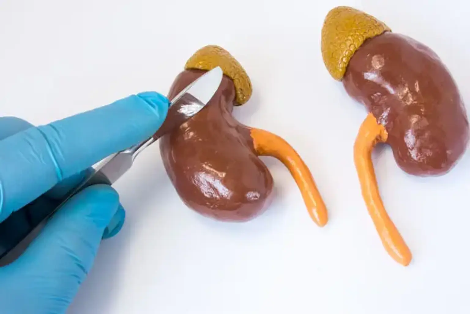
The kidney's collecting system is made up of minor calices (2-4), large calices, and a renal pelvis. The renal pelvis is located in the renal sinus and narrows to produce the ureter at the UPJ. The vein is normally anterior in the renal hilum, and the renal pelvis is posterior, with the artery in between.
Indications for PCNL
Symptomatic calculi in a caliceal diverticulum are another reason for percutaneous nephrolithotomy (PCNL) (dilated cavities connected to the collecting system by a narrow infundibulum). Percutaneous therapy resulted in 88-100 percent stone-free and symptom-free rates in this condition. Large stone loads or those contained within a long-necked diverticulum are best handled in this manner.
PCNL has always been conducted in the prone position, and it is a well-established procedure with a high success rate and minimal morbidity. The prone position, on the other hand, can be difficult and is linked with a significant risk in morbidly obese individuals with cardiopulmonary illness; hence, the supine posture has been designed to assist overcome these difficulties. Theoretical benefits of the supine position include safer and easier patient placement, less cardiovascular change and risk, less need for patient repositioning, and perhaps faster operating time.
The theoretical disadvantages of the supine position include anterior/lateral puncture sites, which may increase the risk of visceral organ injury (e.g., colon, liver, spleen); collapse of the collecting system; and difficulty approaching the upper pole calyx.
The Surgery
Pre-operative evaluation
Prior to any percutaneous renal operation, a comprehensive history and physical examination should be gathered.
Anticoagulation, bleeding disorders, contrast-medium responses, cancer, obesity, spinal cord damage, and a history of urinary tract infections with urine cultures and sensitivities should all be considered.
Coagulation profile
Prothrombin time with international normalized ratio (INR), activated partial thromboplastin time, and platelet count are all included.
An uncorrected coagulopathy is the sole contraindication to percutaneous renal access. Prior to percutaneous access, any anomalies must be corrected. Platelet administration should be used to treat thrombocytopenia. Preoperatively, aspirin and clopidogrel (Plavix) should be discontinued to allow platelet function to be maximized.
Patients taking Coumadin should have their coagulation factors adjusted before undergoing elective surgery.
Overall renal function
Prior to doing percutaneous surgery, this should be examined. A blood urea nitrogen and creatinine level is usually sufficient.
Complete blood cell count
Prior to providing percutaneous access, a full blood cell count is required. Intraoperative and postoperative bleeding are potential complications of percutaneous renal surgery, and understanding the patient's baseline hematocrit is crucial in patient treatment if considerable bleeding develops.
Furthermore, a high white blood cell count may signal a concomitant infectious condition that need more stringent antibiotic prophylaxis.
Urinalysis and urine culture
Before attempting to manipulate the urinary tract, it is critical to rule out urinary tract infection. In the presence of infection and/or blockage, percutaneous manipulation of the kidney can quickly escalate to sepsis.
Appropriate antibiotic coverage is beneficial before to the surgery and should be accessible throughout the procedure to aid in the prevention of intraoperative sepsis.
Patients with known infectious stone loads may require phased therapy with percutaneous tract maturation, particularly if resistant organisms are detected on preoperative urine cultures.
Imaging Studies
A baseline diagnostic renal imaging is essential before installing a percutaneous nephrostomy. The most frequent imaging modalities used to identify renal architecture and disease are intravenous or retrograde pyelography and renal ultrasonography. To characterize unexpected anomalies, computed tomography, nuclear renography, and magnetic resonance imaging can all be employed.
CT scanning may assist in identifying any posteriorly lying loops of colon that may overlie a prospective nephrostomy tract and in establishing a safe angle for nephrostomy tube installation. Before inserting a nephrostomy tube, all radiographic data should be thoroughly scrutinized. CT scanning is especially beneficial in developing a safe percutaneous path into the renal collecting system in patients with renal ectopy, malrotation, or a history of surgery that might modify the perirenal architecture.
When planning and performing nephrostomy implantation, both ultrasonography and fluoroscopy are beneficial. Prior to the surgery, baseline ultrasonography offers helpful information such as the depth and lay of the kidney, the location of stone load, and the degree of hydronephrosis. Real-time imaging is used throughout every percutaneous renal access operation. When precise renal access is necessary, such as with planned intrarenal endoscopic operations, fluoroscopy is recommended.
The intrarenal collecting system must be defined using contrast medium–enhanced imaging. If the kidney operates normally and is not blocked, intravenous pyelography may be used to assist identify the caliceal system and stone burden. Retrograde ureteropyelography can assist define the collecting system if renal function is inadequate. After gaining first access, antegrade research can be conducted.
In difficult circumstances, such as designing a percutaneous approach to a staghorn calculus in a malrotated kidney or exposing crossing arteries in a patient with UPJ blockage, a 3-dimensional CT scan with reconstruction pictures can be employed judiciously. Recently, CT urography has been employed to offer a highly accurate anatomical roadmap.
Technique
In most cases, the sector probe is utilized to acquire access. The puncture is performed either freehand or with the aid of a needle guide. The puncture guide can be either on the side or in the center of the ultrasonic probe, depending on the manufacturer. In most circumstances, the electronic dotted line corresponds to the needle's route. An echo tip needle is useful for accurate needle vision.
Alternatively, the needle's serrated side should be towards the probe. The ultrasound probe should be scanned from back to front. The posterior calyx would be the first to be seen. The probe should then be positioned so that the calyx, infundibulum, and pelvis are visible. The needle should ideally be visible throughout its journey. A sharp needle or a motionless/steady probe would be required for this.
The point at which the needle was inserted into the skin should be indicated. The needle is injected into the subcutaneous tissue with a jiggling motion, then moved through the cup of the calyx into the target calyx. Ideally, the needle's trajectory should be visible throughout the course. The outflow of clear urine confirms the correct perforation.
This technique has been performed on a significant number of patients over the last few years and is now considered the standard of care for patients with kidney stones that are big, particularly hard, or resistant to conventional kinds of stone therapy. As a result, in the great majority of cases, it has replaced open surgeries for kidney stones.
Typically, the procedure lasts three to four hours. A minor 1 cm incision is made in the patient's flank region to execute the procedure. Under x-ray guidance, a catheter is inserted through the incision into the kidney. After that, a tiny telescope is sent down the tube to visualize the stone, break it up, and remove it from the body. A laser or another equipment known as a lithotripter may be used to break up the stone before it can be removed if necessary. When compared to open stone surgery, this method has resulted in much reduced post-operative discomfort, a shorter hospital stay, and an earlier return to work and everyday activities.
This approach also has a greater success rate for eliminating all stones in a single setting than other treatments, such as extracorporeal shock wave lithotripsy (ESWL), which frequently requires many efforts.
Although supine PCNL has become a viable alternative for the removal of heavy stone loads, further prospective, randomized controlled trials are required to evaluate the safety and long-term effectiveness of this method. The choice of patient placement during PCNL should ultimately be guided by the surgeon's preference and expertise.
Potential Complications
Although this technique has shown to be highly safe, there are risks and potential problems, as with any surgical operation. When compared to open surgery, the safety and complication rates are comparable. Among the potential hazards are:
- Bleeding: This treatment will result in some blood loss, however patients will seldom require a blood transfusion. If you want to get autologous blood transfusions (donate your own blood), you must notify your surgeon. When your surgical information packet is given to you, you will also get an authorization form to fill out and return to the Red Cross. This must be coordinated with the Red Cross in your region.
- Infection: To reduce the possibility of infection following surgery, all patients are given broad-spectrum antibiotics. If you develop any indications or symptoms of infection following the operation (fever, discharge from the incision, urine frequency or discomfort, pain, or anything else that concerns you), please call us right once.
- Tissue / Organ Injury: Although uncommon, harm to adjacent tissue/organs such as the colon, vascular structures, spleen, liver, lung, pancreas, and gallbladder may necessitate further surgery. Kidney function loss is uncommon, although it is a concern. Scar tissue may potentially develop in the kidney or ureter, necessitating further surgery.
- Conversion to open surgery: If complications arise during this surgical treatment, it may be necessary to convert to a regular open surgery. This may necessitate a bigger conventional open incision and, as a result, a lengthier recovery period.
- Failure to Remove the Stone: There is a chance that the stone(s) will not be entirely removed, mainly owing to the size or placement of the stone (s). It's possible that you'll need to get some extra help.
Miniature percutaneous nephrolithotomy (Mini-Perc)
Miniature percutaneous nephrolithotomy (Mini-Perc) was used to remove kidney stones that had become resistant to ESWL in a 2-year-old preterm infant. The procedure involves successive dilating to a 16F sheath, followed by the use of a 12F vascular peel-away sheath. To remove the stones, a pediatric endoscope was employed. The Mini-Perc approach was updated by Jackman et al to employ an 11F ureteral access sheath. At a 3.8-week follow-up, the stone-free percentage in their group of 9 patients was 89 percent.
Mini-Perc supporters point to less blood loss, improved mobility, reduced postoperative discomfort, and a shorter hospital stay. The procedure's limitations include the need to fracture stones into small enough bits to slip through a smaller sheath and lengthier operational periods.
A prospective research comparing Mini-Perc to conventional PCNL for stones ranging in size from 1-2 cm. The Mini-Perc group utilized an 18F sheath (range 15-20F), whereas the normal PCNL group used a 26.8F sheath (range 24-30F). The Mini-Perc approach had longer operational periods (45 vs 31 min), less blood loss (0.8 vs 1.3 g hemoglobin), and a shorter hospital stay, according to the research (3.2 vs 4.8 d). At a one-month follow-up, the postoperative stone-free rates were comparable between the two groups.
Cheng et colleagues also conducted a randomized experiment comparing Mini-Perc to regular PCNL. They observed that, because to the smaller sheath, the Mini-Perc was linked with longer operational periods (134 vs 89 min); nevertheless, blood loss needing transfusion was statistically considerably lower (1.4 percent vs 10.4 percent ).
The two groups exhibited comparable stone-free rates for staghorn and simple renal pelvic stones; however, the Mini-Perc group had a statistically substantially greater stone-free rate for numerous calyceal stones. The rates of hospitalization, postoperative discomfort, and stone removal were comparable between the two groups.
Tubeless percutaneous nephrolithotomy
Tubeless PCNL is the technique of doing a nephroscopic operation using a new access track without first inserting a nephrostomy tube.
Previously, an internal ureteral stent was employed for drainage; however, more recent research have concentrated on completely tubeless drainage, in which neither an external nephrostomy nor an internal ureteral stent is left after the treatment.
The tubeless and entirely tubeless procedures have advantages such as less surgical discomfort and a shorter hospital stay. The use of hemostatic medications such as fibrin glue injections to assist seal the nephrostomy tract following PCNL is one modification of the fully tubeless procedure.
Wang et al conducted a meta-analysis of seven trials and found that tubeless PCNL had the advantage of a shorter hospital stay and the requirement for postoperative analgesics in select, uncomplicated patients. There were no differences between the two groups in terms of operation length or hematocrit alteration.
Although tubeless PCNL has been demonstrated to be possible and effective in reducing postoperative pain, it is not recommended in severe situations (e.g., hemorrhage, renal perforation, extravasation, infection) or when a phased PCNL is required. More research is needed to establish the evident therapeutic advantage of tubeless PCNL.
Contraindications
An uncorrected coagulopathy is the sole contraindication to percutaneous renal access. Prior to percutaneous access, any anomalies must be corrected. Platelet administration should be used to treat thrombocytopenia. Patients taking warfarin (Coumadin) should have their coagulation factors balanced before undergoing elective surgery.
Future directions
What will PCNL look like in 5–10 yr?
First and foremost, the search for the ideal entry approach will continue. Preoperative planning may frequently include three-dimensional (3D) CT planning, as well as the use of simple 3D models for navigation. Access may include enhanced technology, such as tablet computers used to superimpose CT scans, or it may rely on tracking systems. In terms of instrumentation, it is probable that we will do more mini-PCNLs because mini-PCNLs rely on the suction effect of higher flow.
However, the question of size remains. This might eventually range between 16 and 22 degrees Fahrenheit. As a result, energy sources will be adapted to the size of the tract. Larger stones, on the other hand, may be better suited for combination ultrasound/ballistic devices, and advancements in auxiliary apparatus will be critical.
Developments in holmium laser technology, particularly the dusting process, may be crucial for smaller size tracts. Multipuncture, multitract PCNL may become uncommon. ECIRS with PCNL might become the standard of treatment in specialist centers where a team of surgeons would support EGA in order to decrease access-related morbidity while increasing SFR. Furthermore, as the PCNL technology improves, the ambulatory approach may become more realistic.
Conclusion
PCNL has emerged as the most effective method among various modalities to stone removal, while it is not without problems and skill requirements. With the passage of time, we have been able to lower tract size, so reducing discomfort while maintaining a clearance rate equivalent to the usual approach.
This was made feasible by the development of better lasing methods, as well as enhanced suction and lithoclast alternatives. Shock pulse technology has recently made an appearance in PCNL for stone lysis. However, the operation is not without difficulties. The most serious consequence is bleeding, and the need for a phased operation with large quantities of disease remains a worry.
In certain circumstances, tubeless PCNL should be done, allowing the patient to be free of nephrostomy-related morbidity. We should be able to determine which type of PCNL to use based on stone density, volume, and patient characteristics.
The study of population-based data sources and registries, which have supplied a plethora of outcome information, has greatly advanced our understanding of PCNL. Although the GSS is the most studied of the several nephrolithometry categorization systems, no single approach has been proved to be superior. Further research and changes may change this in the future.
The growing acceptance of the supine method has paved the way for ECIRS and single-tract surgery. Obtaining good renal access is a critical stage in the learning curve of PCNL, and EGA may reduce this curve. The goal to avoid access-related problems and bleeding has fueled the spread of minimally invasive improvements.
Efforts for initial stone removal should be increased during PCNL to reduce the danger of retreatment from RFs. Even RFs are used post-procedure.