Introduction
Prosthetic Joint Infection (PJI) is a serious complication that can occur after joint replacement surgery, affecting patients who have undergone procedures like hip, knee, or shoulder replacements. Infections of the joint prosthesis can be caused by bacteria that enter the body during or after surgery, leading to symptoms like pain, swelling, and fever. PJI not only threatens the success of the joint replacement but can also lead to severe complications like sepsis if left untreated.
With the increasing number of joint replacement surgeries performed globally, the incidence of PJI has become a growing concern. Early detection and effective treatment are critical to improving outcomes and minimizing the long-term impact on patients' health and mobility. This article will provide a comprehensive look at PJI, its causes, diagnosis, symptoms, and the latest treatment options available.
Understanding PJI: Causes and Risk Factors
PJI is most commonly caused by bacteria, including Staphylococcus aureus and Streptococcus species, which can infect the prosthetic joint during surgery or afterward through bloodborne pathogens. These bacteria can form a biofilm on the surface of the implant, making them difficult to treat with antibiotics alone. The infection can either occur shortly after surgery (acute PJI) or develop months or even years later (chronic PJI).
Several risk factors increase the likelihood of developing PJI, including:
Age: Older adults tend to have weaker immune systems, making them more susceptible.
Diabetes: Patients with diabetes are at a higher risk of infections due to poor wound healing and immune system dysfunction.
Obesity: Obese patients may experience complications from both surgery and infection due to poor blood circulation.
Immunocompromised states: Conditions like HIV or medications that suppress the immune system increase infection risk.
Previous infections or surgeries: A history of infections or multiple surgeries around the joint can predispose a patient to future infections.
Understanding these risk factors is crucial for preventing and managing PJI. Preoperative screenings and careful monitoring after surgery can help reduce the chances of infection.
Surgical Options for Treating PJI
In addition to antibiotics, surgical intervention is often necessary to treat PJI effectively. The two main surgical approaches include:
Debridement, Antibiotics, and Implant Retention (DAIR): This procedure is often the first line of treatment for acute PJI. It involves removing infected tissue and retaining the original implant, followed by intensive antibiotic therapy. DAIR is typically successful if the infection is caught early.
Two-Stage Revision Surgery: For more severe or chronic cases of PJI, two-stage revision is the preferred method. The infected implant is removed in the first stage, and the joint is treated with antibiotics. After several months, a new prosthetic joint is implanted. This method has a high success rate, particularly for difficult-to-treat infections.
One-Stage Revision Surgery: This involves removing the infected implant and replacing it with a new one in the same procedure. It is only suitable for carefully selected cases, especially if the infection is well-controlled and the bacteria are sensitive to antibiotics.
Managing Chronic and Recurrent PJI
Chronic or recurrent PJI can be challenging to manage. In some cases, the infection persists despite initial treatment. Chronic PJI is often associated with biofilm formation, which makes the bacteria more resistant to antibiotics and difficult to fully eliminate.
For long-term management:
Antibiotic suppression therapy may be used to control the infection when surgery is not an option or as a bridging treatment before further surgery.
Repeat surgeries (e.g., another two-stage revision) may be necessary for some patients, especially if there is a recurrence of infection.
Successful management of chronic PJI requires ongoing monitoring and tailored treatment strategies to prevent further complications.
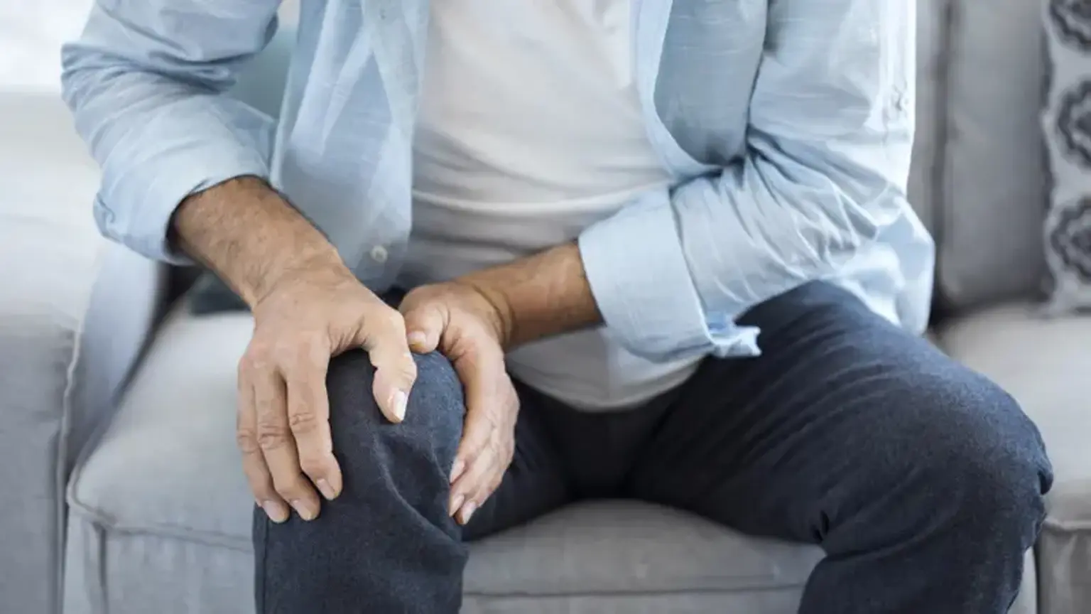
Symptoms of Joint Infection After Surgery
Recognizing the symptoms of PJI early is key to successful treatment. Acute infections usually present with severe pain, swelling, warmth, and redness around the joint. A fever, sometimes accompanied by chills, is also a common sign that the infection is spreading. Patients may experience difficulty moving the joint or increased stiffness.
Chronic PJI may develop more subtly. In these cases, patients might experience persistent joint pain, low-grade fever, or swelling that does not resolve over time. Because the symptoms can mimic other post-surgical complications, such as joint stiffness or inflammation, it’s important to monitor for any changes or worsening of symptoms over time.
If any of these signs develop after joint replacement surgery, immediate consultation with a healthcare provider is essential. Timely intervention can prevent the infection from spreading and reduce the need for more invasive treatments.
Infection Control and Prevention During Surgery
Preventing Prosthetic Joint Infections (PJI) starts before surgery. Infection control protocols are critical in reducing the risk of PJI. Preoperative screening for bacteria, especially Staphylococcus aureus, is essential, as carriers of this pathogen are more likely to develop infections after joint surgery. Antibiotic prophylaxis, typically administered intravenously right before surgery, helps prevent infection during the procedure.
Maintaining a sterile surgical environment is equally important. Surgeons must follow strict aseptic techniques, and hospital staff should adhere to best practices for cleaning, sterilizing instruments, and handling implants. Additionally, using advanced surgical drapes and antimicrobial-coated implants has been shown to reduce infection rates.
The Psychological Impact of PJI on Patients
Dealing with a PJI diagnosis can be emotionally and psychologically taxing. Patients often face extended recovery periods, the possibility of additional surgeries, and uncertainty about their long-term mobility. The fear of recurring infections can also lead to anxiety.
Psychological support is essential for coping. Patients benefit from clear communication with their healthcare team, who can reassure them about treatment options and recovery timelines. Support groups and counseling can also help patients manage the emotional burden, improving their overall well-being during treatment.
Case Studies: Real-World PJI Treatment Examples
Case 1: Successful One-Stage Revision Surgery A 65-year-old patient with acute knee PJI due to Staphylococcus aureus underwent a one-stage revision surgery. After thorough debridement and antibiotic treatment, the patient’s infection cleared up, and the new prosthesis was successfully integrated. The patient was able to return to normal activities within a few months.
Case 2: Chronic PJI with Antibiotic Suppression A 70-year-old patient with a long-standing hip replacement developed chronic PJI. After initial antibiotic therapy failed, they were placed on long-term antibiotic suppression therapy while preparing for a two-stage revision surgery. The infection was eventually eradicated, and the patient achieved a good outcome.
These examples highlight the range of approaches used in PJI management and the importance of timely intervention and customized treatment plans.
Diagnosing Prosthetic Joint Infections
Early diagnosis of PJI is essential to prevent the infection from worsening. The diagnosis typically involves a combination of clinical evaluation, laboratory tests, and imaging studies. Here's a breakdown of the common diagnostic tools used:
Clinical Evaluation: A thorough assessment by a doctor includes reviewing the patient’s medical history, surgical history, and a physical examination. This helps rule out other potential causes of pain or swelling around the joint.
Blood Tests: Elevated levels of C-reactive protein (CRP) and erythrocyte sedimentation rate (ESR) indicate inflammation in the body, which can be suggestive of infection. However, these tests alone are not conclusive for PJI and must be used in conjunction with other methods.
Joint Aspiration: A needle is used to draw fluid from the joint to test for infection. The presence of white blood cells, bacteria, or other pathogens in the fluid is a strong indicator of PJI.
Imaging Studies: X-rays, MRI scans, and positron emission tomography (PET) scans are used to identify changes in the bone or joint that may suggest an infection. MRI and PET scans are especially useful for detecting early-stage infections that may not yet be visible on X-rays.
Microbial Cultures: The most definitive way to diagnose PJI is by culturing bacteria from joint fluid, tissue samples, or blood. This allows the physician to identify the exact pathogen responsible for the infection and tailor the treatment to the specific infection.
A comprehensive approach that combines these diagnostic tools helps ensure that PJI is accurately diagnosed, leading to timely and appropriate treatment.
Advances in PJI Treatment: Latest Research and Techniques
Recent advancements in PJI treatment have significantly improved outcomes. New rapid diagnostic techniques allow for quicker identification of pathogens, enabling tailored antibiotic therapy and reducing the time patients spend on ineffective treatments. Additionally, innovations in implant materials, like antimicrobial-coated prosthetics, are showing promise in reducing infection rates.
Surgical techniques have also evolved. Minimally invasive approaches for debridement and revision surgeries are being explored to reduce recovery times and surgical risks. The role of robotic-assisted surgery in improving precision and reducing complications is also gaining attention.
The ongoing research into biofilm-targeting therapies is another exciting development. Researchers are working on drugs and techniques that can better disrupt the biofilms that bacteria form on prosthetic surfaces, making it easier to treat infections.
Global Trends in PJI Management
PJI management varies across different regions, influenced by healthcare infrastructure, resources, and infection control practices. In high-income countries, there is greater access to advanced diagnostic tools, surgical expertise, and postoperative care, which often leads to better outcomes for patients. Conversely, in low-resource settings, access to specialized care and timely interventions can be more limited, which may result in higher complication rates.
However, there is growing global collaboration in research and knowledge-sharing. Hospitals around the world are improving their infection control protocols, utilizing evidence-based practices, and learning from centers of excellence. Multinational studies and global health initiatives are helping standardize PJI treatment approaches, making cutting-edge care more accessible worldwide.
The Role of Antibiotics in PJI Treatment
Antibiotics are the cornerstone of PJI treatment, especially in the early stages. Once an infection is identified, targeted antibiotic therapy begins. In most cases, intravenous (IV) antibiotics are initially administered, particularly in the hospital setting, to quickly manage the infection. As the infection improves, oral antibiotics may replace IV antibiotics for continued treatment.
The choice of antibiotics is guided by the pathogen identified in microbial cultures. Biofilm—a protective layer formed by bacteria on the implant—can complicate treatment, requiring long-term antibiotic therapy. The goal is to ensure the infection is fully eradicated while preventing antibiotic resistance.
Role of Orthopedic Surgeons in PJI Management
Orthopedic surgeons play a crucial role in diagnosing and treating PJI. A multidisciplinary approach is often required, involving collaboration between orthopedic surgeons, infectious disease specialists, microbiologists, and other healthcare professionals.
Surgeons determine the most appropriate surgical technique based on the patient's specific condition. They also ensure the removal of infected tissue, minimize risks of further infection, and guide post-surgery recovery. Regular follow-up visits are critical to monitor recovery and detect any recurrence of infection.
Patient Education: What to Expect During PJI Treatment
Patient education plays a crucial role in the success of PJI treatment. Understanding the treatment process can alleviate anxiety and ensure better compliance with recovery plans.
Before Treatment: Patients are informed about the infection's seriousness, the risks of recurrence, and the importance of early intervention. Clear communication about surgical options, such as one-stage or two-stage revision surgery, and the expected duration of recovery is provided.
During Treatment: Patients should know the steps involved in their treatment, whether it involves debridement, antibiotic therapy, or surgery. They should also understand the importance of follow-up care to monitor for any signs of infection.
Recovery: After treatment, patients are educated about post-surgery care, including how to manage pain, watch for symptoms of infection, and when to return for check-ups. Encouraging patients to stay active within their capabilities and participate in physical therapy helps with long-term mobility and reduces the risk of complications.
Risks and Complications of Untreated PJI
If left untreated, PJI can result in severe complications. The infection may spread to surrounding tissues, bones, and even the bloodstream, leading to sepsis, which can be life-threatening. Chronic infections can also cause permanent damage to the prosthetic joint, leading to the failure of the implant and the need for multiple revision surgeries.
Joint deformities and severe pain are common long-term outcomes, significantly affecting the patient’s quality of life. Furthermore, untreated infections can increase the risk of further joint infections in the future. It is essential to treat PJI as soon as possible to avoid these complications and improve the chances of a full recovery.
The Role of Antibiotic Stewardship in PJI
Antibiotic stewardship is a key aspect of managing PJI, as improper use of antibiotics can lead to resistance, complicating treatment. It's important for healthcare providers to select the right antibiotic based on the pathogen causing the infection and its sensitivity profile. Overuse or inappropriate choice of antibiotics can lead to adverse outcomes, including the development of antibiotic-resistant strains.
Implementing antibiotic stewardship protocols helps ensure that patients receive the appropriate duration and dosage of antibiotics. It also helps minimize unnecessary treatments and side effects, promoting better recovery outcomes and reducing the burden of antibiotic resistance.
The Importance of Postoperative Care in Preventing PJI Recurrence
After treatment for PJI, effective postoperative care is crucial to prevent recurrence. This includes regular follow-ups, wound care, and monitoring for signs of infection. Surgeons typically recommend frequent check-ups within the first few months to ensure the joint is healing properly and there are no indications of infection.
Patients are also instructed to keep the surgical site clean and dry to avoid introducing bacteria. Activity restrictions may be advised to minimize stress on the joint during the healing process. In some cases, further imaging or blood tests may be required to assess recovery and confirm that the infection has been fully eradicated.
Long-Term Prognosis for PJI Patients
The long-term prognosis for patients with PJI varies depending on factors like the timing of diagnosis, the type of bacteria involved, and the patient's overall health. When treated promptly, many patients can fully recover and regain normal function, with the new prosthetic joint lasting many years.
However, those with chronic or recurrent infections may face a longer recovery period and may need multiple surgeries. For patients with advanced infections or complications, the prognosis can be less favorable, potentially leading to ongoing pain or the need for further revision surgeries.
Ultimately, with proper treatment and adherence to recovery plans, most patients can lead active lives, though continued monitoring is essential for preventing future complications.
Financial Considerations: Cost of PJI Treatment
The cost of treating PJI can be significant, especially for patients requiring revision surgeries or extended hospital stays. In addition to the immediate medical expenses, there may be long-term costs related to physical therapy, follow-up care, and potential loss of work productivity during recovery.
For healthcare systems and insurers, managing the cost of PJI involves balancing the need for effective infection control and timely interventions with cost containment. Preventative strategies, like preoperative screenings and proper surgical techniques, can help reduce the incidence of PJI, ultimately saving costs over time.
Patients should discuss financial concerns with their healthcare providers and insurance companies to understand the full scope of treatment costs, including potential out-of-pocket expenses.
Future Directions in PJI Research and Treatment
As the global healthcare community works to address PJI, ongoing research is exploring innovative ways to improve diagnosis, treatment, and prevention. Advances in biomaterials may lead to the development of implants that are less prone to infection. Additionally, research into genomic-based diagnostics could allow for faster and more accurate identification of the pathogens responsible for PJI.
Emerging treatments such as targeted therapies that directly attack bacterial biofilms and innovations in tissue regeneration hold promise for better outcomes in the future. Furthermore, enhanced rehabilitation techniques and personalized treatment plans are expected to improve recovery times and long-term functionality for patients.
As our understanding of PJI deepens, these advancements may lead to more effective strategies for managing this challenging condition, ultimately improving patient quality of life.
Conclusion
Prosthetic Joint Infection (PJI) remains a significant challenge in orthopedic care, with serious implications for patients’ health and quality of life. However, with ongoing advancements in diagnosis, treatment, and prevention, the outlook for managing PJI is continually improving. Early detection through improved diagnostic methods and timely intervention is crucial in preventing long-term complications like joint failure or systemic infection.
The combination of surgical expertise, appropriate use of antibiotics, and postoperative care ensures that many patients can recover successfully and return to active lives. While the risks of PJI remain, the global medical community is making strides in both the prevention of infection and the refinement of treatment strategies. Patients must stay informed, follow prescribed care regimens, and work closely with their healthcare team to optimize their chances of a successful outcome.
As research progresses, the future of PJI management looks promising, with innovative treatments and preventative measures on the horizon. By embracing these advancements, healthcare providers can offer patients a better quality of life and reduce the burden of infections in joint replacement surgeries worldwide.