PET-CT
The medical community is beginning to recognize positron emission tomography (PET) and PET/computed tomography (CT) as significant imaging procedures, and their use is rising. Although PET has been a valuable research tool for many years, its significant expansion into therapeutic applications has only recently begun. Its primary application today is in oncologic imaging. However, it also has a wide range of clinical uses in psychiatry, neurology, and cardiology. One of the main benefits of PET in oncologic imaging is that it can accurately measure the activity and spread of the disease with just one whole-body scan. PET/CT combines the CT scan's structural details with the PET's functional information, considerably helping in correct staging, therapeutic response evaluation, and early diagnosis of recurring cancer.
What PET Scan?
Positron emission tomography, also known as a PET scan or PET imaging, is a new nuclear medicine imaging technique.
A tiny amount of radioactive material known as radiotracers is used in nuclear medicine. Nuclear medicine is a tool used by doctors to identify, assess, and treat a variety of disorders. These include illnesses like cancer, heart disease, digestive, endocrine, or neurological problems, among others. Exams using nuclear medicine identify molecular activity. They may be able to detect disease in its earliest stages as a result. They can also demonstrate your level of treatment response. It is non-invasive to use nuclear medicine. It normally isn't painful, except for intravenous injections. These tests employ radioactive substances known as radiopharmaceuticals or radiotracers to diagnose and evaluate medical disorders.
Radiotracers are molecules that have a little proportion of radioactive material attached to them or labeled with them. They concentrate on inflammatory or tumorous areas. They can bind to particular proteins in the body as well. F-18 fluorodeoxyglucose (FDG), a chemical similar to glucose, is the most widely used radiotracer. Because they have more active metabolisms, cancer cells may take up glucose more quickly. PET scans can show this greater rate. Your doctor is now able to identify the disease before it is shown on other imaging tests. One of the various radiotracers in use or development is FDG.
The radiotracer is typically administered through an injection. Or, depending on the exam, you might ingest it or breathe it in as a gas. In the area that is being examined, it accumulates. Gamma-ray emissions from the radiotracer are picked up by a specialized camera. Pictures and molecular data are provided by the camera and computer.
To create unique viewpoints, several imaging facilities mix nuclear medicine images with CT or MRI scans. Doctors refer to this as co-registration or image fusion. The doctor can combine and interpret data from two different exams on one image by using image fusion. This results in more accurate information and a diagnosis. Both the positron emission tomography/CT (PET/CT) and single photon emission CT (SPECT/CT) exams can be performed simultaneously. The upcoming imaging technology is PET/MRI. Right now, not everywhere has access to it. A PET scan detects vital body processes like metabolism. It aids medical professionals in assessing how well organs and tissues are operating.
CT imaging creates numerous images of the inside of the body using specialized x-ray equipment and, occasionally, a contrast agent. On a computer screen, a radiologist evaluates and analyzes these images. Anatomical information is very well provided by CT imaging. Nearly all PET scans today are done on combined PET/CT machines. In comparison to the two independent scans, the combined scans may help identify aberrant metabolic activity and offer more precise diagnoses.
PET-CT Benefits
- Exams in nuclear medicine offer distinctive information that is frequently impossible to obtain using conventional imaging techniques. This information may also include details about the anatomy and function of certain body parts.
- For many disorders, nuclear medicine offers the most helpful diagnostic or therapeutic information.
- Compared to exploratory surgery, a nuclear medicine scan is less expensive and might provide more accurate results.
- PET imaging has the potential to identify early illness beginnings before it is seen on other imaging tests like CT or MRI since it identifies changes in the body at the cellular level.
- Increased accuracy and detail; less room for error because both scans are carried out simultaneously without requiring the patient to change positions.
- When CT and PET are performed together rather than separately, the patient benefits from greater convenience.
PET-CT Indications
PET and PET/CT scans are done by doctors to:
- Make a diagnosis of cancer and detect it.
- To check if cancer has spread throughout the body.
- Evaluate the success of the therapy.
- Identify whether cancer has come back after therapy.
- Assess the prognosis.
- Assess the viability and metabolism of the tissue
- Find out which parts of the heart are affected by myocardial infarction.
- The cardiac muscle regions that would improve from angioplasty or coronary artery bypass surgery (in conjunction with a myocardial perfusion scan).
- Examine any abnormalities of the brain, such as tumors, memory problems, convulsions, and other conditions of the central nervous system.
- Map the heart and brain in a healthy person.
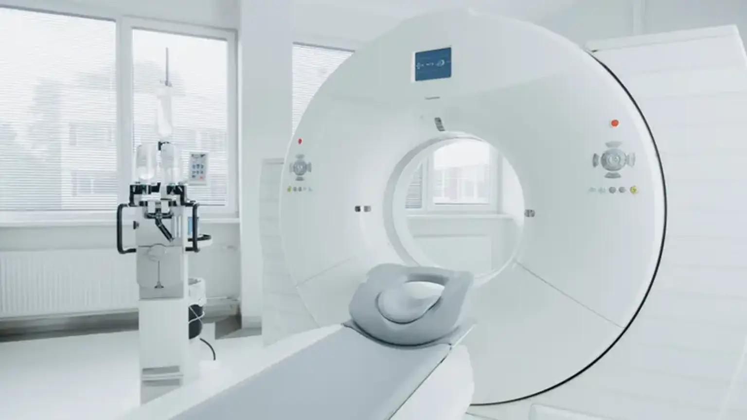
PET-CT Equipment
A large device called a PET scanner has a hole in the middle that resembles a circular donut. It resembles a CT or MRI machine. The machine records the energy emissions from the radiotracer in your body using many rings of detectors.
Typically, the CT scanner is a sizable donut-shaped device with a small tunnel in the middle. In this little tunnel, you will lay on a small table that moves in and out. The electronic x-ray detectors and x-ray tubes are placed next to one another in a ring, or gantry, which revolves around you. A separate control room houses the computer workstation that analyzes the imaging data. Here, the technologist controls the scanner while keeping a close eye on your exam. Using a speaker and microphone, the technician will be able to hear you and communicate with you.
PET/CT scanners that combine the two technologies have a similar appearance. The images are produced by a computer utilizing information from the gamma camera.
PET-CT Preparation
During the exam, you can either wear a gown or your clothes. Women should always disclose whether they are pregnant or nursing to their doctor and the technician.
Any drugs you are taking, such as vitamins and herbal supplements, should be mentioned to the doctor and the exam technologist. Any allergies, recent infections, and other medical issues should be reported.
Depending on the kind of PET scan, you will be given specific instructions. For this exam, diabetic individuals will receive additional instructions.
Ask your radiologist or physician how to proceed if you are nursing during the exam. It might be beneficial to pump breast milk in advance and save it for use once the PET radiotracer and CT contrast agent have left your body.
Metal things, such as jewelry, eyeglasses, dentures, and hairpins, should be left at home as they could interfere with CT imaging. Hearing aids and removable dental work may need to be taken out.
Usually, a whole-body PET/CT scan will urge you to avoid eating for several hours before. Eating can affect how the PET tracer is distributed throughout your body and result in a less-than-ideal scan. Following the dietary advice is very crucial because failing to do so may result in you having to repeat the scan on a different day. For many hours before the scan, you should refrain from consuming any liquids that contain sweets or calories. You are advised to sip water instead. Your doctor might give you specific advice if you have diabetes. All the medications you are taking should be mentioned to your doctor. List any allergies, especially those to iodine or contrast materials.
Your doctor will examine you for any conditions that can make getting intravenous contrast material riskier.
How Does PET-CT Work?
The body is imaged using x-rays during routine x-ray tests. Radiopharmaceuticals or radiotracers, which are radioactive substances, are used in nuclear medicine. This substance is often injected into your bloodstream by your doctor. You might also ingest it or breathe it in as a gas. In the area being examined, the material builds up and emits gamma rays. These energies are detected by specialized cameras, which then produce images of your organs and tissues' appearance and function with the help of a computer.
Only radiotracer injections are used in PET scans. Nuclear medicine focuses on internal bodily processes, as opposed to other imaging modalities. These could be metabolic or other chemical activity levels. Hot spots are regions with higher intensity. These might exhibit high levels of radiotracer concentrations as well as intense chemical or metabolic activity. Less intense regions, or cold spots, signify a lower radiotracer concentration and less activity.
PET-CT Scan
Nuclear medicine exams are performed by doctors on inpatients and outpatients.
On an exam table, you will lie down. An intravenous (IV) catheter will (if required) be inserted into a vein in your hand or arm by a nurse or technologist.
The radiotracer is normally absorbed by the area being examined after traveling through your body for 30 to 60 minutes. You will be asked to remain still, without moving or speaking.
A contrast agent that localizes in the intestines and aids the radiologist in interpreting the test might be given to you to drink.
You'll be positioned within the PET/CT scanner so that imaging may start. For the duration of the imaging, you must stay still. The CT examination is conducted before the PET scan. Sometimes a PET scan is followed by a second CT scan with intravenous contrast. Less than two minutes pass during the CT scan. It takes 20 to 30 minutes for the PET scan.
Typically, the entire scanning process takes 30 minutes.
Additional tests with other tracers or drugs may be done, depending on the area being examined. The operation can take three hours as a result of this. For instance, if your heart is being evaluated, you might get a PET scan before and after working out or before and after getting an IV medication that improves blood flow to the heart.
You might have to wait until the technician decides whether more images are required after the examination. The technician may occasionally take more images to clarify or enhance the visualization of particular locations or structures. The need for more pictures does not always indicate that the exam was flawed or that something is out of the ordinary. You shouldn't be concerned about it.
Your technician will typically remove any intravenous (IV) lines you may have been given for the treatment. If you need an IV line for another treatment that day, the technologist will leave it in place.
What Will I Experience During and After PET-CT?
Most nuclear medicine procedures, except intravenous injections, are painless. Significant discomfort or negative effects are rarely reported.
When the technician places the needle for the intravenous line in your vein, you may feel a little pinprick. During the radiotracer injection, you can experience a chilling sensation traveling up your arm. There are typically no further negative effects.
The technician might insert a catheter into your bladder during some procedures. This could lead to brief discomfort.
During the exam, it's essential to keep calm. Nuclear imaging is painless. However, staying still or in one place for a prolonged period might be uncomfortable.
If you're scared of being in enclosed spaces, you can experience anxiety during the exam.
You can continue with your regular activities after your checkup unless your doctor advises you differently. Before you leave, a technologist, nurse, or doctor will give you any necessary specific instructions.
Through the process of radioactive decay, the tiny amount of radiotracer in your body will gradually lose its radioactivity. In the hours or days following the test, it may also leave your body through your urine or feces. To help the material leave your body, drink a lot of water.
Who Interprets PET-CT Results?
The images will be interpreted by a radiologist or another physician with specialized training in nuclear medicine, who will then send a report to your referring physician.
A radiologist with specific experience in interpreting CT scans will provide a report to your referring doctor if your physician has requested a diagnostic CT.
PET-CT Risks
- Nuclear medicine exams provide a comparatively low radiation exposure because they only use a little dosage of the radiotracer. For diagnostic tests, this is acceptable. The very modest radiation risk is therefore outweighed by the potential advantages of an exam.
- Nuclear medicine diagnostic techniques have been used by doctors for more than 60 years. Such low-dose exposure has no known long-term negative effects.
- Your doctor will constantly balance any potential hazards with the advantages of nuclear medicine treatment. Before starting treatment, your doctor will go over the important risks with you and provide an opportunity for questions.
- Radiotracers seldom cause allergic reactions, which are often minor. Always report any allergies you may have to the nuclear medicine staff. Describe any issues you might have encountered during prior nuclear medicine tests.
- Minor discomfort and redness could result from the radiotracer injection. This ought to be quickly resolved.
- Women should always report to their doctor and the radiology technician if they think they could be pregnant or nursing.
What are the PET-CT limitations?
Procedures in nuclear medicine can take a while. The radiotracer may take several hours or days to build up in the desired area. Additionally, imaging procedures could take up to several hours. Some procedures can be significantly shortened by improved equipment.
Nuclear medicine images might not have the same level of image resolution as CT or MRI images. Nuclear medicine scans, on the other hand, are more sensitive for several reasons. Other imaging methods frequently cannot produce the functional information they produce.
Patients with diabetes or those who have eaten recently may get negative test results due to altered blood sugar or blood insulin levels. A person who is extremely obese might not fit through the entrance of a typical PET/CT unit.
Conclusion
In oncology, cardiology, and neurology, PET has become a significant molecular imaging method with practical therapeutic uses throughout time. Combining anatomical and functional imaging in the form of PET/CT has marked a significant advancement in medical imaging. It is quickly becoming a potent imaging tool and has taken center stage in oncologic imaging. Future developments in the clinical use of more advanced PET tracers will enable more potential clinical applications. Understanding these approaches' clinical applications, benefits, and limitations is crucial for making the best use of them. Ultimately, proper diagnosis, therapy evaluation, surveillance, and prognostication will be achieved through the best use of multimodality imaging systems and particular imaging agents.