Peyronie's disease

Last updated date: 13-May-2023

Originally Written in English

Peyronie's disease

Overview

Peyronie's Disease (PD) is a penile fibrosing illness that causes plaque accumulation and penile deformity. Once thought to be rare, Parkinson's disease has lately been discovered in up to 13% of males, and it can have a significant impact on both patients' and their partners' sexual and psychological function. While the cause of Parkinson's disease is unknown, it is assumed to be caused by a stressful event followed by abnormal fibrosis or dysregulated wound repair.

A complete history and physical exam, emphasizing on the penis in both flaccid and erect phases, are performed on males who come with PD. Erectile dysfunction (ED) and various other comorbidities are frequently connected with Peyronie's Disease (PD). 

Oral, topical, intralesional, mechanical, and surgical therapy are all available for Parkinson's disease. In general, there is minimal evidence to support the effectiveness of oral, topical, and mechanical therapy.
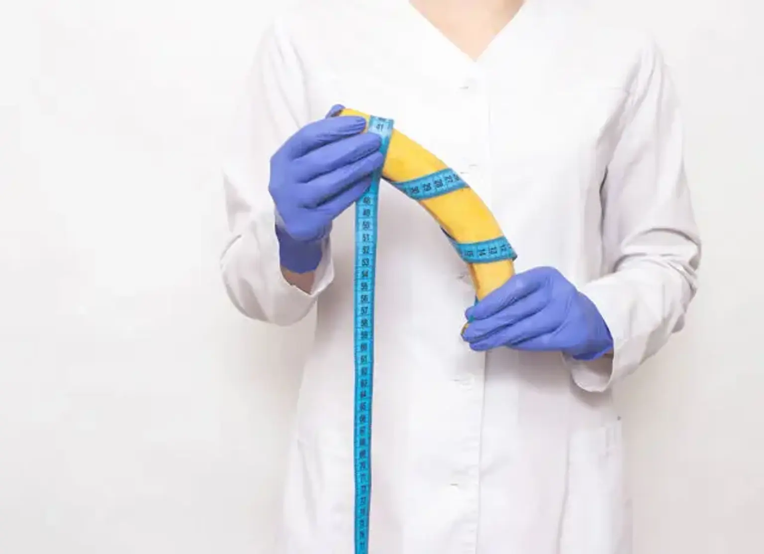
 

Peyronie's disease definition

Peyronie's disease definition

Peyronie disease (PD) is named after François Gigot de la Peyronie, a French physician who diagnosed the ailment in 1743. PD is distinguished by penile shaft curvature , which is frequently preceded by painful erections and is accompanied by a fibrotic region.

The distinctive angulation is usually associated with erectile dysfunction (ED), either as a result of penile shaft buckling with intromission or due to a lack of stiffness distal to the region of related fibrosis. The compromise to the distal penile blood supply appears to be the cause of this lack of stiffness.

Peyronie disease, which is common in many sexual problems, can obviously cause great distress in sufferers. Problems emerge not just from the deformity and the resulting decreased appearance and function, but also because PD is connected with erectile dysfunction, which is an unpleasant disease in and of itself. This chapter will discuss the etiology and pathophysiology, epidemiology, history and examination results, and typical evidence-based therapies.


Epidemiology

The prevalence of Peyronie's Disease ranges from 0.3 to 20.3 percent. There is variation by nation, cohort, and age and race of patients in the research. Some of the variation can also be attributed to the fact that studies may employ various diagnostic criteria or data gathering procedures, such as patient or physician reporting strategies.

the frequency of Parkinson's disease among 11,420 males in the United States, with reported rates of up to 13% Due to the shame of the condition, it would be impossible to precisely establish the incidence of PD in any particular group; it is probable that the true prevalence is closer to the upper end of what has been recorded.


Etiology 

To recognize the genesis and pathophysiology of Peyronie disease, a fundamental understanding of penile anatomy is essential. The penis is made up of two erectile chambers known as the corpora cavernosa, which span the length of the penis. These chambers fill with blood during an erection, increasing the size and stiffness of the penis.

Each corpus cavernosum contains an elastic fiber sheath called the tunica albuginea, which is mostly made up of type 1 collagen fibers. A condensation of the tunica albuginea in the midline separates the two corpora cavernosa, generating a septum that joins to the top and bottom of the penis.

A fibrous plaque arises in the tunica albuginea of the corpora cavernosa in Peyronie disease. The smooth upward curve of the erect penis is changed to a more kinked look by this inelastic plaque. The precise method by which the plaque develops has yet to be determined; nevertheless, various research and ideas have sought to throw some light on the pathophysiology.

The plaque is said to form as a result of damage to the penis, according to popular belief. Buckling pressures cause overstretching and ultimate delamination of the tunica albuginea fibers where the septum connects when the penis is excessively compressed or twisted. Damage to the microvasculature also occurs, leading to blood extravasation and the activation of an inflammatory cascade.

The inflammatory process begins with a shift in collagen type from type I to type III in the tunica albuginea, followed by the deposit of fibrin. Invading macrophages produce elastase, which breaks down the elastic fibers in the sheath of the corpora cavernosa, diminishing the tunica albuginea's elastic qualities.

This idea helps to explain why PD arises after penile trauma. Unfortunately, many individuals have no recollection of any major trauma. Microvasculature damage, chromosomal instability of fibroblasts implicated in plaque development, abnormalities in the inducible nitric oxide pathways, and patient human leukocyte antigen (HLA) subtype have all been proposed as explanations for plaque formation. 

The truth is that we cannot predict who will get PD; nonetheless, it is more probable in vulnerable men who participate in strong sexual or nonsexual activities (such as certain sports) that produce micro-injuries to the penis. The following modifiable and non-modifiable risk factors have been documented with differing degrees of evidence:

  • Penile injury

Previous penile damage is a major predictor of developing PD. Iatrogenic damage, such as catheterization, cystoscopy, and TURP, has been associated with an increased incidence of PD. There is also some evidence that RP is connected with a 15.9 percent likelihood of acquiring Parkinson's disease.

  • Connective tissue disorders

PD is frequently linked with other fibroproliferative illnesses, including Dupuytren contracture and plantar fasciitis, implying a major pathophysiologic and genetic commonality between these superficial fibrosing disorders. 

Numerous investigations have found a relationship between Parkinson's disease and various systemic fibrotic disorders such as idiopathic pulmonary fibrosis, Paget's disease of the bone, retroperitoneal fibrosis, scleroderma, polyfibromatosis, and systemic sclerosis.

  • Diabetes

Men with diabetes-related erectile dysfunction were four to five times more likely to develop Peyronie disease than the general population. Other studies have found a comparable greater frequency of Parkinson's disease in this cohort. Diabetes is thought to exacerbate the fibrotic process involved in Parkinson's disease.

  • Smoking and alcohol

There is some evidence that smoking is linked to Peyronie's Disease, while the relationship between the quantity of smoking and risk is unclear. Similarly, the literature on alcohol remains divided.

  • Age

Men in their 60s are the most typically affected by Peyronie's Disease, with an average age at diagnosis ranging from 52 to 57 years old. Although Peyronie's Disease can strike at any age in adulthood, some people as young as 21 years old have been reported to be afflicted. 


Pathophysiology

As previously stated, the tunical fibrosis of the corpora cavernosa, which results in plaques, is the hallmark of Peyronie's Disease. When the penis is flaccid or erect, the tunica is responsible for its pliancy and stiffness. Animal and cadaveric investigations have reported ultrasound and histological alterations in the tunica. The major characteristics are disorganized, fractured, and sparse elastin fibers and collagen deposition in exceptionally dense clusters.

Other results include the aberrant presence of fibrin, which was found in 95 percent of PD plaques but not in control samples in one investigation. The increased cellularity around the tunica is hypothesized to be caused by abnormal inflammation and wound healing, which results in perivascular lymphocytic infiltration around or inside the tunica itself.


Clinical picture

Clinical picture

Peyronie's Disease has two stages: acute and chronic. The penile deformity progresses in the first 6-18 months, with concomitant discomfort in either the erect or flaccid states. This is the acute phase, and no therapy is advised at this time.

The chronic phase is characterized by a symptom plateau lasting at least 3-6 months; the deformity stays stable with partial or total pain relief. Treatment is more effective during the chronic period. The pain appears to be the differentiating feature between the stages. They documented pain relief in all 246 patients and pain elimination in the majority within a year of presentation; after 18 months, 89 percent were pain-free.

History

A comprehensive history and examination are critical in forming an accurate diagnosis of Parkinson's disease. This may be a sensitive and unpleasant issue for patients since the effects of Peyronie's Disease on a man's sex life and relationships can be severe. As a result, physicians must be compassionate and understanding when investigating the patient's symptoms and extracting their views, fears, and expectations about their condition. The condition's psychological effect may necessitate the involvement of counselors and therapists.

A full presenting complaint, as well as previous medical and surgical history, should be included in a complete history. Sexual history is very important in Parkinson's disease. The following items are deemed necessary while taking a PD history:


Timing: Onset and progression of symptoms. Is the patient in the acute or chronic phase?

  • Deformity: How would the patient describe the penile deformity? What is the direction and degree of curvature? Is there an hourglass deformity, hinge effect, or any other concerning abnormality?
  • Erection: the degree of rigidity, ability to sustain and maintain an erection, presence of nocturnal erections.
  • Pain: If pain is present, is it associated with the flaccid or erect state or both?
  • Trauma: History of penile trauma/fracture, urologic procedures or surgeries
  • Family history: Any family history of Peyronie’s disease or Dupuytren's disease?
  • Medical/Surgical history: Diabetes, hypertension, and cardiovascular disease.
  • Social history: Sexual history, smoking, and recreational drug use.
  • Psychosocial factors: Impact of the condition on the patient’s mood, relationships, and self-esteem.


Examination

Examination

An accurate assessment of penile deformity is essential for establishing a baseline and planning therapy. The penis should be checked while it is flaccid and erect. This offers a better grasp of the amount of the malformation as well as confirmation of what the patient is experiencing. Because patient estimations are notoriously incorrect, an objective measurement of the degree of curvature is also essential for disease monitoring and therapy development.

The penile stretch length is measured with the penis flaccid. The penis is gripped at the glans and gradually pushed away from the body at 90 degrees. According to the American Urology Association standards, examination in the erect state can be conducted following intracavernosal injection of vasoactive drugs.

In fact, ultrasound following intracavernosal injections is superior to photos or vacuum erectile device-assisted erection in diagnosing the kind and degree of PD deformity. Duplex Doppler penile ultrasonography is the most objective way to examine plaque size, location, and calcification. This approach can also help determine the etiology of ED if it is present.

Because Parkinson's disease involves a clinical diagnosis, diagnostic laboratory testing has a limited function. They are useful, however, when a hypogonadal etiology is suspected. Given the close association with other illnesses, no workup would be complete without evaluating the patient for comorbidities including diabetes, cardiovascular disease, and other fibroproliferative ailments like Dupuytren's disease, plantar fasciitis, and scleroderma, among others.


Diagnosis

Diagnosis

The history and physical exam are the most crucial aspects of assessment. A Peyronie disease workup does not require any laboratory or imaging testing.

Thin-section, high-resolution T2 MRI without fat suppression has also been demonstrated to be a good imaging technique for penile pathology, including Parkinson's disease. Plaques will appear as thicker tunica albuginea with low signal intensity. Calcifications are undervalued. Given the high cost and limited availability, the value of MRI in the regular workup for Peyronie's Disease is debatable.


Management

Clinicians should only analyze and treat a man with Peyronie disease if they have the necessary skills and diagnostic tools to diagnose, advise, and treat the problem. Clinicians should next go over all of the available therapy choices, as well as the recognized benefits and risks or costs associated with each one.

Comprehensive counseling about the nature of Parkinson's disease and the normal illness course may be enough to allay some patients' fears. Before the intervention, there is no agreed-upon minimum curvature. In addition to any objective measurements of curvature and erectile function, the patient's anguish over symptoms and degree of worry, as well as his readiness to pursue various methods of therapy, should be thoroughly examined in the decision-making process.


Nonsurgical Management

In the non-surgical treatment of Peyronie's Disease, a range of oral and injectable medications are used. However, only a small number of these interventions have been backed by well-designed, double-blind, placebo-controlled, randomized studies. A limited number of patients recruited in research, resulting in poor power, variability of treatments and duration of follow-up, and a diversity of study outcomes are all obstacles to having adequate literature to support therapy. The defining symptoms of the active period include discomfort with or without erections.

Nonsteroidal anti-inflammatory drugs (NSAIDs) may be prescribed by doctors to assist control discomfort during this time. Oral treatment using vitamin E, tamoxifen, procarbazine, omega-3 fatty acids, or a vitamin E/L-carnitine combination. There have been studies on all of these potential drugs, some with positive results, but the most majority are non-randomized, uncontrolled case series of varying sizes.

Intralesional injections are a feasible non-surgical approach for the treatment of Parkinson's disease. Intralesional administration of collagenase Clostridium hystolyticum, interferon-a-2b, and verapamil. This is frequently done in conjunction with modeling by both the therapist and the patient in order to lessen penile curvature. It is advised for people with a curvature of more than 30 degrees but less than 90 degrees and normal erectile function.


Surgical Management

Surgical Management

A deformity that interferes with normal sexual relations, a stable deformity without discomfort for at least three months, significant plaque calcification, and failed nonsurgical treatments are all indications for surgical treatment of Peyronie's Disease. Procedure selection should take into account the kind and location of the deformity, the degree of the deformity, baseline erection function, penile dimensions, the surgeon's experience, and the patient's desire. Penile plication, plaque incision or excision with graft, and penile prosthesis insertion are surgical alternatives.

Patients considered for penile plication should have appropriate preoperative penile stiffness with or without medication, adequate penile length for good intercourse, simple curvature of fewer than 60 degrees, and no hinge defect or hourglass deformity. The Nesbit operation was the first to be utilized for plication, and versions of this approach have since been employed to repair Parkinson's disease.

The surgical procedure entails creating a midline or circumcising incision and inserting plicating sutures in the tunica albuginea with permanent, synthetic braided sutures contralateral to the location of maximum curvature. This technique has several variations; please read the list of all potential penile operations below. The breadth of this article does not allow for a description of each approach. After 4 to 6 weeks of healing, patients can resume sexual activity.

Potential complications include perceived loss of penile length due to shortening of the long side of the penis, unstable penis, persistent pain, persistence or recurrence of penile curvature, penile hematoma, urethral injury, and sensation loss for neurovascular bundle injury during dorsal plication procedures.

Plaque incision or excision and grafting: indications for this technique include full to near-complete pre-procedural rigidity, which may be accompanied by or without oral pharmacotherapy, complex penile deformity, simple deformity greater than 60 degrees, large plaque, destabilizing hourglass, or hinge effect, and short penile length.

An incision is created on the plaque at the point of maximum curvature on the convex side of the penis for an incision treatment. The graft material is then put into the defect to assist extend the penis's shorter side. Excision entails removing part or all of the plaque and inserting a graft into the defect. Autologous grafts, allografts, xenografts, and synthetic grafts are some of the graft materials available.

Penile prosthesis placement. This method is excellent for men who have ED, severe deformity that is resistant to medical treatment, or substantial penile instability. Plaque and corporal fibrosis can make prosthesis insertion more difficult. This can make corpora dilatation difficult and raise the risk of corporal perforation. Prosthesis options include 2 or 3 pieces inflated prosthesis and malleable prosthesis. For further information about prosthesis installation surgery, please check the erectile dysfunction section.


Differential Diagnosis

  • Balanitis:

In urology clinics, 11 percent of adult men and 3 percent of boys have glans penis inflammation. It is caused by bacterial invasion of the soft tissue in boys. In males, it is caused by a combination of poor genital hygiene, intertrigo, irritating dermatitis, maceration damage, and bacterial or candidal overgrowth, and it is treated with better hygiene practices, avoidance of genital irritants, and better glycemic management in diabetics.

  • Chordee

With or without hypospadias, ventral penile curvature occurs. Chordee is thought to be a halt in normal embryological development. Surgical treatment is often undertaken after six months of age.

Intraoperative artificial erection tests should be performed at the time of repair to determine the point of maximum curvature and then penile plication should be performed. If there is also a penile fracture: trauma or contusion, or a fracture of the tunica albuginea during sexual intercourse, hypospadias should be repaired at the time of surgery.

 The typical scenario involves the sound of a "pop" followed by an immediate beginning of intense pain and penile detumescence. On examination, penile edema, ecchymosis, and a potential palpable defect in the corpora cavernosa are common. Physical exam and history can be used to make a diagnosis, as can an MRI of the penis, which has a high sensitivity for detecting penile fractures.


Conclusion 

Peyronie disease (PD) is defined by a curvature of the penile shaft, which is commonly associated with plaque or fibrosis and is preceded by painful erections. PD is characterized as a wound-healing condition of the tunica albuginea, resulting in tunica albuginea fibrosis or scarring.The precise etiology is unknown, however, several explanations have been proposed. 

There are several management alternatives. In the non-surgical care of Parkinson's disease, a wide range of oral and injectable medications are used. However, only a small number of these treatments are backed by well-designed randomized studies. There are other injectable and surgical treatments for removing the plaque.

PCPs must be able to have an open and nonjudgmental talk with their patients about their sexual activity and happiness with their sexual capacity, as this is frequently the first checkpoint when it is revealed that a patient has a problem such as PD. From that point on, PCPs must realize that penile curvature or plaques are curable issues that should be referred to a urologist for further therapy.

At that time, the urologist can provide a number of remedies to the condition, and the patient's care can go ahead. The capacity to identify the patient with the illness and get them examined by the appropriate medical expert will be at the heart of the problem. This will mostly fall under the purview of the patient's primary care physician.