Pituitary tumors
Overview
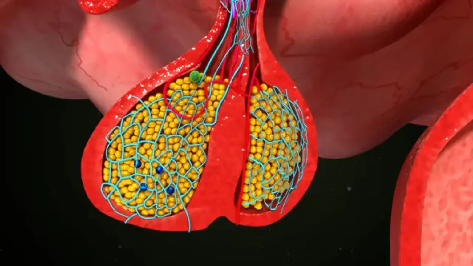
Different hormone gene products are expressed by anterior pituitary cell types. ACTH, GH, PRL, TSH, FSH, and LH are among them. Pituitary adenomas are distinguished by both excessive proliferation of one of these differentiated cell types and abnormally high levels of specific hormone hypersecretion. In tightly regulated regulatory loops, the six pituitary trophic hormones govern respective target hormone synthesis and endocrine gland function. Adenoma hypersecretion is generally caused by a malfunction in hormone production or secretion.
Each of these cell-specific adenomas is linked with a benign sellar mass as well as a distinct clinical condition caused by the hypersecretion of the hormone. Thus, lactotroph adenomas cause hyperprolactinemia with hypogonadal characteristics and galactorrhea.
Corticotroph adenomas hypersecrete ACTH, resulting in hypercortisolism symptoms such as central obesity, hypertension, hyperglycemia, infections, and psychological disorders. Somatotroph adenomas oversecrete GH, resulting in acromegaly symptoms such as acral alterations, arthritis, hypertension, headache, soft tissue edema, and hyperglycemia. Thyrotroph adenomas that produce excessive TSH are exceedingly uncommon and may be linked with moderate hyperthyroidism and goiter.
Tumors derived from gonadotroph cells seldom hypersecrete complete FSH or LH, preferring to produce glycoprotein subunits or no extra hormone. Because of the compressive effects of the increasing bulk, these adenomas are frequently detected accidentally and are commonly linked with hypogonadism and pituitary failure.
Pituitary Adenoma definition
Pituitary adenomas are anterior pituitary tumors. The majority of pituitary tumors are slow-growing and non-cancerous. They are categorised according to their size or cell of origin. Pituitary adenoma is classified into three types based on size: microadenoma, macroadenoma, and gigantic tumors. A microadenoma is a tumor that is less than 10 mm in size, whereas a macroadenoma is a tumor that is greater than 10 mm in size.
Pituitary tumors that are larger than 40 mm are known as giant pituitary tumors. There are functional pituitary adenomas in which the cell type that makes them induces an increase in the production of one or more anterior pituitary hormones. Nonfunctioning adenomas, on the other hand, do not release hormones but might possibly compress the surrounding parts of the anterior pituitary, resulting in hormonal deficits.
Pituitary adenoma patients should be assessed by a multidisciplinary team that includes endocrinology, ophthalmology, and neurosurgery.
Nonfunctional adenomas (null cell adenomas)
These are the most prevalent sort of tumor. They do not produce additional hormone. You may not have any symptoms until the tumor has reached a specific size. When a tumor becomes large enough, it can cause headaches and visual issues. Normal pituitary cells can be crushed by large pituitary tumors. This results in symptoms caused by low hormone production.
Prolactin-producing tumors (prolactinomas)
These benign tumors are also rather prevalent. They produce an excessive amount of prolactin. If you are a woman, elevated prolactin levels might cause your menstrual cycle to be erratic or possibly halt. Even if you are not pregnant or breastfeeding, some tumors can cause you to produce breastmilk. If you are a guy, you may suffer from erectile dysfunction or a lack of sexual interest. You may also have larger breasts, a low sperm count, or less hair on your body. You may get headaches and eyesight difficulties over time.
ACTH-producing tumors
The adrenal gland is stimulated by ACTH (adrenocorticotropic hormone) to produce hormones that alter metabolism. These are known as glucocorticoids. They alleviate redness and swelling (inflammation) throughout the body. They also suppress your immune system. Cushing's disease can be caused by an excess of ACTH. Fat accumulates in your face, neck, back, belly (abdomen), and chest as a result of this disorder. Your arms and legs will also slim out. Purple stretch marks and high blood pressure are other possibilities. These tumors can also cause bone deterioration.
Growth hormone-producing tumors
These tumors produce an excessive amount of growth hormone. Too much growth hormone accelerates the development of practically all bones in youngsters. When this happens, the condition is known as gigantism. Gigantism can manifest as excessive height (over 7 feet), rapid growth, joint discomfort, and excessive perspiration. Acromegaly is a disorder caused by having too much growth hormone in adults.
Epidemiology
Pituitary adenomas are typically discovered by chance on imaging modalities collected for other reasons. Given the insidious nature of pituitary adenomas, their lower size, and accidental diagnosis, estimating the frequency of pituitary adenomas in the general population is difficult. Autopsy and radiological data are used to infer the predicted prevalence of pituitary adenomas.
The prevalence varies depending on the study and the source of information. Pituitary adenomas were found in 16.7 percent of autopsies, 14.4 percent of autopsies, and 22.5 percent of radiology tests, according to a meta-analysis. Several population-based studies from various geographic locations have been conducted over many years to characterize the epidemiology of pituitary adenomas. The most widely cited research from Iceland found a frequency of 115 per 100,000 people.
Etiology
Pituitary adenoma's etiology is uncertain. The majority of pituitary adenomas are sporadic. In an Icelandic investigation of 410 pituitary adenomas, 43 percent were non-functioning adenomas, 40 percent secreted prolactin, 11 percent secreted growth hormone (GH), and 6 percent secreted adrenocorticotropic hormone (ACTH)
Pituitary adenoma is seldom characterized by genetic mutation. Pituitary adenomas in families account for 5% of all pituitary tumors. Mutations in the following genes have been linked to the formation of pituitary adenomas.
MEN1 (multiple endocrine neoplasia): MEN1 is a tumor suppressor gene. This gene's loss of function mutation causes tumors to grow in the parathyroid, pancreatic, and pituitary glands.
Multiple endocrine neoplasia type 4 (MEN4): MEN 4 is caused by a mutation in the cyclin-dependent kinase inhibitor 1 B gene (CDKN1B), and symptoms include pituitary tumors, hyperparathyroidism, testicular neuroendocrine tumors, and cervical neuroendocrine tumors.
Familial isolated pituitary adenomas (FIPA): Aryl hydrocarbon receptor-interacting protein (AIP) mutation is observed in around 15% of all FIPA in adolescence or early adulthood. These tumors are often aggressive, and they frequently release growth hormone, resulting in acromegaly.
History and Physical
The appearance of pituitary adenoma is determined by tumor size and functional state. Pituitary microadenoma is frequently discovered by chance on an MRI of the head. Unless the tumor is hormonally active, patients are asymptomatic.
Pituitary macroadenoma manifests as a tumor with probable hormonal deficit or excess. Pituitary apoplexy is characterized by a sudden bleeding into a pituitary adenoma. It is quite rare. It manifests as a mass effect, with symptoms such as abrupt headaches and eyesight problems, as well as a hormone shortage.
1. Symptoms from Mass Effect:
- Visual impairment: Visual impairment is observed in around 40% to 60% of patients. The pituitary adenoma's suprasellar extension compresses the optic chiasm, resulting in vision field abnormalities. The most common pattern is bitemporal deficiency, followed by homonymous faults. Diplopia can result from oculomotor nerve involvement, and invasive cancers may also impact the fourth, fifth, and sixth cranial nerves.
- Headache: Headache is a frequent symptom of pituitary adenoma, however it is a non-specific symptom.
- Hormonal deficiency: Patients with pituitary macroadenoma may have one or more anterior pituitary hormonal deficits.
Amenorrhea in females and erectile dysfunction in males are symptoms of gonadotropin insufficiency. Adults who are deficient in growth hormone (GH) experience weariness and weight gain. Weight gain, tiredness, cold sensitivity, and constipation are indications of thyroid-stimulating hormone (TSH) insufficiency.
Fatigue, arthralgia, weight loss, low blood pressure, dizziness, nausea, vomiting, and stomach discomfort are symptoms of adrenal corticotropic hormone (ACTH) insufficiency.
2. Functioning or secreting adenomas. The clinical presentation depends on the hormone secreted as described below:
- Prolactin-secreting adenoma: Increased prolactin levels reduce gonadotrophin levels, resulting in infertility, decreased libido, and osteoporosis in both sexes. Females complain of amenorrhea and galactorrhea, while men complain of erectile dysfunction and gynecomastia.
- GH secreting adenoma (acromegaly): Headaches, eyesight problems, an increase in ring or shoe size, arthritis, carpal tunnel pain, and excessive perspiration are among symptoms. Patients' clinical characteristics include coarse facial features, frontal bossing, an enlarged nose, prognathism, an enlarged tongue, and skin tags. At the time of diagnosis, other comorbidities such as hypertension, cardiomyopathy, obstructive sleep apnea, and numerous colonic polyps may be present.
- ACTH-secreting adenoma (Cushing disease): Weight gain, physical weakness, emotional issues, easy bruising, and multiple fractures are all symptoms. A round face, facial plethora, supra-clavicular fat, ecchymoses, and purple striae on the abdomen and armpits are all clinical characteristics.
- TSH secreting adenoma: Palpitations, arrhythmias, and weight loss are among the symptoms experienced by patients. They may have tremors and a goiter on examination.
Pituitary tumors diagnosis
The majority of pituitary adenomas are discovered by chance during normal CT imaging. An MRI with gadolinium is required to distinguish a mass from an aneurysm and to evaluate for bleeding into the mass. It is also necessary to test for hypopituitarism and check for hypersecretion.
Even in asymptomatic individuals, the Endocrine Society clinical practice guidelines advocate a comprehensive biochemical examination. Prolactin, TSH, free T4, follicle-stimulating hormone (FSH), IGF-1, GH, ACTH, estradiol, testosterone, BMP, and fasting early morning cortisol are all measured as part of this examination.
- Prolactin: Prolactin levels are frequently related to the size of the adenoma. Because of the "stalk effect" in macroadenoma or treatments, serum prolactin levels in microadenoma are fewer than 200 ng/ml. A blood prolactin level of more than 200 ng/ml indicates the presence of a prolactin-secreting macroadenoma. Pregnancy, breastfeeding, nipple stimulation, chest wall injuries, hypothyroidism, renal failure, and some drugs, such as antipsychotics, antidepressants, opiates, and antiemetics, cause an increase in prolactin. Macroprolactin is the inactive form of prolactin, commonly known as "large -prolactin." To avoid needless medical therapy, it is critical to diagnose macroprolactin. The serum is precipitated with polyethylene glycol to diagnose macroprolactin (PEG). The detection of free prolactin in excess of 60% indicates prolactinoma.
- IGF-1/GH: Serum IGF-1 levels are used to screen for acromegaly and GH insufficiency. IGF-1 and GH levels can be affected by conditions such as poorly managed diabetes, malnutrition, sepsis, hypothyroidism, hepatic and renal failure. In individuals with acromegaly, the blood GH level can be determined following a 75gm oral glucose challenge test if the IGF-1 level is not found to be high or inconclusive. Acromegaly is confirmed by a non-suppressed GH level of more than 1 ng/dl in the presence of hyperglycemia. Adults with isolated GH deficit should be evaluated further using provocative testing such as the Insulin-induced hypoglycemia test, the glucagon stimulation test, or the recently designed macimorelin stimulation test.
- Cortisol: Fasting cortisol levels in the early morning can be used to detect hypothalamic-pituitary-adrenal (HPA) axis insufficiency. A morning cortisol level of more than 14 mcg/dl indicates a healthy HPA axis. If morning cortisol levels are ambiguous or low, the doctor should order a cosyntropin stim test. Cortisol levels measured at random are ineffective in detecting patients with cortisol excess.
Cushing disease screening tests include late-night salivary cortisol (about midnight), 24-hour urine free cortisol, and a dexamethasone suppression test (DST). If done correctly, late-night salivary cortisol testing has a sensitivity and specificity of more than 90%. DST entails measuring cortisol levels in the early morning after ingesting 1 mg of dexamethasone from 11:00 p.m. to midnight. Hypercortisolemia is indicated by a cortisol level of 1.8 mcg/dl or greater.
Urine free cortisol necessitates 24-hour urine collection accuracy. The interpretation of the cortisol excess screening test should be done with caution because various variables, such as exogenous steroids, depression, excessive alcohol use, and oral contraceptives, might impact cortisol levels.
Once hypercortisolemia has been biochemically verified, the next step is to determine the cause by testing ACTH. A corticotroph adenoma is indicated by hypercortisolemia and increased ACTH. ACTH-producing adenomas are typically tiny, and an MRI of the head may be normal in up to 50% of patients. In the event of a normal MRI head or a pituitary microadenoma smaller than 0.6 cm in size, inferior petrosal sinus sampling (IPSS) is advised to distinguish between ectopic and pituitary Cushing.
IPSS is an invasive operation in which catheters are placed bilaterally into the petrosal sinuses. ACTH levels are measured before and after corticotropin-releasing hormone stimulation (CRH). An rise in ACTH levels that is threefold implies a pituitary source.
- TSH/FreeT4: A low free T4 level combined with a normal or low TSH level indicates secondary (central) hypothyroidism. TSH-producing adenoma will present with high T4 and T3, as well as TSH that is abnormally normal or elevated.
- Low estradiol or testosterone levels along with normal or low LH/FSH levels imply hypogonadotropic hypogonadism. If a woman is taking oral contraceptives, her sex hormones cannot be appropriately understood. FSH levels in postmenopausal women will be biologically increased.
Treatments for Pituitary tumors
Pituitary adenomas necessitate a tight collaboration between an endocrinologist and a neurosurgeon in order to establish a "individualized patient-centric" strategy.
Treatment of Non-Functioning Adenomas
Transsphenoidal resection is recommended in patients with macroadenomas and the following scenarios:
- Visual field deficit due to tumor
- Other visual abnormalities as ophthalmoplegia
- Compression of the optic nerves or chiasm on imaging
- Pituitary apoplexy with visual disturbance
- Loss of endocrine function
- Significant growth of pituitary tumor over time
Most patients see an improvement in their visual problems and hormone abnormalities after surgery. Radiotherapy is an option for people who have a persistent residual or recurring tumor.
Annual endocrine follow-up is necessary in nonfunctional adenomas that do not require surgical care to monitor tumor progression and the development of hypopituitarism. An MRI of the head is performed once every three years for the first three years, and thereafter less frequently if the patient is stable.
Treatment of Individual Functioning Tumors
Prolactin-Secreting Adenoma
The therapy objective is to restore gonadal function and shrink the tumor. In individuals with asymptomatic microadenoma, observation with periodic monitoring of prolactin levels may be an option.
Medical Therapy
Dopamine agonists (DA) are the first-line therapy for tumors that secrete prolactin. Cabergoline and bromocriptine are the only DAs that are currently accessible. Cabergoline is more than 90% effective in lowering tumor growth and regulating prolactin levels. Dizziness owing to postural hypotension, valvular heart problems, and the development of obsessive behavior or mood disorders are all side effects of DA.
If an MRI of the head did not reveal a visible tumor after two years of therapy, DA might be stopped. In these individuals, serum prolactin levels must be monitored periodically since there is a possibility of return or growth after quitting DA.
Surgery
Transsphenoidal surgery is frequently reserved for prolactin-secreting tumors that are resistant to medical therapy, individuals who have adverse reactions to dopamine agonists, and patients wanting pregnancy who have tumors larger than one centimeter in size.
Radiation Therapy
Radiotherapy is seldom utilized in situations with aggressive prolactinomas where surgery and medicinal treatment have failed to limit the adenoma's growth.
GH Secreting Adenoma
The goal is to reduce growth hormone levels to less than 1ug/L while increasing IGF-1 levels to normal age-adjusted levels.
Surgery
The first-line therapy for GH-secreting tumors is trans-sphenoidal surgery. Normalization of IGF-1 is accomplished in 80 to 90 percent of patients with microadenomas and 40 to 60 percent of patients with macroadenomas in the hands of an expert surgeon.
Medical therapy
Patients with persistently increased IGF-1 and GH levels three months after surgery, or non-surgical candidates with invasive tumors, are examined for medical therapy. The first-line therapy for acromegaly is somatostatin analogs (SSA). SSAs that are now available include octreotide, lanreotide, and pasireotide.
Gallbladder sludge and stones, abdominal cramps, gas, diarrhea, and baldness are all side effects of SSA. Pasireotide can cause hyperglycemia in 50 to 70% of people. DA, like cabergoline, is also used to treat modestly increased IGF-1 levels following surgery or as an adjuvant treatment in conjunction with SSA. If the GH level remains elevated, pegvisomant, a GH receptor blocker, can be given alone or in conjunction with SSA to treat acromegaly.
Radiotherapy
Radiation therapy may be utilized as an adjuvant following surgery in individuals with increased IGF-1 levels, but it will take several years to be beneficial.
ACTH Secreting Adenoma
The goal of therapy is to reduce cortisol levels quickly while also reducing complications and death.
Surgery
The first-line therapy for Cushing's disease is trans-sphenoidal surgery. In the hands of a skilled surgeon, first and subsequent procedures have a cure rate of 70% to 90%.
Medical therapy
DA (cabergoline) and SSA (pasireotide, pasireotide LAR) are pituitary medications that reduce ACTH production. Ketoconazole, metyrapone, mitotane, and etomidate all reduce cortisol synthesis in the adrenal gland. Ketoconazole can cause liver damage and QT interval prolongation. Metyrapone is 50 to 60% effective in lowering cortisol levels.
Mitotane is an adrenolytic medication that is most commonly used in individuals with adrenocortical carcinoma. In critically sick patients with severe hypercortisolemia, etomidate is administered intravenously as a bridge to other therapy measures. Mifepristone, a glucocorticoid receptor blocker, can be utilized in some individuals with hypercortisolemia and diabetes.
Bilateral adrenalectomy
Bilateral adrenalectomy can lead to an immediate cure of hypercortisolemia with resultant adrenal insufficiency requiring lifelong treatment. Nelson syndrome, which is radiological pituitary tumor enlargement, can occur in 50 % of patients after adrenalectomy.
Radiotherapy
Radiation therapy is used as a supplement to surgery and medicinal treatments.
TSH Secreting Adenoma
Trans-sphenoidal surgery is the initial favored treatment choice, with cure rates ranging from 50% to 90%. To avoid a thyroid storm, it is critical to treat hyperthyroidism before surgery. Anti-thyroidal medicinal treatment, such as methimazole or SSA, is used to achieve presurgical euthyroidism. Patients who are not cured by surgery can be treated with SSA alone or in conjunction with radiation therapy to reduce TSH levels and tumor growth.
Differential Diagnosis
Differential diagnosis includes other sellar masses as:
- Arachnoid cyst
- Basilar artery thrombosis
- Brainstem glioma
- Cavernous sinus syndrome
- Cerebral venous thrombosis
- Craniopharyngioma
- Dermoid cyst
- Ependymoma
- Glioblastoma multiforme
- Leptomeningeal carcinomatosis
- Low-grade astrocytoma
- Meningioma
- Primary CNS lymphoma
- Rathke cleft cyst
- Tuberculous meningitis
Prognosis
The prognosis of pituitary adenomas is determined by whether or not they are functional. If non-functioning adenomas and prolactinomas are treated quickly with surgery and/or medication treatment, they have a great prognosis. Adenomas that function, such as Cushing's disease and acromegaly, are linked with a number of additional co-morbidities and consequences. There is an increase in mortality, particularly in people with Cushing's disease, when medical or surgical therapy is delayed.
Conclusion
A pituitary tumor is a growth that occurs in the pituitary gland. The pituitary gland is a tiny gland located in the brain. It can be found behind the back of the nose. It produces hormones that influence numerous other glands and processes in your body. The majority of pituitary tumors are not malignant (benign). They are not contagious to other regions of your body. They can, however, cause the pituitary gland to produce too few or too many hormones, producing difficulties in the body.
Pituitary tumors that produce an excess of hormones will drive other glands to produce an excess of hormones. This will result in symptoms associated with each of the various hormones. Many pituitary tumors will also push on the optic nerves nearby. This can lead to eyesight issues.
The majority of pituitary tumors do not produce symptoms. As a result, they do not receive a diagnosis. Alternatively, they are discovered only during a normal brain imaging exam. Around 25% of people may develop minor pituitary tumors without realizing it.