Pleural Disease

Last updated date: 09-Jun-2023

Originally Written in English

Pleural Disease

Overview

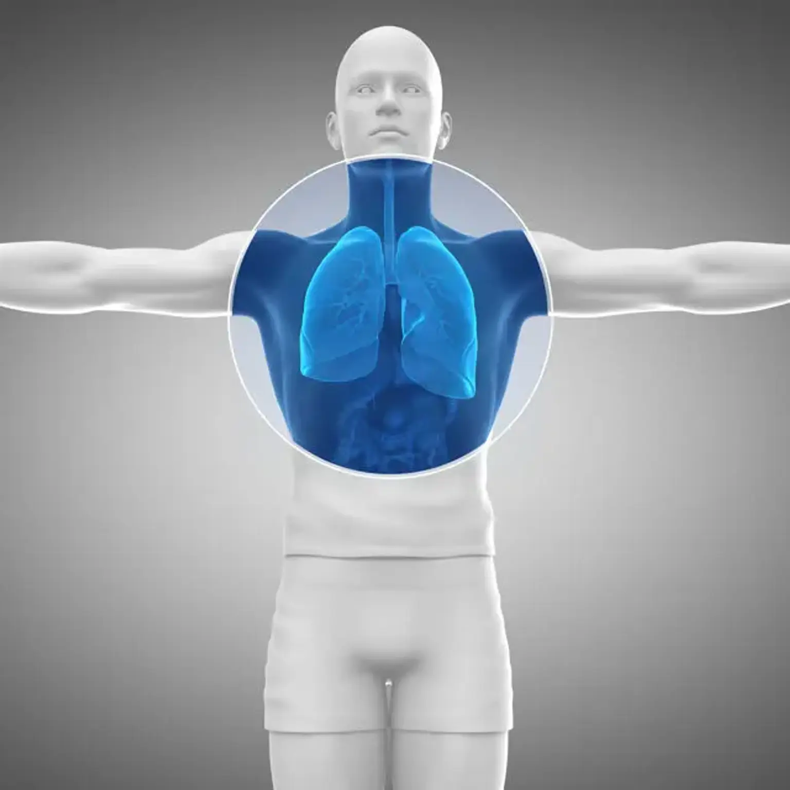
The chest cavity is a compartment bounded by the spine, ribs, and sternum (breast bone), and divided from the abdomen by the diaphragm. Among other vital organs, the chest cavity houses the heart, thoracic aorta, lungs, and esophagus (swallowing route). The rib cage and diaphragm form the wall of the chest cavity.

The pleura, a thin glossy membrane that covers the inside surface of the rib cage and stretches across the lungs, lines the chest cavity. During breathing, the pleura normally generates a little quantity of fluid that acts as a lubricant for the lungs as they move back and forth against the chest wall.

The pleura and pleural cavity are involved in a number of illnesses, each with its own set of origins, symptoms, and therapies.
 

Pleural Anatomy

Pleural Anatomy

The lung and the inner chest wall are separated by two layers of pleura. The visceral pleura is a single mesothelial cell layer that surrounds the lung tissue. They are supplied with blood by the bronchial arteries, which supply the lungs. In contrast to the visceral pleura, the parietal pleura surrounds the inner chest wall and includes stomata that drain pleural fluid to lymphatic capillaries in the loose connective tissue, which also contains systemic blood arteries and nerves.

The lymphatics drain into their respective regional lymph nodes along the sternum or vertebra, finally draining into the thoracic and right lymphatic ducts. The lymphatics have a diameter of 10um-12um, which is large enough to hold intact erythrocytes.

Pleural fluid accumulates when the intake exceeds the removal rate (increased capillary plasma filtration) or when the removal rate is slowed (blockage of lymphatic drainage). Except at the hilum of the lung, where they are continuous, the usual quantity of pleural fluid divides the two pleura by 10 to 20 micrometers. The parietal pleura has connective tissue and is innervated by sensory nerve fibers, but the visceral pleura lacks connective tissue and is innervated by the vagus nerve, making it pain insensitive.

The phrenic nerve supplies the parietal pleura in the central diaphragmatic area, and its involvement may produce referred discomfort to the ipsilateral shoulder. The intercostal nerves of the appropriate intercostal gaps supply the various sections of the parietal pleura.

The pleural membranes and pleural fluid's principal role is to facilitate frictionless movement/sliding of the lung relative to the chest wall. On inspiration, a negative pressure is delivered to the pleural space by the outward movement of the chest cavity and the downward excursion of the diaphragm, resulting in lung expansion. It creates a negative pressure in comparison to air pressure. This gradient permits ambient air to enter the lungs.


Pleurisy

Pleurisy

Pleurisy is a sign of a disease-causing inflammation of the pleura that causes localized chest discomfort. Pleurisy can occur as a result of a primary pleural disease or as a result of a systemic sickness. In the fifth century B.C., Hippocrates defined "pleuritis" as "pain in his side, fever, and shivering" followed by "orthopnea" and tachypnea. Pleuritis was classified into three types: "bilious," "sanguineous," and "dry."


Epidemiology

The epidemiology of pleuritis or pleurisy is determined by the cause. Pleurisy causes vary according to regional, demographic, occupational, comorbid, and other host characteristics.


Etiology 

Pleural inflammation can arise as a result of a number of disorders. Pleurisy develops rapidly (within minutes to hours) in crises such as pneumothorax, acute coronary syndromes, pulmonary emboli, acute pericarditis, and chest wall injuries. Tachypnea and dyspnea are common symptoms of acute and hyperacute causes.

Synpneumonic pleurisy caused by viral and bacterial pneumonia can potentially develop over hours to days. Parapneumonic effusions occur in 20 to 40% of hospitalized pneumonia patients. Thoracentesis is advised for any new onset effusion, particularly those accompanied with probable pneumonia. Familial Mediterranean fever, thoracic endometriosis, and repeated spontaneous pneumothoraces can all cause recurrent pleuritis.

Subacute or chronic pleurisy is commonly associated with rheumatoid arthritis, cancer, or TB (days to weeks). Pleural lymphomas, a single fibrous tumor of the pleura, angiosarcoma of the pleura, pleuropulmonary blastoma, and synovial sarcoma are more prevalent than primary pleural tumors, all of which can cause pleuritic discomfort. Mesothelioma is an uncommon type of pleurisy that affects males over the age of 60 with a history of occupational asbestos exposure. It produces persistent, severe, and refractory pleural discomfort.


History and Physical

Pleuritis History and Physical

Pleuritis causes acute and localized thoracic or shoulder discomfort. Respiratory movements, such as coughing, sneezing, or chest wall/trunk movement, aggravate it. Pain might manifest as dull aching, searing, or simply as a "catch." The duration and frequency of symptoms may help determine the reason. Pleurisy, like other causes of chest discomfort, is diagnosed primarily by a history and physical exam.

The sharpness of the onset (e.g., hyperacute - pulmonary embolism, primary spontaneous pneumothorax, and traumatic pleural inflammation/traumatic pneumothorax), length, and development of symptoms can help narrow down the differential diagnosis. A thorough medical history may aid in the discovery of underlying systemic illnesses such as systemic lupus erythematosus, human immunodeficiency virus infection, and TB infection.

Travel history, tobacco/electronic cigarette usage, alcohol use history, and illicit (particularly intravenous) drug use history may all give indications to the underlying etiology of pleurisy. The afflicted hemithorax has dullness to percussion, reduced breath sounds, and vocal/tactile resonance, which distinguishes effusion from pneumothorax (resonant to percussion). A pleural rub may be detected in auscultation during inspiration and may also be felt, as opposed to a pericardial friction rub, which is audible throughout both inspiration and expiration and can still be heard after respiratory movements have stopped.


Diagnosis

Pleural Disease Diagnosis

Because chest pain is the most prevalent symptom at presentation, it is left-sided, so examination for an acute coronary syndrome is required. If clinical suspicion prevails, a detailed history, ECG, and serum troponin should be considered. Although pleuritic chest pain is not a common symptom of ischemic heart disease, it can occur in acute pericarditis and aortic dissection, both of which can induce localized substernal discomfort or transferred pain to the shoulder.

Because chest pain is the most prevalent symptom at presentation, it is left-sided, so examination for an acute coronary syndrome is required. If clinical suspicion prevails, a detailed history, ECG, and serum troponin should be considered. Although pleuritic chest pain is not a common symptom of ischemic heart disease, it can occur in acute pericarditis and aortic dissection, both of which can induce localized substernal discomfort or transferred pain to the shoulder.

A chest X-ray can be useful, although it can miss tiny nodules and loculated or minor pleural effusions. For a more accurate diagnosis of pneumothorax, acute pulmonary embolism (with intravenous contrast), and pulmonary nodules/masses, a computed tomography (CT) of the chest may be required. The involvement of the pleura should elicit a cytological examination of the pleural fluid for malignant cells.


Management

Pleural Disease Management

Treatment is determined on the underlying cause. Acute coronary syndromes should be handled under the supervision of a local experienced cardiologist. Aortic dissections, particularly type A (ascending aorta), should be treated urgently by a cardiovascular surgeon. If a pneumothorax is present, patients with large pneumothorax (>2 cm from the chest wall at the level of the hilum), hemodynamic instability, new oxygen requirement, significant dyspnea, or those without a known underlying lung disease (secondary spontaneous pneumothorax) or trauma should have immediate air evacuation (traumatic pneumothorax).

For hemodynamically stable initial spontaneous pneumothoraces, needle aspiration (non-inferior to tube thoracostomy) is an option, as is tube thoracostomy for hemodynamically unstable secondary spontaneous pneumothoraces, traumatic pneumothoraces, or hemopneumothoraces.

Pulmonary embolism can be treated in a variety of ways, including at-home anticoagulant therapy, inpatient anticoagulation start, systemic fibrinolytic drugs, and catheter-directed fibrinolytic therapies.

The primary determinants of in-hospital versus out-of-hospital management are oxygen requirement, pain severity, signs of right heart strain on CT pulmonary angiography or echocardiogram, impending or apparent hemodynamic compromise despite volume resuscitation, social conditions that may impede immediate follow-up, and comorbid acute or chronic conditions that may warrant hospitalization.

The treatment and analysis of pleural fluid in the case of pleural effusion is dependent on the effusion's size and imaging properties. Based on the radiographic features, culture, and chemical values of the pleural fluid, classifications of pleural effusion have been proposed.

  • On lateral decubitus film, Category 1 pleural effusions are smaller than 10 mm in diameter. Category 1 pleural effusions have a very minimal probability of bad outcome and should not be sampled.
  • Pleural effusions in category 2 are greater than 10 mm in diameter but do not cover more than half of the hemithorax. It is advised that pleural effusions in categories 2 to 4 undergo diagnostic thoracentesis and empyema be ruled out (must have negative culture, gram stain, and a pH more than 7.20). A category 3 effusion involves more than half of the hemithorax, pleural loculations, or thickening of the parietal pleura.
  • A pH of 7.20 or a positive gram stain/culture can also be used to define a category 3 effusion (empyema)
  • Purulent substance on thoracentesis defines Category 4 effusions.

Thoracostomy drainage is suggested for effusions in categories 3 and 4. Empyema, also known as category 4 effusions, may necessitate surgical evacuation and decortication.

Because of the high mortality rate of category 3 and 4 pleural effusions (30%), additional procedures for full draining of contaminated pleural fluid include intrapleural fibrinolytic followed by drainage and/or surgical decortication.


Prognosis

The prognosis of pleurisy and pleuritic chest discomfort is determined by the etiology and the efficacy of therapy. If untreated, acute coronary syndromes, aortic dissection, pneumothorax, and pulmonary embolism have a significant morbidity and fatality rate. If left untreated or treated ineffectively, category 3 and 4 pleural effusions (also known as complex parapneumonic effusion or empyema) have a significant death risk (30%) and morbidity.

Although lupus pleuritis does not have a high fatality rate, it does have severe morbidity. Lupus pleuritis affects 43 percent of people with systemic lupus erythematosus at some stage in their disease's progression. Malignant pleural illness has a relatively dismal prognosis, with one study having a median survival span of 13 months following diagnosis.


Pleural Effusion

Pleural Effusion

Pleural effusion is the buildup of fluid in the pleural cavity, which is located between the parietal and visceral pleura. It can arise on its own or as a consequence of other parenchymal illness such as infection, cancer, or inflammatory diseases. One of the leading causes of lung mortality and morbidity is pleural effusion.

Every healthy person has a little quantity of pleural fluid, which lubricates the area and allows regular lung motions during respiration. This delicate fluid balance is maintained by oncotic and hydrostatic pressure, as well as lymphatic drainage; disruptions in any of these systems can result in pleural fluid buildup.


Epidemiology

Pleural effusion is the most prevalent pleural illness, affecting 1.5 million individuals in the United States each year. Pleural effusions can be caused by a variety of diseases, including lung diseases such as pneumonia and asbestos exposure, systemic diseases such as lupus and rheumatoid arthritis, or the pleural manifestation of diseases that affect other organs such as congestive heart failure, pancreatitis, or diseases local to the pleura such as pleural infections and mesothelioma.


Etiology 

Based on modified Light's criteria, pleural fluid is classed as a transudate or an exudate. If at least one of the conditions is satisfied, pleural fluid is termed an exudative effusion. 

  1. Pleural fluid protein/serum protein ratio more than 0.5
  2. Pleural fluid lactate dehydrogenase (LDH)/serum LDH ratio of more than 0.6
  3. Pleural fluid LDH is more than two-thirds of the upper limits of normal laboratory value for serum LDH.

Conditions that affect the hydrostatic or oncotic pressures in the pleural space, such as congestive left heart failure, nephrotic syndrome, liver cirrhosis, hypoalbuminemia leading to malnutrition, and the commencement of peritoneal dialysis, are common causes of transudates.

Exudates are commonly caused by pulmonary infections such as pneumonia or tuberculosis, cancer, inflammatory disorders such as pancreatitis, lupus, rheumatoid arthritis, post-cardiac injury syndrome, chylothorax (due to lymphatic obstruction), hemothorax (blood in the pleural space), and benign asbestos pleural effusion.

A pulmonary embolism, which can be exudate or transudate, drug-induced (e.g., methotrexate, amiodarone, phenytoin, dasatinib, generally exudate), post-radiotherapy (exudate), esophageal rupture (exudate), and ovarian hyperstimulation syndrome are some of the less common causes of pleural effusion (exudate).


History and Physical

Depending on the degree of thoracic excursion impairment, a patient with pleural effusion may be asymptomatic or present with exertional dyspnea. Pleurisy is an active pleural inflammation that causes intense, severe, localized discomfort with breathing or coughing. When an effusion forms, discomfort may lessen, giving the impression that the patient's health has improved. Constant discomfort is also a defining feature of malignant illnesses such as mesothelioma. The patient may also experience a cough, fever, and systemic symptoms depending on the origin of the effusion.

Physical examinations might be deceptive. There would be fullness of intercostal gaps and dullness on percussion on that side in significant effusion. Auscultation showed reduced breath sounds as well as reduced tactile and vocal fremitus. The superior side of the effusion has the most marked egophony.

Because pleural effusion can be caused by a variety of diseases, the history and physical examination should include an emphasis on the underlying pulmonary or systemic source of the effusion. In congestive heart failure (CHF), look for jugular venous distension, S3, and pedal edema; in cirrhosis leading to hepatic hydrothorax, look for ascites and other liver disease symptoms.


Diagnosis of effusion 

To confirm the existence of effusion, chest radiographs are important. The effusion results differ depending on the quantity of effusion. A minimum of 200ml of fluid is necessary to obliterate the costophrenic angle, often known as the meniscus sign of a pleural effusion, on an upright posteroanterior (PA) view. This indication, however, can be identified in a lateral view with 50 ml of fluid.

Chest ultrasound is more sensitive and useful for diagnosing pleural effusions, and it also aids in the planning of thoracentesis. To diagnose the etiology of pleural fluid in adults, all unilateral effusions require thoracentesis. This is also known to help the patient's symptoms and speed up recovery.

The disparity is narrowed by determining whether the fluid is an exudate or a transudate. Light's criteria, on the other hand, should be read clinically because they misdiagnose 20% of transudates as exudative. A patient who has been persistently diuresed for heart failure, for example, might raise the pleural fluid protein level and be categorized as an exudate.

Fluid pH, fluid protein, albumin, and LDH, fluid glucose, fluid triglyceride, fluid cell count differential, fluid gram stain and culture, and fluid cytology are all common pleural fluid tests used to diagnose etiology. Exudates are distinguished by increased protein, increased LDH, and reduced glucose.

LDH levels in the pleural fluid can be more than 1000 U/L in TB, lymphoma, and empyema. Low pH (below than 7.2) implies complicated pleural effusion in the setting of pneumonia and nearly invariably necessitates the placement of a chest tube for drainage. Low pH can also be caused by an esophageal rupture or rheumatoid arthritis.

Fluid cell counts in transudates demonstrate that mesothelial cells predominate. Cell counts in parapneumonic effusions, lupus pleuritis, and acute pancreatitis are dominated by neutrophils. Malignancy, lymphoma, TB, sarcoid, chronic rheumatoid pleural effusion, and malignancy are some of the causes of lymphocyte-predominant effusions. Eosinophilia in pleural effusion is uncommon and occurs most often in the presence of air (pneumothorax), blood (hemothorax), a parasite illness, or drug-induced effusion.

The presence of organisms by gram stain or culture leads to the diagnosis of empyema and needs the insertion of a chest tube for pus drainage. The presence of malignant cells in the pleural fluid must be determined by cytology. The sensitivity of pleural fluid cytology in the presence of malignant effusion is about 60% in the initial thoracentesis, and the yield improves with further tries, achieving 95% by three samples on successive days. If a malignant effusion is highly suspected and cytology is negative, medical thoracoscopy with pleural biopsy can be done after two to three thoracenteses to confirm the diagnosis.

Other tests that may be conducted on pleural fluid to ascertain etiology include adenosine deaminase (ADA), which when raised is suggestive for TB in places where tuberculosis is prevalent. The presence of amylase in pleural fluid is indicative of esophageal rupture. An increased NT-proBNP level in pleural fluid may be detected in heart failure. A chylothorax is indicated by the presence of more than 110 mg/dL of triglycerides in the pleural fluid. Pleural fluid is normally straw-colored, but if it is milky white, a chylothorax is suspected. A hemothorax can be diagnosed if the pleural fluid hematocrit is greater than 0.5 times that of the serum hematocrit.

An x-ray of the chest may demonstrate a mediastinal shift to the contralateral chest cavity. If the bronchus is clogged, the trachea may also be displaced to the ipsilateral side. A CT scan can help determine the cause of a problem, such as a malignancy.


Management

Once the etiology of pleural effusion has been recognized, the underlying cause must be addressed. Chest tube drainage, combined with antibiotics, is frequently needed in situations of complicated parapneumonic effusions or empyema (pleural fluid pH less than 7.2 or presence of organisms). Small-bore drains work just as well as large-bore drains for this purpose.

If proper antibiotics and sufficient drainage are not effective, thoracoscopic decortication or debridement may be required. Intrapleural fibrinolytics and DNAse instillation may be utilized to enhance drainage in patients who do not respond to adequate antibiotic treatment and are not surgical candidates.

If a patient has malignant pleural effusion but is not experiencing symptoms, drainage is not always necessary unless an underlying infection is suspected. Pleurodesis (where the pleural space is obliterated physically or chemically by introducing irritants into the pleural space) and tunneled pleural catheter implantation are alternatives for managing malignant pleural effusions that require regular drainage.

Chylous effusions are initially managed conservatively but most require surgery.

It is not recommended to remove more than 1500 ml of fluid in a single try since this might result in re-expansion pulmonary edema. Following thoracentesis, a chest x-ray is required to assess leftover fluid and the existence of a pneumothorax.


Conclusion 

Pleural diseases are ailments that affect the tissue that borders the inside of your chest cavity and covers the exterior of your lungs. The pleura is the tissue, and the pleural space is the narrow gap between its two layers. The pleural area is filled with a little quantity of fluid, and when you breathe in and out, this fluid helps the pleural layers slide smoothly against each other.
Pleural illnesses are classified into three groups based on their causes: pleurisy, pleural effusion, and pneumothorax. Pleurisy is defined as inflammation of the pleura. Pleural effusion and pneumothorax occur when fluid, pus, blood, air, or other gases accumulate in the pleural space as a result of an infection, medical condition, or chest injury.

Chest discomfort, shortness of breath, and coughing are frequent symptoms of all forms of pleural illnesses, although therapy differs based on the nature and severity of the disorder.