Pneumomediastinum
Overview
Pneumomediastinum is described as free air or gas within the mediastinum that nearly always comes from the alveolar space or the conducting airways. Pneumomediastinum has complex pathogenesis.
Pneumomediastinum is more common in children because their mediastinum is comprised of loose tissue. Our mediastina become fibrous as we age. Air may pass through loose tissue more easily than fibrous tissue.
Pneumomediastinum definition
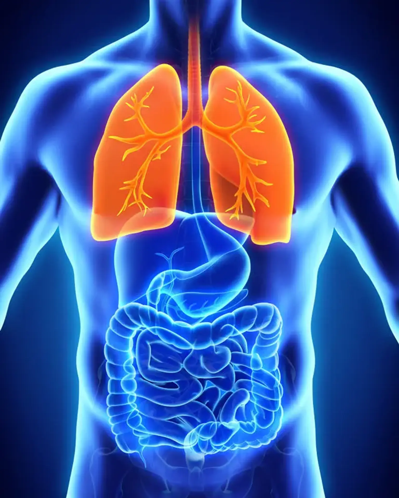
Pneumomediastinum is described as air in the mediastinum and is also known as mediastinal emphysema. The mediastinum is a space in the central thorax that is bounded laterally by the lungs' parietal pleura, superiorly by the thoracic outlet, inferiorly by the diaphragm, anteriorly by the sternum, and posteriorly by the thoracic vertebral column.
The mediastinum contains vital organs such as the heart, tracheobronchial tree, and esophagus. When air extravasates from the airways, lungs, or esophagus and migrates into the mediastinal region, pneumomediastinum occurs. Extravasated air may then dissect adjacent cervical subcutaneous tissues, the epidural space, the pericardium, or the peritoneal cavity.
Air in the mediastinum is also regarded spontaneously appeared when no mechanical ventilation is used or no Valsalva maneuver is done. This phenomenon is seen in a considerable proportion of individuals in whom no cause for the pneumomediastinum can be established. When a causal cause is recognized, the presence of air in the mediastinum is called secondary pneumomediastinum.
Respiratory illnesses, such as asthma or respiratory infections, can cause pneumomediastinum. Asthma, interstitial lung disease, COPD, bronchiectasis, lung cysts, lung cancer, frequent vomiting, and trauma are common causative causes (including iatrogenic).
Recreational drug use, such as cocaine, marijuana, and methamphetamine, has recently been identified as an additional risk factor for pneumomediastinum. Other causes of pneumomediastinum include the use of a powerful Valsalva maneuver, delivery, fast ascent of scuba divers, the presence of foreign materials in the airway with air trapping, anorexia neurosum, sports activities, and inhalation of poisonous gases.
It should be emphasized, however, that in numerous cases, the broader term SPM has been permitted even when a putative causal element has been found. The lack of such explanation has sparked debate among writers, as it is evident that SPM has a better result than secondary pneumomediastinum.
Epidemiology
Pneumomediastinum is extremely uncommon, with a reported frequency as low as 0.002%. Spontaneous pneumomediastinum is particularly frequent in younger guys of tall stature with a low BMI (76 percent of cases). One explanation for this preference for younger adult patients is that their mediastinal tissues are less fibrotic than those of older patients, allowing for simpler air extravasation and subsequent dissemination. Except in newborns, it is not well reported in the pediatric literature.
Air leaks of any form, excluding pneumothorax, occurred in 3.7 percent of patients with sepsis-induced acute respiratory distress syndrome. Pneumomediastinum was seen in up to 10% of adult patients who had had acute chest trauma.
Etiology
Spontaneous or primary pneumomediastinum develops in otherwise healthy people for no apparent reason. Secondary pneumomediastinum refers to any condition in which an underlying cause, such as trauma, can be identified. Alveolar rupture is the most prevalent cause of pneumomediastinum.
Risk Factors for Spontaneous Pneumomediastinum:
- Smoking or tobacco use
- Recreational drug inhalation (cocaine, methamphetamine, marijuana, ecstasy)
Causes of Secondary Pneumomediastinum
Intrinsic Lung and Airway Causes
- Asthma
- Chronic obstructive pulmonary disease (COPD)
- Bronchiectasis
- Interstitial lung disease
- Lung cancer
- Foreign body in the airway
- Increased intrathoracic pressure
- Childbirth
- Excessive vomiting ( anorexia nervosa)
- Excessive coughing (i.e., asthma exacerbation)
- Strenuous physical activities
Iatrogenic Causes
- Endoscopy
- Intubation
- Central venous access procedures
- Thoracostomy
- Chest or abdominal surgeries
Traumatic Causes
- Blunt trauma
- Penetrating trauma
- Blast injuries
Pathophysiology
The Macklin phenomenon describes how spontaneous pneumomediastinum develops in three steps:
- Increased intra-alveolar pressure results in alveolar rupture
- Air dissects into peribronchial and perivascular sheaths
- Air progressively spreads into the mediastinum and surrounding tissue
Excessive vomiting or coughing, hard physical activity, or Valsalva movements can all cause a rise in intra-alveolar pressure (e.g., childbirth). Asthma and COPD produce bronchial constriction, which predisposes individuals to airway damage due to elevated baseline alveolar pressures. Forceful breathing during illicit drug use may result in intra-alveolar pressure alterations or direct barotrauma, both of which raise the risk of pneumomediastinum.
Trauma to the trachea or esophagus is the most common cause of secondary pneumomediastinum. Penetrating trauma (such as stab wounds) or blunt trauma (such as a car collision) can result in tracheal or esophageal injuries. Blasts may potentially produce secondary pneumomediastinum, albeit this is not well documented. Similar injuries can occur as a result of iatrogenic trauma (e.g., endoscopy or endotracheal intubation).
History and Physical
It is critical to collect a history that includes any current risk factors for pneumomediastinum as well as any prior episodes. The following are important historical elements:
- History of smoking or recreational drug use
- History of airway or lung disease
- History of excessive vomiting or coughing
- Recent history of instrumentation
- History of blunt or penetrating trauma
The most prevalent symptom of pneumomediastinum is retrosternal chest discomfort, which can radiate to the neck or back in 60 to 100 percent of patients. Pain is usually severe and is accompanied by considerable coughing or vomiting. Other signs and symptoms include:
- Dyspnea (75% of patients)
- Coughing spells (80% of patients)
- Neck pain (36% of patients)
- Odynophagia
- Dysphagia
- Emesis
- Abdominal pain
Subcutaneous emphysema is the most prevalent physical exam finding (70 percent of patients). The Hamman sign, a mediastinal crunch or click synchronous with heart sounds auscultated above the cardiac apex, is an uncommon but distinctive sign that may be present. Other indicators include:
- Rhinolalia (nasal tone of speech)
- Dysphonia
- Hoarseness
- Neck swelling
- Tachycardia
- Tachypnea
In rare situations, malignant pneumomediastinum, which is severe pneumomediastinum that obstructs the major veins of the heart, can develop, leading in cardiac tamponade physiology.
Subcutaneous emphysema is seen in 70% of individuals with diagnosed pneumomediastinum. Other presenting symptoms include rhinolalia (a nasally sounding voice caused by air dissection into the soft palate), hoarseness, and neck swelling, depending on the underlying condition.
Clinical examination may also reveal tachycardia, tachypnea, or anxiety, with the majority of individuals appearing to be in good health. The existence of a mediastinal crunch or click on auscultation across the cardiac apex and the left sternal boundary synchronous with the heart beat is known as the Hamman's sign, and it should be sought for.
Malignant pneumomediastinum is defined as the buildup of a considerable volume of air in the mediastinum, producing vascular or tracheal blockage and causing tamponade and reduced venous return symptoms. Only a few occurrences with this unfavorable aspect have been documented, and it is possible that this occurrence is unusual.
As previously stated, pneumothorax may be the primary presenting symptom, with a frequency of 40% in certain studies. Again, there have only been a few reports of tension pneumothorax.
Diagnosis
A chest X-ray is generally used to identify pneumomediastinum. Air outlining mediastinal structures is the most prevalent finding (90 percent of cases). Lateral chest x-rays are almost never required. Other symptoms shown on a chest x-ray include:
- Subcutaneous emphysema
- Elevation of the thymus in pediatric patients (spinnaker sign)
- Air surrounding the pulmonary arteries of the lung (ring sign)
- A hyperlucent V shape between the descending aorta and the left hemidiaphragm
- Double bronchial wall
- Continuous diaphragm sign
- Pleural effusion (which may indicate esophageal injury)
If the results of a chest x-ray are ambiguous, a computed tomography (CT) scan of the chest can definitely rule in or rule out pneumomediastinum. CT scans may detect extremely minute quantities of air in the mediastinum or subcutaneous tissues and distinguish between pneumomediastinum and pneumopericardium, which a chest X-ray cannot.
In an emergency, ultrasonography may be useful in detecting pneumomediastinum. The "air gap indication," which appears as a spectrum of sonographic echoes that hide the heart structures underneath, may be present. Other signs of air bubbles in the soft tissue include air near to the heart or diaphragm and subcutaneous hyperechoic white foci.
In most cases with pneumomediastinum, further tests such as bronchoscopy, esophagoscopy, or esophagogastroduodenoscopy are not necessary. Given the clinical situation, obtaining these extra tests is necessary. The lack of aerodigestive damage on multidetector CT in the setting of pneumomediastinum in blunt trauma is sufficient to rule out further traumatic sequelae involving the aerodigestive system. However, after blunt trauma, posterior mediastinal air, involvement of all mediastinal compartments, and simultaneous hemothorax are related with greater mortality.
In an effort to diagnose pneumomediastinum in situations of high urgency, ultrasound of the mediastinum has recently been used in the accident and emergency department.
Many patterns of air outlining intrathoracic structures have been described in the literature, based on chest X-ray or CT findings: thymic sail (elevation of the thymus due to air, most commonly seen in children), ring sign (caused by air surrounding the pulmonary artery or one of its main branches), double bronchial wall, continuous diaphragm sign, or air adjacent to hemidiaphragm or spine.
Rising white blood cells or C-reactive protein are examples of inconclusive test findings. ECG anomalies have also been documented, however they are not clinically significant. Based on their high suspicion and higher prevalence of pulmonary illness, several writers have proposed that pulmonary function tests be conducted in spontaneous cases with no clear aggravating conditions, particularly in youngsters.
Management
Because spontaneous pneumomediastinum is frequently self-limited and rarely recurs, most patients with pneumomediastinum detected on imaging examinations are found to have no significant mediastinal organ impairment and simply require symptomatic therapy. These individuals are usually healthy-looking and hemodynamically stable.
Bed rest, oxygen treatment when required, antitussives, and analgesics are all used to treat these individuals. Prophylactic antibiotics are not usually required in spontaneous pneumomediastinum, although they may be necessary in individuals who have had aerodigestive damage as a result of trauma or instrumentation. Most patients with spontaneous pneumomediastinum can be released with intensive outpatient monitoring or hospitalized for brief inpatient surveillance.
Similarly, blunt trauma patients with isolated pneumomediastinum may be handled conservatively as well. In patients in distress, who are febrile, or who are at risk of subsequent pneumomediastinum, hospitalization is recommended for observation, further diagnostic tests, or surgery.
A contrast swallow study should be performed on patients who have emesis, dysphagia, concomitant traumatic injuries, hemodynamic instability, pleural effusion, symptoms of infection, or pneumoperitoneum. Patients with severe subcutaneous emphysema may require surgical decompression, while those with pneumothorax may require a chest tube. Finally, in cases with malignant pneumomediastinum, video-assisted thoracoscopic surgery or thoracotomy may be required.
Any underlying aggravation of a pre-existing disease, such as asthma or COPD, should be treated appropriately. Most writers believe that pain management and pneumomediastinum stability, as well as the absence of complications such as pneumothorax, should be regarded enough for dismissing such patients.
The buildup of a substantial amount of air in the mediastinum is a rare sequllae of pneumomediastinum that occurs in the majority of instances owing to undiagnosed esophageal/pulmonary trauma that causes significant air leak. Although uncommon, simple pneumomediastinum can progress to malignant pneumomediastinum, resulting in tamponade and airway compression. VATS or even thoracotomy may be required in such circumstances for decompression. Tamponade caused by pneumopericardium is extremely uncommon and may necessitate surgical evacuation through subxiphoid incision or VATS.
Differential Diagnosis
Pneumomediastinum's clinical aspects are similar to those of other disorders of the respiratory, cardiovascular, and gastrointestinal systems. When examining a patient with pneumomediastinum, the following differentials should be examined.
Cardiac
- Acute coronary syndrome
- Pericarditis
- Myocarditis
- Pneumopericardium
Pulmonary
- Pleuritis
- Pulmonary embolism
- Pneumothorax
- Pneumonia
- Traumatic tracheal rupture
Gastrointestinal
- Gastroesophageal reflux disease
- Pancreatitis
- Esophageal rupture
Musculoskeletal
- Costochondritis
- Sternal and rib contusions
- Rib fractures
Others
- Malignancy
- Sickle cell crisis
Prognosis
Although recurrent pneumomediastinum is a danger, it is typically harmless, and morbidity and death are mostly due to the underlying cause. The majority of pneumomediastinum patients resolve on their own, with just symptomatic therapy or monitoring.
Recurrences have been documented, thus more investigation is advised. Spontaneous pneumodediastinum normally resolves on its own, however long-term instances (>2 months) have been observed. Few recurrences of pneumomediastinum have been documented in general, confirming its benign nature.
In situations of recurrence, further diagnostic testing should be performed to rule out any overlooked underlying illnesses, such as pulmonary or esophageal pathology. As previously indicated, several writers have proposed doing i.e., lung function tests, in order to discover missing pulmonary underlying illnesses.
Complications
Other air-leak syndromes, such as pneumothorax, severe subcutaneous emphysema, malignant pneumomediastinum, and pneumopericardium, can aggravate pneumomediastinum. Tension pneumomediastinum can arise, causing considerable vascular compression and restricting venous return, resulting in hypotension and hypoxemia due to a ventilation-perfusion mismatch. Other difficulties may arise as a result of the cause.
Pneumomediastinum-related mortality and morbidity are typically due to underlying illness conditions. Spontaneous pneumomediastinum is typically a self-limiting condition with few significant or life-threatening symptoms.
As with Boerhaave syndrome, the death rate linked with pneumomediastinum may be as high as 50-70 percent (esophageal rupture following vomiting). the development of an air leak is not related with an elevated death rate in patients with sepsis-induced ARDS. Trauma (blunt and penetrating, especially high velocity damage), asthma, and tracheobronchial perforation are further risk factors for high fatality rates.
Symptoms such as chest discomfort, voice alteration, and cough are the most prevalent morbidities associated with pneumomediastinum. In rare cases, tension pneumomediastinum can cause a reduction in cardiac output. It has been noted that laryngeal compression causes stridor. Gas embolism is an uncommon occurrence.
Conclusion
The presence of air in the mediastinum is referred to as pneumomediastinum. This disorder can occur as a consequence of physical trauma or other circumstances that cause air to escape from the lungs, airways, or intestines into the chest cavity. Pneumomediastinum is an uncommon condition that arises when air enters the mediastinum.
A chest X-ray or CT scan of the thorax can be used to confirm the diagnosis. The most common symptom is acute central chest discomfort. Other symptoms include labored breathing, vocal distortion (similar to helium), and subcutaneous emphysema of the face, neck, and chest. Pneumomediastinum can also be distinguished by the normal shortness of breath associated with a respiratory system dysfunction.
On auscultation, it is frequently identified by a "crunching" sound synchronized with the cardiac cycle . As a result of the increased intrapulmonary pressure on venous flow to the heart, pneumomediastinum may potentially manifest with symptoms resembling cardiac tamponade. Because the tissues of the mediastinum slowly resorb the air in the cavity, most pneumomediastinum are treated cautiously.