Posterolateral Endoscopic Lumbar Decompression
Overview
The spine is composed of 24 bones called vertebrae that are placed on top of one another. These bones join together to form a canal that protects the spinal cord.
Intervertebral disks Flexible intervertebral disks exist between your vertebrae. These disks are flat and spherical, and roughly a half-inch thick.
When you walk or run, your intervertebral disks absorb force. They are composed of two parts:
- Annulus fibrosus: This is the disk's strong and flexible outer ring.
- Nucleus pulposus: The disk's soft, jelly-like core.
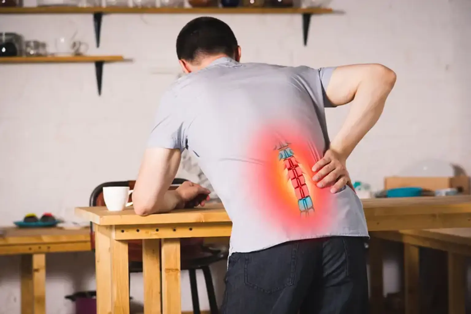
A herniated disk can develop anywhere along the spine, although it most commonly happens in the lower back (lumbar region). It is also known as a bulging, protruding, or ruptured disk. It is one of the most prevalent causes of lower back pain and leg pain, sometimes known as sciatica.
What is Posterolateral Endoscopic Lumbar Decompression?
A lumbar discectomy (posterolateral endoscopic lumbar decompression) is a surgical operation used to remove herniated disc material from the lower back (lumbar) that is pushing on a nerve or the spinal cord. When this surgery is done with an endoscope, it is referred to as a lumbar endoscopic discectomy/decompression.
Because just a little incision is required, it is called a minimally invasive technique. Furthermore, the endoscope (a short metal tube with a camera and light on the end) allows direct visibility through enlarged video pictures, as well as a passageway for the surgical equipment, avoiding the need to tear or cut the patient's muscles.
Most individuals who have a lumbar endoscopic discectomy have less surgical stress and a shorter recovery period than those who have more extensive conventional back surgery because of the minimal damage to bone and muscle tissue.
The treatment of choice for surgical therapy of lumbar disc herniations is posterolateral endoscopic lumbar decompression (PLELD).
Endoscopic discectomy procedures have yielded surgical results comparable to those of other discectomy techniques while providing several advantages such as the avoidance of general anesthesia, preservation of paravertebral soft-tissues, speedier rehabilitation, and overall superior clinical results.
Foraminal disc herniation (FDH) cases provide a unique surgical challenge in terms of nerve root retraction and decompression. Regardless of the surgical technique utilized, the clinical result can be considerably impacted by both the technique of exiting nerve visualization/retraction and the adequacy of decompression.
Who are the best candidates for Posterolateral Endoscopic Lumbar Decompression (PLELD)?
Although endoscopic spine surgery techniques offer a great clinical outcome with little invasiveness, one of their major drawbacks is the limited number of surgical candidates.
Candidates for Posterolateral Endoscopic Lumbar Decompression often have ruptured or bulging discs that cause pain, weakness, or numbness and have not responded to conservative treatment measures such as exercise, stretching, corticosteroids, pain medication, or physical therapy.
This surgery may also be used to alleviate pain and discomfort in those who have damaged discs or persistent radiculitis (pain traveling down the leg). A PLELD is frequently used to treat patients who have progressively deteriorating numbness or weakness in their legs, as well as substantial mobility limitations as a result of their disease.
The following are the primary indications for PLELD:
- Soft LDH as shown on MRI and CT images.
- Probable lumbar radiculopathy based on radiological evidence.
- Nonoperative therapy failure for at least 6 weeks.
The following are contraindications to Posterolateral Endoscopic Lumbar Decompression:
- Extreme central canal stenosis
- Vertebral segmental instability
- Painless weakness.
- Cauda equina syndrome
- Pathologic issues such as tumors or infections.
What are the Advantages of Posterolateral Endoscopic Lumbar Decompression?
You may wonder why I would pick endoscopic versus conventional, and how they compare?
The patient is fully anesthetized during traditional surgery, whereas endoscopic surgery can be conducted with a combination of local and concurrent sedation. While unusual, traditional has the risk of nerve injury. Endoscopic incisions are substantially smaller (7mm as compared to 4-8 cm). Scar tissue grows very slowly when the prolapsed disc tissue is removed, and your chances of recurrence are quite low.
Another significant advantage of endoscopic over traditional surgery is that there is less waiting time. These types of surgeries might have lengthy wait times. That implies extra time in pain and discomfort, whereas endoscopic procedures may be scheduled very fast. With less discomfort and faster recovery (not in a hospital), and less waiting time in agony, it's easy to see why patients prefer this choice.
What conditions can be treated with PLELD?
Endoscopic spine surgery has a long history of growing surgical indications. What endoscopes can perform has expanded as surgical methods have evolved. The primary purpose for endoscopic spine surgery in the early days was confined lumber disc herniation without disc migration or stenosis. Complex instances, such as highly migrating, far-lateral, or recurring LDH, may now be addressed using the novel concept of PLELD.
Posterolateral endoscopic lumber decompression can be used to treat a wide range of spine disorders, including:
- Disc Bulge
- Herniated Disc
- Disc Tear
- Radiculitis
- Radiculopathy
- Brachial Neuritis
- Cervical Disc Bulge
- Cervical Disc Tear
- Cervical Herniated Disc
- Spinal stenosis
How is Posterolateral Endoscopic Lumbar Decompression is performed?
While lumbar disc herniation is the most frequent spinal issue treated by neurosurgeons, foraminal stenosis has always been a challenging task to control. To solve this problem, former open procedures highlighted the necessity to entirely remove the facet, intrinsically weakening the spine and mandating spinal fusion.
PLELD surgery consists of two processes: (1) a percutaneous transforaminal approach under fluoroscopic guidance and (2) selective removal of herniated disc pieces under the endoscopic visual field. Under local anesthetic, the surgery is performed using the usual PLELD approach. To monitor for adverse events, patients are positioned prone on a radiolucent operating table and kept under conscious sedation. The skin entrance is decided by the goal of the treatment. The patient's body size, foraminal measurements, and intended landing place on the disc surface all play a role.
Preoperative MRI and computed tomography (CT) images should be examined to define the skin entrance point, a surgical tract, and the goal landing spot. In addition, intraoperative fluoroscopic imaging can guide transforaminal endoscopic access in accordance with the preoperative strategy. Following local anesthetic infiltration, an 18-gauge needle is placed under fluoroscopic supervision. On the anteroposterior (AP) fluoroscopic view, the needle tip is positioned between the medial and lateral pedicular lines and the posterior vertebral line on the lateral fluoroscopic view, or at the disc surface. To avoid approach-related or procedural pain, a 1 percent lidocaine epidural block is indicated prior to the intradiscal injection of the needle.
The discography is then done using an indigo carmine and contrast combination (1 mL:6 mL). Pathological disc material is stained blue and distinguishable from surrounding neural or soft tissues. This discography procedure is useful for precisely removing herniated disc pieces. The needle is then replaced with a guidewire, and a stab skin incision is created for obturator insertion. The cannulated obturator is introduced into the disc through the foraminal keyhole after sliding over the guidewire.
Significant approach-related pain may arise during the obturator insertion phase due to irritation to the exiting nerve root or any irritated epidural tissues. To alleviate the approaching agony, a serial dilatation process using dilators of varying diameters is used before to obturator insertion. Finally, a bevel-ended working sheath is inserted over the obturator. After carefully inserting the working sheath into the disc, a rigid ellipsoidal endoscope with an eccentrically formed working channel and two irrigation channels is introduced.
The surgeon must first view the intradiscal space and indigo carmine-stained disc material using the endoscopic visual field. The surgeon can validate anatomical alignment by performing first sub-annular decompression using forceps and a radiofrequency coagulator.
It is possible to confirm anatomical layers such as the dural sac, adherent nerve root, surrounding epidural fat, congested annulus, and herniated nucleus. Through an endoscopic view, it is crucial to distinguish herniated disc fragments and diseased soft tissues from compressed neural tissues. The herniated disc has detached from the maternal disc and is firmly attached to the dural sac and nerve root.
Because the fragment is frequently held in place by a fibrotic annular fissure, the annular anchoring should be freed before the herniated disc is removed. It is difficult to remove the attached disc material without first performing an annular releasing process. Micro-forceps and other supplementary devices can be used to grab the released and loosened primary herniated piece.
The surgeon must carefully dissect tissues while keeping the neural tissues safe and decompressed during selective discectomy. The angle of the endoscope and the viewing field can be adjusted as needed throughout these selective releasing and removal operations. One of the advantages of the transforaminal endoscopic method over the traditional open posterior approach is the flexibility of the endoscopic angle and vision.
The surgeon can locate the decompressed neural tissues after removing the primary bulk of the ruptured disc. The procedure's end point can be detected by looking for a robust pulse of the dural sac and free mobility of the nerve root. If the end-point condition is confirmed, certain remnant disc materials that are strongly adherent to the neural tissue can be left. Excessive dissection of the neural tissue may result in unanticipated dural trauma, the most serious consequence of PLELD. The endoscope is then removed, and a one-point subcutaneous stitch is done, followed by a sterile dressing. After screening for any surgical problems, patients can be released within 24 hours.
What is the recovery timeline following Posterolateral Endoscopic Lumbar Decompression?
PLELD is a relatively short treatment that takes about 30-45 minutes from start to finish. Your incision (entry site) is covered with a tiny patch before you go to recuperation. You'll be observed in the recovery room for 2-3 hours before being released home if you pass the initial examination. Several follow-up appointments will be required, including one the day following surgery and three months afterwards.
It is critical that you maintain the care and follow-ups that have been recommended in order to recover fast and, more critically, to avoid returning to previous injury. You'll be able to exercise/play sports in 6 weeks and return to work in about two. The success rate is approximately 90-95 percent.
What are the complications of Posterolateral Endoscopic Lumbar Decompression?
PLELD has crucial and intrinsic problems, despite the good benefits of minimally invasive decompression and its efficiency. To achieve clinical success, surgeons should keep these considerations in mind.
Irritation of the Exiting Nerve Root:
The initial technical challenge of the posterolateral technique is nerve root irritation. To avoid neurological damage, the access needle, serial dilators, and working cannula should be inserted into the disc through the intervertebral foramen.
Inadequate care may cause harm to the intact exiting nerve root, dorsal root ganglion, or even the furcal nerve. If the patient is awake, neural irritation might result in significant approach-related pain and treatment failure. If the patient was unaware of the existing nerve root irritation, it might result in postoperative dysesthesia or possibly severe neurological sequelae.
Dural Tear:
The most critical complication of PLELD may be a dural tear during tissue dissection and discectomy. If unrecognized, an intraoperative dural tear may cause disastrous sequelae. There are certain distinct characteristics of dural tears in PLELD patients that should be noted. First, because the treatment is frequently conducted under continuous saline irrigation, detecting dural tears might be difficult. The surgical field is flooded with fluid, making it impossible to detect cerebrospinal fluid leaking or nerve root herniation.
Second, a dural rip is frequently found on the dural membrane's ventral or ventrolateral side. As a result, a future neurological impairment in the form of motor weakness may ensue. Third, the likelihood of an undiagnosed dural rupture may be greater than that of a typical open microdiscectomy, making postoperative flaring or neurologic impairments more significant. Finally, even if it is recognized during the process, intraoperative closure of the defect is difficult, and open revision surgery is frequently necessary immediately.
The most dependable strategy to avoid this essential problem is to maintain a precise anatomical layer throughout the whole dissection and decompression procedure. The surgeon's ability to achieve technical skill and overcome the learning curve is mostly based on how to prevent dural tears.
Infection (Epidural Abscess or Discitis):
Surgical site infection following PLELD is said to be uncommon when compared to open surgery. PLELD, like any other percutaneous intradiscal technique, has the possibility of causing postoperative spondylodiscitis. Post-PLELD spondylodiscitis may have a more difficult clinical course than predicted. First, infection frequently spreads quicker than in open surgery. Infection symptoms and signs may appear many days after the operation. Typical symptoms include atypical back discomfort, radicular pain, fever, wound swelling, and perhaps meningeal irritation indications.
Concurrent clinical signs include the increase in infection markers such as C-reactive protein, erythrocyte sedimentation rate, neutrophil count, and procalcitonin levels. MRI is not a good early diagnostic tool for septic spondylodiscitis. Signs of infection may arise on imaging examinations such as MRI, CT, and plain radiography after the early postoperative phase. Percutaneous or open disc culture and biopsy can provide a definitive diagnosis.
The use of proper antibiotics and bed rest for many weeks is the first line of treatment. If medication therapy is ineffective, a major surgical incision and drainage may be recommended. Continuous irrigation with prophylactic antibiotics during the surgery is required to avoid this problem. Because of the tiny size and complex constructions of various components, surgical instruments should be properly cleaned. It is best to use disposable tools, such as radiofrequency or other supplemental devices.
Early Recurrence/Incomplete Decompression:
Despite the lack of proof, some detractors may argue that the postoperative recurrence rate is greater than in open microdiscectomy. After a pain-free interval, some individuals may have brief recurring pain. This temporary flare-up normally goes away 4 to 6 weeks following the operation. However, certain individuals with prolonged radicular pain may require a second assessment endoscopic treatment or open surgery.
There are various possible causes for the early return of symptoms. First, due to the existence of hidden disc pieces, endoscopic discectomy and decompression may be insufficient. Inflammatory tissue is present when the herniated component attaches to the dural sac or nerve root. Second, removing the disc may not be enough to decompress the neural tissues. There might be loose disc pieces in the central nucleus that the endoscopic visual field missed. The postoperative rise in disc pressure may result in central core herniation.
Finally, throughout the recovery phase, there is minimal buffering capacity for the increasing intradiscal pressure. PLELD solely involves removing the herniated disc; no laminectomy is performed. When opposed to open surgery, the PLELD approach has an inherent restriction in the lack of posterior decompression. To avoid incomplete decompression, accurate understanding of endoscopic anatomy and detection of the decompression endpoint are required. Reducing recurrent disc herniation requires complete excision of the whole fragment, including the base and tip of the herniated disc.
Hematoma:
The PLELD procedure may cause two forms of vascular insult: retroperitoneal and epidural hematoma. Retroperitoneal hematoma is a rare vascular complication associated with the percutaneous posterolateral approach. A large hematoma in the open retroperitoneal space or psoas muscle may result from a mechanical trauma to the radicular lumbar artery or its branches.
Depending on the volume of hematoma and the patient's condition, an emergency vascular intervention or open surgical evacuation may be necessary if this occurrence happens. To avoid vascular damage, the transforaminal landing should be posterior to the posterior vertebral line before entering the disc. Furthermore, the surgeon should use caution while doing foraminal or extraforaminal exploration near the radicular lumbar artery.
Unlike in open surgery, postoperative epidural hematoma is uncommon during PLELD. In most situations, epidural hematomas are self-limiting or asymptomatic. However, if the endoscopic treatment includes bone excision or extensive spinal investigation, substantial epidural hematoma may ensue. Depending on the decompression procedure, an epidural drain may be required in such circumstances.
How much does PLELD Costs?
Endoscopic discectomy costs between $15,000 and $25,000 USD in the United States, however expenses may vary based on geographical location, method, and the amount of disk injury. Costs may also increase as a result of the tests, imaging, drugs, or treatments used in therapy. Patients who fly to India, South Korea, or South Africa for the same operation may save thousands of dollars. In South Korea, for example, Posterolateral Endoscopic Lumbar Decompression (PLELD) costs $10,700 USD.
Every year, 500,000 lumbar discectomy procedures are conducted in the United States. The entire annual expense of low back pain in the United States exceeds $100 billion.
According to the Spine Patient Outcomes Research Trial (SPORT) for lumbar disc herniation, while surgery costs ($27,273) were greater than nonoperative expenses ($13,135), the cost per QALY ($34,355) of surgery ($34,355) was lower than that of nonoperative therapy ($69,403).
Surgery improves the quality of life tenfold and is more cost-effective than nonsurgical therapy. When the willingness to pay per gained QALY is between $50,000 and $100,000, surgical therapy is considered cost-effective.
Conclusion
A herniated disk is an issue with one of the rubbery cushions (disks) that reside between your spine's bones (vertebrae). A herniated disk, which can develop anywhere in the spine, is most commonly found in the lower back. Depending on the location of the herniated disk, it might cause pain, numbness, or weakness in an arm or leg. A herniated disk might cause no symptoms in many people. Symptoms tend to improve over time in persons who do have them. Surgery is typically not required in most of the cases.
If surgery is required, a posterolateral endoscopic lumbar decompression is the best option. It is a surgical procedure that uses a tiny incision and an endoscope to remove herniated disc material from the lower back (lumbar) that is pressing on a nerve or the spinal cord. Because of the little damage to bone and muscle tissue, most people who have a lumbar endoscopic discectomy experience less postoperative stress and a shorter recovery period than those who undergo more invasive traditional back surgery.
Endoscopic discectomy treatments have produced surgical outcomes equivalent to other discectomy techniques while offering various advantages such as the avoidance of general anesthesia, preservation of paravertebral soft tissues, faster rehabilitation, and overall improved clinical results. Following surgery, it is essential that you follow the suggested treatment and follow-up instructions in order to heal quickly and avoid re-injury. You'll be able to exercise or participate in sports in 6 weeks and return to work in about two weeks. The success rate is roughly 90-95 percent.
Complications of posterolateral endoscopic lumbar decompression surgery include:
- Irritation of the Exiting Nerve Root
- Dural Tear
- Infection such as Epidural Abscess or Discitis
- Early Recurrence/Incomplete Decompression
- Retroperitoneal or epidural hematoma