Primary cell culture
Last updated date: 15-Aug-2023
Originally Written in English
Primary cell culture
What is primary cell culture?
Primary cell culture is the cultivation of cells acquired directly from a multicellular organism. The first stage in primary culture is to get tissues from a human or an animal; these are often tissue masses that must be disaggregated using chemical, mechanical, or enzymatic procedures to break apart tissues and get viable cells. These live, disaggregated cells are then stimulated to proliferate in culture containers using sophisticated, specialized media.
Primary cell culture can be divided depending on cell properties such as differentiation, adhesion to surfaces, and morphological structure. Cells are grown and proliferated in a suitable growth media until they attain confluence - a phrase used to describe the condition in which the cells occupy the whole growth area of the culture container - in all methods of primary cell culture.
Primary cell cultures are more closely related to the physiological condition of cells in vivo and produce more meaningful data regarding live systems. Primary cultures are cells that have been newly obtained from a living creature and are grown in vitro (outside the living body and in an artificial environment). Primary cells are classified based on the genus from which they were obtained, as well as the species or tissue type.
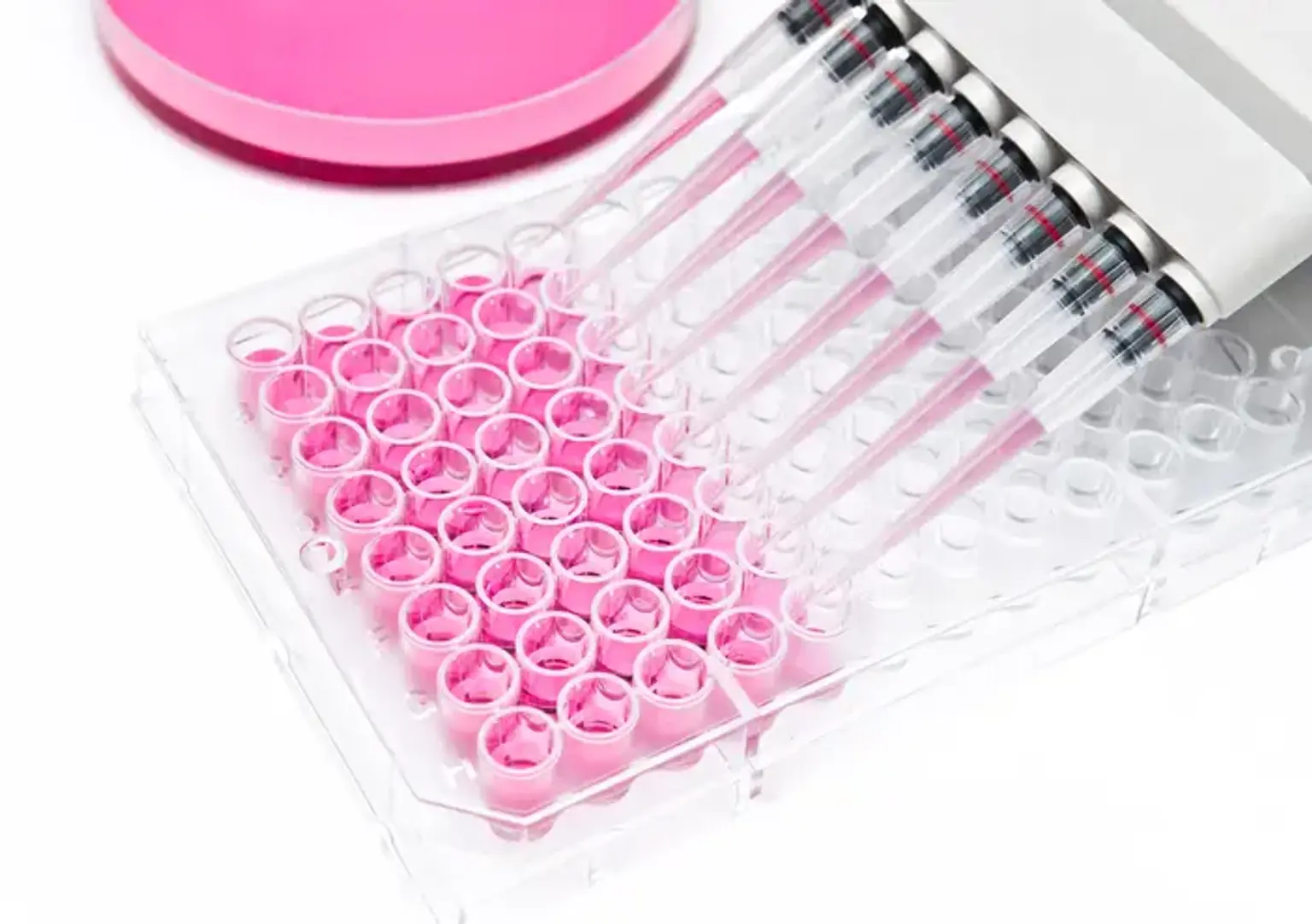
Each mammalian tissue type is derived from the embryonic germ layer, which consists of ectoderm, endoderm, and mesoderm cells that differentiate into the numerous cell types that organize into tertiary structures such as skin, muscle, internal organs, bone and cartilage, the nervous system, blood, and blood vessels.
Epithelial cells, fibroblasts, keratinocytes, melanocytes, endothelial cells, muscle cells, hematopoietic and mesenchymal stem cells are the most common cell types identified in primary cell culture. A cell line is defined as the first-time cells are sub-cultured from a primary culture. Secondary culture is described as further passaging of the cell line, in which cell cultures are sequentially moved across growing media.
What are the applications of primary cell culture?
Cell culture is a popular method in cellular and molecular biology research, and it is frequently used in therapeutic settings. Primary cell culture can be an effective model system for researching cell physiology and biochemistry, medication toxicity and metabolism, and other medicinal uses. Primary cell culture is employed by researchers all over the world because it helps to preserve uniformity, efficiency, and reproducibility of results, as well as being a good mimic model of in vivo physiological settings. The following are examples of primary cell culture applications:
3D Model System:
Primary cell culture can be used to explore cell biology and biochemistry, cell interactions, pathological interactions, medication effects and toxicology, aging, and a variety of other research topics.
Cancer Research:
The difference between normal and cancer cells may be examined utilizing primary cell culture as a model for screening of over-expressed or under-expressed markers leading to malignant activation. Furthermore, primary cells are often used as control models in anti-cancer research.
Virology:
Primary cell cultures are used to replicate viruses and can also be used to detect and isolate viruses for studying their growth and development cycle. Primary cells are also used in virology to study the mode and mechanism of virus infection.
Toxicity Testing:
Primary cell culture is used to study the effects of new drugs, cosmetics and chemicals on cells. Primary cells are also used to determine the maximum permissible dosage of the drugs.
Vaccine Production:
Cultured primary cells are utilized in viral replication to create vaccines. Primary cell culture is used to create vaccines for polio, rabies, chicken pox, and hepatitis B.
Genetically Engineered Protein:
Primary cell cultures are used to create genetically modified proteins such as monoclonal antibodies, insulin, hormones, and so on. Many treatments are being reshaped using recombinant technology.
Replacement Tissue or Organ:
In wound healing and other tissue engineering applications, primary cell culture can be employed as replacement tissue or organs. Artificial skin, for example, may be created from primary cells to heal patients with burns and ulcers. Artificial organ cultivation, such as the liver, kidney, and pancreas, is being researched. Many researchers are interested in stem cell cultivation because these cells have the ability to self-proliferate and differentiate into various cell lineages. Many therapeutic uses of adult stem cells for tissue/organ regeneration and replacement technologies are now being tested.
Drug Screening and Development:
Primary cell cultures are used to investigate cytotoxicity and safe medication doses. As a result, primary cell cultures play a significant role in the pharmaceutical sector.
Basic properties of primary cells
Primary cells undergo a limited number of cell divisions before approaching senescence after being adapted to in vitro culture conditions. The Hayflick Limit, food needs, culture conditions, and the competence with which they are managed and sub-cultured limit the number of times a basic cell culture may be passaged. Cell lines immortalized via viral or tumorigenic transformation, on the other hand, often undergo endless cell division and have an indefinite longevity. Furthermore, unlike tumor cell lines, primary cell cultures are fastidious, necessitating tailored growth conditions, such as the inclusion of tissue-specific cytokines and growth factors for proliferation in serum-free or low-serum growth media.
What is the difference between primary and secondary cell culture?
Before the cells achieve confluence, they need to be sub-cultured because nutritional fatigue, toxic metabolite buildup, and accessible room for growth all restrict proliferation. Contact inhibition is an extra component in the case of adherent cells, when cells cease multiplying when they attain confluency and occupy the surface of a culture container. Confluency can be more difficult to assess in suspended cell cultures, however, the cells tend to form clumps in the medium.
Primary cell culture cells have a limited lifetime since they can only divide a certain number of times. To stimulate continued development and proliferation, they must be sub-cultured into fresh growth media or onto new growth substrate before reaching confluence. This is referred to as secondary cell culture. A similar mechanism highlights primary and secondary cell cultures, but they differ in terms of lifetime, biological importance, and cell origin.
As the culturing process progresses, cells with the greatest growth potential predominate, and the degree of genetic and morphological homogeneity increases. Cells can develop immortalized secondary cell lines as a result of genetic change. Immortalized cell lines, which are created by viral or chemical addition, are commonly utilized in research because their immortality (the capacity to divide endlessly) allows researchers to run studies on genetically identical cells several times.
Similarly, malignant cells can be employed to generate continuous cancer cell lines with short production periods and the ability to be passed on endlessly. When cells are not transformed, they go through a limited number of population doublings and are thus referred to as finite cell lines.
Working with primary and secondary cultures has advantages and disadvantages, and the decision is determined by the experiment's purpose. Primary cell cultures are frequently favored over cell lines because they are more representative of the tissue of origin. In turn, primary cultures are the greatest experimental platform for in vivo investigations because they have the same genetic makeup as their corresponding parent tissue and display traits that cultured cells do not. The main constraint of primary cell culture is its limited lifetime. Furthermore, it is critical to evaluate the consequences and procedure for getting a meaningful biopsy or animal model.
Secondary cell cultures, on the other hand, can last indefinitely since they are replenished with fresh nutrients and substrate at regular intervals. Cell lines, however, mutate. Secondary cell cultures may become entirely indistinguishable from their parent tissue as a result of continuous cell passaging due to genetic and morphological alterations.
Another constraint in primary culture is the generation of sufficient primary cells for analysis. In general, this can only be solved by beginning with a larger amount of tissue, which might be difficult to do. However, developments in analytical tool sensitivity are providing a solution. Single-cell technologies, such as sequencing, western blots, and mass cytometry, for example, requires relatively tiny amounts of beginning material, decreasing the requirement to cultivate vast numbers of original cells.
3D cell culture, on the other hand, permits cells to grow and interact with an extracellular framework in all three dimensions. As a result, 3D cultures more nearly resemble their natural physiological condition, allowing cells to interact with one another and with the extracellular matrix. Because of its accuracy in predicting in vivo reactions, this technique has proved revolutionary in fields such as drug discovery and development. This has resulted in cutting-edge technologies giving highly contextual models for drug screening and development, such as patient-derived organoids and organ-on-a-chip.
What are the benefits of using primary cells?
Primary cell cultures are frequently utilized as in vitro tools for preclinical and investigative biological research, such as investigations of inter- and intracellular communication, developmental biology, and disease mechanism elucidation in diseases such as cancer, Parkinson's disease, and diabetes.
Historically, researchers have used immortalized cell lines in tissue function studies; however, the use of cell lines with severe mutations and chromosomal abnormalities gives poor markers of normal cell phenotypic and early-stage disease development. The usage of primary cells, which are only maintained in vitro for brief periods of time, is presently the best representative of the major functional component of the tissue (in vivo) from which they are produced.
How is Primary cell culture done?
Isolation of primary cells:
Differential centrifugation or positive sorting using magnetic beads can be used to easily isolate and purify peripheral blood cells. The separation of a pure population of cells from primary tissue, on the other hand, is frequently challenging and needs understanding of how the cell strata should be peeled apart into a suspension containing just one major cell type.
Growth requirements:
Except for those produced from peripheral blood, primary cells are anchorage-dependent, adherent cells, which means they require a surface to develop correctly in vitro. Most initial cells are cultivated in a flat, uncoated plastic vessel, however a microcarrier, which may dramatically increase surface area, can be employed in some situations. A complete cell culture media is required, consisting of a basic medium supplemented with suitable growth factors and cytokines.
It may be advantageous to incorporate an antibiotic in the growth media during the creation of primary cultures to limit contamination brought from the host tissue. These may comprise a combination of gentamicin, penicillin, streptomycin, and amphotericin B. However, long-term antibiotic usage is not recommended since certain compounds, such as amphotericin B, may be harmful to cells over time.
Maintenance:
When cells have connected to the surface of the culture plate, the maintenance phase starts. Attachment normally begins approximately 24 hours following culture initiation. When starting a culture of cryopreserved primary cells, it is critical to remove the wasted medium after the cells have adhered since the cryoprotectant is toxic to primary cells and may reduce post-thaw viability.
It is time to subculture when cells have attained the necessary percentage of cellular confluence and are actively multiplying. Because post-confluent cells may undergo differentiation and demonstrate reduced proliferation following passaging, it is ideal to subculture primary cell cultures before they reach 100% confluence.
Cellular confluence:
The fraction of the culture container occupied by connected cells is referred to as cellular confluence. For example, 100% cellular confluence indicates that the whole surface area is covered by cells, whereas 50% confluence means that nearly half of the surface area is covered. It is a critical metric to monitor and evaluate in primary cell culture since various cell types require different confluence end points, after which they must be sub-cultured.
Subculture:
Anchorage-dependent cells develop in monolayers and require repeated subcultures to maintain exponential growth. Sub-cultivation of monolayers necessitates the dissolution of both intercellular and intracellular cell-to-surface connections. Most adherent primary cells require protein attachment bond breakdown using a low dose of a proteolytic enzyme such as trypsin/EDTA. After being dissociated and dispersed into a single-cell solution, the cells are counted and diluted to the right concentration before being transferred to fresh culture vessels where they will reattach and divide.
Cell counting:
Hemocytometers are frequently used to evaluate cell number and viability using an exclusion dye such as Trypan Blue or Erythrosin B. A hemocytometer is a glass slide that has two counting chambers, one on each side. Each counting chamber has a mirrored surface with a grid of 9 counting squares measuring 3 × 3 mm2. The chambers have elevated sidewalls that allow a coverslip to be held exactly 0.1 mm2 above the chamber floor. Each of the nine counting squares has a capacity of 0.0001 mL.
Cryopreservation and recovery:
To reduce cell damage and death during the cryopreserve and thaw processes, special care must be taken. Cryopreservation of human cells works best when a cryoprotectant, such as DMSO or glycerol, is used. The majority of primary cell cultures may be cryopreserved in a combination of 80% complete growth media, 10% FBS, and 10% DMSO. To avoid the production of ice crystals within the cells, the freezing process should be gradual, at a pace of -1°C per minute. Cultures are frozen and kept in the vapor phase of liquid nitrogen, or at temperatures below -130°C.
Characteristics for efficient development of primary cultures
Primary culture refers to the culturing procedures used after cell isolation but before the first subculture. Large tissue masses are typically used to prepare primary cultures. As a result, these cultures may contain a wide range of differentiated cells, such as fibroblasts, lymphocytes, macrophages, and epithelial cells.
According to the experiences of employees working in tissue culture laboratories, the following criteria/characteristics are recommended for efficient primary culture development:
- Embryonic tissues are favored over adult tissues for primary cultures. This is because embryonic cells may be quickly disaggregated and yield more viable cells, in addition to rapidly proliferating in vitro.
- The number of cells utilized in primary culture should be increased because their survival rate is significantly lower (when compared to subcultures).
- For use in primary culture, the tissues should be treated with minimal cell injury. Dead cells should also be eliminated.
- Choosing an adequate medium (ideally one rich in nutrients) is recommended. Fetal bovine serum is favored over calf or horse serum for serum addition.
- Centrifugation is required to eliminate the enzymes utilized in cell disaggregation.
Cell culture contamination
Microbial contamination is a significant problem in cell culture, however there are several approaches that may be used to avoid or eradicate contamination. Contamination can occur as a result of the operator and the laboratory environment, as well as other cells utilized in the laboratory and reagents. Some pathogens pose a risk to laboratory personnel; containment and aseptic procedures are the primary lines of defense against such threats. Control of suspected infection may entail only removing a single potentially contaminated culture.
If a more widespread problem is discovered, all contaminated cultures and associated unused media opened during this time should be discarded, equipment should be inspected and cleaned, cell culture operations should be reviewed, and isolation from other laboratories should be implemented until the problem is resolved. Staff training, laboratory structure, proper quarantine for new cultures or cell lines, cleaning and maintenance, and quality control are all significant aspects in preventing contamination in cell culture facilities.
Conclusion
A primary cell culture is created by directly inoculating cells from animal or human tissue into growing media. The tissue is often broken into minute pieces before being treated with Trypsin, a proteolytic enzyme that disaggregates the tissue into individual cells.
Primary cell cultures maintain the majority of the features of the cells from which they were derived, and they are often anchorage dependent. These cells display "contact inhibition," which causes cells to line up in tightly orientated parallel strands. These cells replicate in any container until they reach a maximum density, at which point they cease growing. In terms of chromosomal number, these cells often preserve their diploid karyotype.
The utilization of primary cells involves a variety of difficulties. One of the most significant challenges for primary cell culturists is limited cell accessibility owing to donor tissue supply constraints, difficulties with cell isolation/purification, quality assurance and consistency, and contamination threats. Data comparability is also a significant concern with primary cell utilization, owing to variations in chemicals utilized and processes employed by various scientists and laboratories to identify and cultivate primary cells.
Primary cultures are newly isolated cultures from mammalian systems until they are sub-cultured. At this stage, they are often heterogeneous, but still closely resemble the parent cell types and display tissue-specific features.
After the initial subculture or passage, the main culture transforms into cell lines that may be propagated or sub-cultured many times. The cell line will either die off or change into a continuous cell line after multiple subcultures on fresh medium. Many differences exist between these cell lines and original cultures, including modifications in
- Cytomorphology
- Increased growth rate
- Increase in chromosome variation
- Tumorigenicity
In vitro transformation is primarily concerned with acquiring an unlimited life span. Animal cells can be grown in two ways: in suspension or adhered to a solid surface. They lack contact inhibition, and some are anchorage independent and may be grown in suspension cultures. The karyotype of cell lines varies greatly. Continuous cell lines are often aneuploid, with chromosomal numbers ranging between diploid and tetraploid.
The cell culture technique has become a standard and popular approach due to its numerous uses in cell biology, biotechnology, and medical research. Isolation of primary cells from cancer cells is an important component of cell culture technology because it provides a dependable source for understanding normal physiological, morphological, and molecular processes in human cells.
Many disease entities and histogenesis are connected to fibroblasts since they are the main cells of the connective tissue of the oral mucosa. Oral fibroblast cell culture allows mouth biologists and researchers to explore the morphological and molecular processes in oral disorders.