Raynaud’s phenomenon
Overview
The body adjusts to cold conditions by reducing blood flow to the skin. This is done as a thermoregulating process to prevent further heat loss and to keep the core body temperature stable.Blood-flow restriction occurs during cold temperatures and mental stress in Raynaud's phenomenon. Raynaud's syndrome is characterized by vasoconstriction of the digital arteries and cutaneous arterioles. Maurice Raynaud initially identified this phenomenon in 1862, and Sir Thomas Lewis studied it in 1930.
Raynaud's phenomenon (RP), in general, is a transient and peripheral vasoconstrictive reaction to cold temperatures or emotional stress. Raynaud's phenomenon is classified as either primary or secondary.
Raynaud's syndrome is characterized by hypersensitive blood vessels in the fingers and toes. They are more sensitive to cold and stress. The blood vessels constrict significantly, resulting in a reduction in blood flow and a color change. A decrease of blood flow frequently causes a pale, or white, discolouration. As a result of the quick flow of blood into the fingers following the incident, the digits might become blue and eventually red.
A history and physical examination are used to diagnose RP. Patients with primary Raynaud's disease usually have a normal physical exam and blood testing. Secondary Raynaud's patients usually have an abnormal physical and/or blood testing. A method known as nailfold capillaroscopy can also be used to examine the blood veins beneath the fingernails.
Raynaud's disease can be treated with both lifestyle changes and drugs. Keeping the body warm, especially the center of the body, is one of the lifestyle changes that may be made. Hand warmers and mittens/gloves are frequently used by patients to keep their fingers warm. Stress reduction and smoking cessation are also suggested as ways to reduce Raynaud's episodes.
Many patients can control their symptoms simply by changing their lifestyle. However, drugs are occasionally required, and there are several alternatives available. Calcium channel blockers (amlodipine, nifedipine, felodipine, and others) and angiotensin-receptor blockers are examples of blood pressure drugs. These drugs work by boosting blood flow to the fingers and toes. Other drugs, such as sildenafil or prostacyclins, can be used for people with more severe symptoms or who have acquired problems such as finger ulcers.
Types of Raynaud's phenomenon
This condition was called after the French doctor who discovered it in 1862. It may be referred to by a variety of names. There are two kinds of Raynaud's phenomenon:
Primary Raynaud's: occurs in the absence of any other sickness. The symptoms are frequently modest.
Secondary Raynaud's: it is caused by another condition. It is frequently associated with a disorder that targets your body's connective tissues, such as lupus or rheumatoid arthritis. It is less prevalent, but it has a higher risk of causing major health issues. Skin sores and gangrene are examples of this. These occur when cells and tissue in your toes and fingers die as a result of a lack of blood.
What causes Raynaud's disease?
Secondary Raynaud phenomenon is caused by a variety of causes. Scleroderma, systemic lupus erythematosus, Sjogren syndrome, and antiphospholipid syndrome are the most prevalent conditions linked with it.
Secondary Raynaud phenomenon can be caused by drugs such as antimigraine medications, interferon alpha and beta, cyclosporine, and nonselective beta blockers.
Males are disproportionately affected by occupations that expose them to overt vibrational exposure from vibrating machines. This is referred to as hand-arm vibration syndrome. Other occupational-related causes of secondary Raynaud phenomenon include polyvinyl chloride exposure, cold damage from employment, and munitions work.
Obstructive vascular disease is a common cause of Raynaud's phenomenon in adults over the age of 60. Thromboangiitis obliterans, microemboli, diabetic angiopathy, and atherosclerosis are all causes of obstructive vascular disease.
Infections associated with secondary Raynaud phenomenon include parvovirus B19, cytomegalovirus, hepatitis B, and hepatitis C.
Other causes of secondary Raynaud phenomenon include fibromyalgia, polycythemia, arteriovenous fistula, myalgic encephalitis, or malignancy.
How common is Raynaud's phenomenon?
Raynaud's phenomenon is more common in women (approximately 20% to 30%), especially in younger populations (teens to 20s). The male to female ratio is nine to one.
How Raynaud's phenomenon develops?
Raynaud's phenomenon is caused by three processes. These include reduced blood flow, constriction of blood vessels, neurogenic reactions, and inflammatory and immunological responses.
Based on environmental stimuli, the somatosensory system aids in temperature perception. Cold temperatures stimulate afferent nerve fibers, which activate A-delta and unmyelinated C-fibers. This ultimately activates the cold receptor TRPM8 (transient receptor potential ion channel), which detects changes in cold temperatures. TRPM8 activation causes cutaneous vasoconstriction, thermogenesis, and cold avoidance.
The sympathetic nervous system releases vasoconstricting neuropeptides and norepinephrine in response to cold temperatures, resulting in vasoconstriction of arteriole smooth muscle and reduced blood flow to the skin. Endothelial cells secrete endothelin-1, which induces vasoconstriction in secondary Raynaud's phenomenon.
An increase in alpha-2 adrenergic sensitivity in the digital and cutaneous arteries causes a vasoconstrictive response to cold temperatures and emotional stress in primary Raynaud's phenomenon. The sympathetic nervous system affects alpha-2 adrenergic receptors on the distal artery smooth muscles of the digits. The use of alpha-2 adrenergic receptor inhibitors in individuals with cold-induced episodes reduced the intensity of the attack, according to studies.
The underlying condition is the cause that affects normal vascular response to cold temperatures in secondary Raynaud phenomenon. Typically, the endothelial function of the digital and cutaneous arteries is disturbed, resulting in vasoconstriction and tissue ischemia. In systemic scleroderma, for example, vascular fibrosis leads to endothelial dysfunction and tissue ischemia.
Symptoms and physical signs of Raynaud's phenomenon
When evaluating a patient with probable Raynaud phenomenon, doctors should inquire about the patient's age of onset, the location of afflicted regions, the existence of symmetry, the presence of digital ulcerations, and the intensity of the episodes.
To rule out secondary Raynaud's phenomenon, other systemic symptoms should be elicited. Once Raynaud's phenomenon has been confirmed, the triggering variables should be determined. Exposure to cold temperatures, sympathetic nervous system stimulation from mental stress, or being startled can all be triggers.
Certain variables enhance the risk of an increase in the frequency of attacks, as well as an increase in severity and discomfort. Stress, female gender, low temperature, and the existence of a concomitant connective tissue disease are examples of these. The later Raynaud phenomenon appears, particularly in the 30s and 40s, the greater the chance of developing a connective tissue condition.
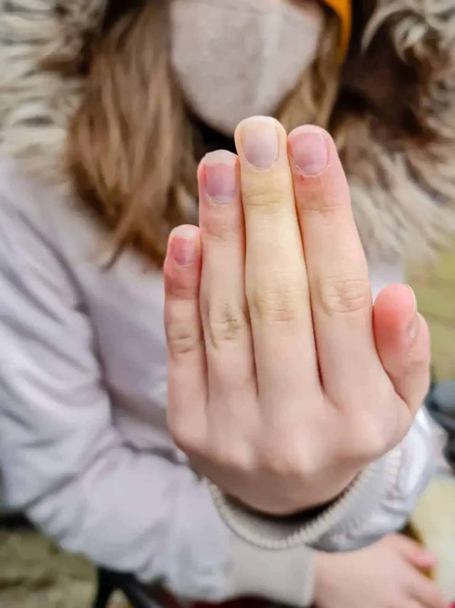
Raynaud's phenomenon most commonly affects the fingers (distal areas). A Raynaud phenomenon attack is characterized by the appearance of cold digits and the demarcation of "white" areas, often known as skin pallor or white attack. Instead of a white attack, cyanotic skin changes, often known as a blue attack, may occur. The white or blue attack normally lasts around 20 minutes. Following a flare-up of the Raynaud phenomenon, vascular reperfusion with subsequent rewarming ensues. This causes erythema in the region where the flare occurred. It is referred to as reactive hyperemia.
A Raynaud phenomenon attack begins with a single digit and then spreads symmetrically to additional digits on both hands. Typically, the thumb is spared. If the thumb is afflicted, this might indicate secondary Raynaud's phenomenon. It can also cause cutaneous vasospasm in the face, ears, knees, or nipples.
Raynaud phenomenon episodes are commonly associated with pins and needles feeling, numbness, or finger pains or discomfort as a result of vasoconstriction. Digital ulceration of the tips of fingers and toes may occur owing to prolonged vasoconstriction with subsequent tissue ischemia if the Raynaud phenomenon episode is severe, as is common with secondary Raynaud phenomenon.
In extreme cases of Raynaud's phenomenon episodes, gangrene or digit loss may occur. Raynaud phenomenon attacks are often less severe in initial Raynaud phenomenon than in secondary Raynaud phenomenon.
Another skin condition noticed during a Raynaud's episode is livedo reticularis. Due to minor blood vessel clots, this causes purple mottling or a reticular pattern on the skin. Rewarming can reverse this result, however secondary reasons such as antiphospholipid syndrome, vasculitis, cold agglutinin disease, or peripheral vascular disease cannot.
How Raynaud's phenomenon is evaluated?
It is critical to distinguish Raynaud's disease (primary Raynaud's) from Raynaud's phenomenon (secondary Raynaud's). Examining for signs of arthritis or vasculitis, as well as a series of laboratory testing, may help distinguish them.
Nail fold capillary examination, often known as "capillaroscopy," is one of the most sensitive ways for objectively diagnosing RP with connective tissue problems and distinguishing a secondary from a primary variant. If secondary to systemic sclerosis, thermography is a technology that may aid in the prediction of systemic sclerosis. A thorough medical history and the following diagnostic work-up will attempt to identify or rule out any secondary causes.
- Digital artery pressures are measured in the arteries of the fingers before and after the hands have been cooled. A decrease of at least 15 mmHg is diagnostic (positive).
- Doppler ultrasound to assess blood flow.
- Full blood count may reveal a normocytic anaemia suggesting the anaemia of chronic disease or kidney failure.
- Blood test for urea and electrolytes may reveal kidney impairment.
- Thyroid function tests may reveal hypothyroidism.
- Tests for rheumatoid factor, erythrocyte sedimentation rate, C-reactive protein, and autoantibody screening may reveal specific causative illnesses or an inflammatory process. Anti-centromere antibodies are common in limited systemic sclerosis (CREST syndrome).
- Nail fold vasculature (capillaroscopy) can be examined under a microscope.
Treatment of Raynaud’s phenomenon
Secondary Raynaud's is generally controlled by addressing the underlying cause, and, as with primary Raynaud's, by avoiding triggers such as cold, emotional and environmental stress, vibrations and repetitive motions, and smoking (including passive smoking) and sympathomimetic drugs.
Home Treatment for Raynaud’s
These steps can also help you control your condition:
- Avoid smoke: Don't smoke, and avoid secondhand smoke as well. It might cause your blood vessels to constrict, lowering your skin temperature.
- Exercise: It will boost your circulation. If you have secondary Raynaud's disease, see your doctor before doing an outdoor workout in cold weather.
- Manage stress: Keeping it under control could help cut the number of attacks.
- Keep your temperature constant: If possible, avoid going from a cold environment to a warm space. As much as possible, avoid the frozen food department of the grocery store.
- Dress for the cold: Wear layers, gloves, and heavy socks. Buy chemical warmers for your pockets, gloves, and socks.
- Avoid some medications: Decongestants with phenylephrine, diet pills, migraine medications with ergotamine, herbal medications with ephedra, and the blood pressure medication clonidine (Catapres) can all narrow your blood vessels.
- Soak your hands: Or run warm water over them when you feel an attack starting.
Medications
Medications can be helpful for moderate or severe disease.
Vasodilators: Calcium channel blockers, such as the dihydropyridines nifedipine or amlodipine, ideally in slow-release formulations, are frequently used as first-line therapy. They have the frequent adverse effects of headache, flushing, and ankle edema, although these are usually not severe enough to justify therapy discontinuation. The little evidence suggests that calcium-channel blockers are only marginally beneficial in lowering the frequency of attacks. Other studies, however, suggest that CCBs may be useful in reducing the intensity of attacks, discomfort, and impairment associated with Raynaud's phenomenon. People whose illness is linked to erythromelalgia are frequently unable to utilize vasodilators for treatment because they cause 'flares,' which cause the extremities to turn a bright red owing to an excess of blood flow.
Aspirin: People with severe disease prone to ulceration or large artery thrombotic events may be prescribed aspirin.
Sympatholytic agents: such as the alpha-adrenergic blocker prazosin, may provide temporary relief to secondary Raynaud's phenomenon.
Losartan: with topical nitrates may, reduce the severity and frequency of attacks, and the phosphodiesterase inhibitors sildenafil and tadalafil may reduce their severity.
Angiotensin receptor blockers or ACE: these inhibitors may aid blood flow to the fingers, and some evidence shows that angiotensin receptor blockers (often losartan) reduce frequency and severity of attacks, and possibly better than nifedipine.
The prostaglandin iloprost: it is used to manage critical ischemia and pulmonary hypertension in Raynaud's phenomenon, and the endothelin receptor antagonist bosentan is used to manage severe pulmonary hypertension and prevent finger ulcers in scleroderma.
Statins: they have a protective effect on blood vessels, and SSRIs such as fluoxetine may help symptoms, but the data is weak.
PDE5 inhibitors: they are used off-label to treat severe ischemia and ulcers in fingers and toes for people with secondary Raynaud's phenomenon; as of 2016, their role more generally in Raynaud's was not clear.
Surgery
An endoscopic thoracic sympathectomy treatment might be performed in extreme situations. The nerves that cause the blood vessels in the fingers to contract are surgically severed here. Microvascular surgery of the afflicted regions is another option, although it should only be used as a last resort.
Botulinum toxin is a relatively modern therapy for severe Raynaud's disease. A prior study of 19 individuals with severe Raynaud's phenomenon ranging in age from 15 to 72 years found that 16 patients (84 percent) had pain alleviation at rest; 13 patients reported immediate pain relief, and three more had progressive pain improvement over 1–2 months. All 13 individuals with persistent finger ulcers recovered in less than 60 days. Only 21% of the patients required further injections.
A group of rheumatology and dermatology specialists has produced International Consensus Criteria for the diagnosis of primary Raynaud's syndrome.
Can Raynaud’s disease kill you?
No, however severe cases might block off blood supply to your skin, causing tissue damage. Skin sores (ulcers) or dead tissue can result from a totally blocked artery (gangrene). It's uncommon, but if this happens, the doctor may have to amputate a finger or toe.
Prognosis of Raynaud's phenomenon
The prognosis of primary Raynaud syndrome is often very favorable, with no mortality and little morbidity. However, a minority develops gangrene. The prognosis of secondary Raynaud is dependent on the underlying disease, and how effective blood flow-restoring maneuvers are.
Conclusion
Raynaud syndrome, also known as Raynaud's phenomenon, is a medical disorder in which tiny artery spasm produces episodes of decreased blood flow to end arterioles. It is named after the physician Auguste Gabriel Maurice Raynaud, who originally reported it in his PhD thesis in 1862. Fingers and, less often, toes are typically involved.
The nose, ears, and lips are rarely affected. The afflicted region of the body often becomes white, then blue as a result of the episodes. Numbness or discomfort are frequently experienced. The region gets red and burns as blood flow resumes. The bouts usually last a few minutes but might stay for several hours.
The disorder affects around 4% of the population. The primary form usually appears between the ages of 15 and 30 and is more common in females. The secondary type mainly affects the elderly. In colder climates, both kinds are more prevalent.
Cold or mental stress are common triggers for episodes. Primary Raynaud's disease, also known as idiopathic, indicates that it occurs on its own, has no known etiology, and is unrelated to any other condition. Secondary Raynaud's arises as a result of another condition and manifests at a later age; episodes are excruciatingly painful, asymmetric, and accompanied with skin lesions.
Secondary Raynaud's syndrome can arise as a result of a connective-tissue disorder such as scleroderma or lupus, hand injuries, continuous vibration, smoking, thyroid disorders, and certain drugs such as birth control pills. Symptoms are often used to make a diagnosis.
The primary treatment is avoiding the cold. Other measures include the discontinuation of nicotine or stimulant use. Medications for treatment of cases that do not improve include calcium channel blockers and iloprost. Little evidence supports alternative medicine. Severe disease may in rare cases lead to complications, specifically skin sores or gangrene.