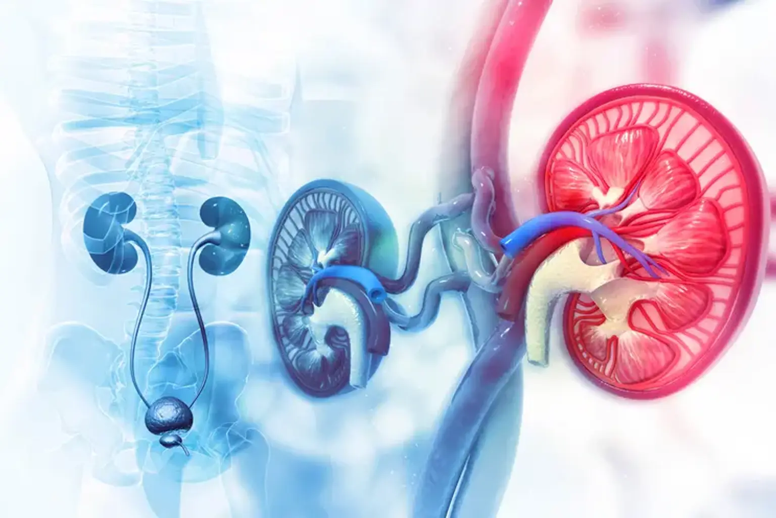
Renal hypertension
Overview
In the United States, high blood pressure affects 75 million persons and accounts for 8.6 percent of all primary care visits. Renal disease is one of the most prevalent causes of treatable hypertension, yet it only accounts for 2% of all hypertension cases. It is mostly caused by blood artery constriction in the kidney.
What is Renal hypertension?
Renal hypertension, also known as renovascular hypertension by health professionals, is a kind of high blood pressure that begins in the kidneys. It is caused by a blockage in the arteries that transport blood to the kidneys.
Stenosis or blockage of a major renal artery, an accessory renal artery, or any of its branches can induce hypertension by increasing the release of renin from the damaged kidney's juxtaglomerular cells. Before stenosis is expected to contribute to blood pressure (BP) increase, the area of the artery lumen must be reduced by 70% and a considerable poststenotic gradient must be present. Renovascular hypertension is substantially less frequent in blacks than in whites for unclear causes.
Around 80% of instances are caused by atherosclerosis, whereas 20% are caused by fibromuscular dysplasia. Atherosclerosis is more frequent in males over 50 and mostly affects the proximal one-third of the renal artery. Fibromuscular dysplasia is more frequent in younger individuals (mostly women) and often affects the distal two-thirds of the major renal artery and its branches. Embolism, trauma, unintentional ligation following surgery, and tumor extrinsic compression of the renal pedicle are all rare causes. Renovascular hypertension is distinguished by elevated cardiac output and peripheral resistance.
What is Hypertension, and how does it affect the kidneys?
Blood pressure is described as an increase in the force exerted by blood on the vessels as it circulates throughout the body. Blood pressure can cause damage and weakening of blood arteries throughout the body, including those in the kidneys, over time. When blood arteries in the kidneys are damaged, it can have an impact on how well the kidneys work.
When the kidneys do not work correctly, excess fluid and waste accumulation in the circulation occurs. This, in turn, can lead to high blood pressure, causing additional kidney injury. A person's kidneys might fail over time.
Symptoms and Signs of Renovascular Hypertension
Asymptomatic renovascular hypertension is common. A systolic-diastolic bruit in the epigastrium, frequently transmitted to one or both upper quadrants and occasionally to the back, is nearly pathognomonic, but it is present in only approximately half of fibromuscular dysplasia patients and is uncommon in renal atherosclerosis patients.
Renovascular hypertension should be suspected if:
- Diastolic hypertension appears suddenly in patients aged 30 to 50.
- Over a 6-month period, new or previously stable hypertension quickly worsens.
- Hypertension is initially severe, linked with decreasing renal function, or is drug-resistant.
A history of back or flank trauma, or acute pain in this area with or without hematuria, implies renovascular hypertension (perhaps related to artery damage), however these results are uncommon. Recurrent bouts of unexplained acute pulmonary edema or heart failure, as well as asymmetric renal size (> 1 cm difference) detected accidentally during imaging studies, imply renovascular hypertension.
Diagnosis of Renovascular Hypertension
Renovascular hypertension patients frequently undergo a thorough assessment to determine the reason of their uncontrolled hypertension.
Laboratory Tests
- Urine analysis: Proteinuria, hematuria, and casts should all be checked. The presence of proteinuria implies a renal parenchymal disease, whereas the presence of hematuria or red blood cell (RBC) casts indicates glomerulonephritis.
- Blood urea nitrogen and serum creatinine: To assess baseline kidney function.
- Basal metabolic profile: To assess for electrolyte disturbances and acid-base balance.
- Complement levels and autoimmune profile: In suspected cases of autoimmune diseases affecting the renal vasculature.
- Plasma free metanephrines or 24-hour urinary fractionated metanephrines and normetanephrine to rule out pheochromocytoma
- Plasma renin-aldosterone ratio to rule out hyperaldosteronism
- To rule out Cushing's syndrome, a 24-hour urine free cortisol or low-dose dexamethasone suppression test is performed.
Imaging
Renovascular hypertension may be assessed using a variety of imaging methods. Because renal artery stenosis is the most prevalent cause of renovascular hypertension, renal arteriography remains the gold standard diagnostic test. Catheter angiography, on the other hand, is intrusive, expensive, and time-consuming, and it might result in problems such as renal artery dissection or cholesterol embolization.
Duplex ultrasonography, computed tomography with angiography (CTA), and magnetic resonance angiography (MRA) are further imaging methods that can be used to assess the renal vessels. The type of imaging test employed is frequently determined by the presence of high-grade lesions and the requirement for intervention.
The initial imaging test of choice for evaluating the renal arteries is duplex ultrasonography. It is relatively affordable, non-invasive, and does not entail the injection of contrast or radiation exposure. The association between a duplex scan and contrast-enhanced angiography has been demonstrated. Though there are various criteria for determining the existence of renal artery stenosis, peak systolic velocity (PSV) is the most essential indicator. A PSV greater than 180 cm/s indicates the existence of more than 60% stenosis.
The resistive index (RI) may also be measured using duplex ultrasonography. A score more than 0.7 shows the existence of pathological flow resistance, and studies demonstrate that a value greater than 0.8 predicts poor response to revascularization therapy. The most notable disadvantages of duplex ultrasonography are its decreased sensitivity in obese individuals, interference from overlaying intestinal gas, and operator dependency.
CT angiography is the injection of intravenous contrast and the acquisition of detailed pictures of blood arteries or tissues by moving the beam in a helical fashion across the area under study. Wittenberg et colleagues discovered that the sensitivity and specificity for hemodynamically significant RAS (>50%) by CTA were 96 and 99 percent, respectively. CTA has a similar negative predictive value to MRA in excluding renal artery stenosis.
It can also be used to identify extrinsic renal artery compression, Fibromuscular dysplasia (FMD), arterial dissection, and to assess adjacent tissues. CTA, on the other hand, can only offer an anatomical assessment of the lesion and cannot measure the degree of restriction to renal blood flow. Other disadvantages of CTA include radiation exposure, contrast allergy, and acute renal damage.
MRA evaluates the renal blood arteries and surrounding structures using a strong magnetic field, radio wave pulses, and intravenous gadolinium. Several investigations have demonstrated that MRA has a sensitivity and specificity of roughly 97 percent and 92 % in identifying renal artery stenosis, respectively. MRA does not utilize radiation, and gadolinium contrast is less likely to trigger an allergic reaction than iodine contrast, which is used in CTA. However, MRA has been proven to overestimate the severity of stenosis and is frequently influenced by motion abnormalities or opacification of renal veins, making visualization of the renal arteries problematic.
In comparison studies, MRA was found to have a higher positive predictive value than CTA due to higher false-positive rates with CTA. Both CTA and MRA have significant negative predictive values (>98% for both). Both techniques provide a high degree of reliability in ruling out major renal artery abnormalities. Both MRA and CTA have been demonstrated to be useful for the diagnosis of Fibromuscular dysplasia (FMD), with CTA having the highest sensitivity (84.2 percent) when compared to angiography.
Nuclear medicine ACE-inhibitor (ACE-I) renography is another non-invasive, reasonably safe imaging approach that evaluates for renovascular hypertension using radioactive material, a specific camera, and a computer. It entails administering an ACE-I to establish whether the cause of hypertension is constriction of the renal arteries. The sensitivity and specificity of this test have been demonstrated to be varied, with results ranging from 74% to 94% for sensitivity and 59% to 95% for specificity.
It is a time-consuming process with the danger of radiation exposure and discomfort or pain from the radiotracer injection. Ultrasound has been demonstrated to be more sensitive than captopril renography, making it a preferable alternative for an initial diagnostic test.
Catheter angiography is the gold standard diagnostic for evaluating renovascular hypertension because it has the highest temporal and spatial resolution. Catheter angiography offers the extra benefit of assessing the hemodynamic relevance of anatomically severe lesions by monitoring translesional pressure gradients. It is especially beneficial in the following situations:
- Patients with a disparity between imaging modalities
- Patients with a high index of suspicion and negative imaging findings
- Patients anticipated of needing an intervention
It may also assess anatomical anomalies of the kidney, renal arteries, and aorta, and it can be followed by endovascular intervention to address major lesions. Furthermore, the surrounding tissues and bones can be eliminated or deleted from the final picture, leaving only the artery framework visible. This is referred to as digital subtraction angiography (DSA). However, the radiation dosages are larger than in CTA, and because it is an intrusive treatment, problems such as artery dissection, tear, rupture, or thromboembolic event are possible.
Management of Renal hypertension
The goal of renovascular hypertension therapy is to address the underlying cause. There are several methods available, including pharmaceutical and invasive therapy.
- Atherosclerotic renal artery stenosis
Angioplasty with stent implantation was formerly thought to be effective for many individuals with atherosclerotic renal artery stenosis. When compared to medical care alone, stent implantation had no effect on outcomes. Although stent implantation reduced systolic blood pressure somewhat, there was no meaningful therapeutic benefit for stroke, myocardial infarction, heart failure, mortality from cardiovascular or renal illness, or progression of kidney disease.
- Fibromuscular dysplasia
Percutaneous transluminal angioplasty (PTA) is suggested for the majority of individuals with renal artery fibromuscular dysplasia. The insertion of a stent lowers the risk of restenosis; antiplatelet medications (aspirin, clopidogrel) are administered thereafter. Only when significant illness in the renal artery branches renders PTA technically impossible is saphenous vein bypass grafting indicated.
Complete surgical revascularization may necessitate microvascular procedures that can only be performed ex vivo with kidney autotransplantation. In adequately chosen patients, the cure rate is 90%; surgical mortality is 1%. In young patients whose kidneys cannot be revascularized for technical reasons, medical therapy is always preferred to nephrectomy.
Differential Diagnosis
The differential diagnosis for renovascular hypertension includes secondary hypertension factors such as:
- Pheochromocytoma: A cluster of symptoms include flushing, headache, tachycardia, and periodic uncontrolled hypertension.
- Primary hyperaldosteronism: is characterized by chronic hypokalemia and metabolic alkalosis.
- Obstructive sleep apnea: is more common in obese men with a larger neck circumference and a history of snoring. Polysomnography/sleep study was used to diagnose.
- Aortic coarctation: is characterized by a systolic murmur, radio-femoral delay, and upper extremity hypertension. MRA or CTA is used to make the diagnosis.
- Cushing syndrome: This condition is characterized by moon facies, buffalo hump, proximal myopathy, glucose intolerance, abdominal striae, and central obesity.
Prognosis
Atherosclerotic renal artery stenosis is a degenerative disease that can lead to stenosis worsening and, eventually, renal failure. Previous research indicated that the 3-year cumulative incidence of disease progression was 18%, 28%, and 49% for renal arteries categorized as normal, 60% stenosis, and >60% stenosis, respectively. High blood pressure (systolic >160 mm hg), diabetes, and high-grade stenosis (>60 percent blockage) have all been linked to a faster rate of development. Untreated renovascular hypertension can also result in end-stage renal failure, with a median survival time of 25 months and a death rate of 35% after 4 years.
The primary goal of renovascular hypertension treatment should be to address the underlying cause. Renovascular hypertension caused by atherosclerotic renal artery stenosis should be treated medically first and foremost, as several trials have failed to demonstrate renal or cardiovascular advantages with invasive treatment.
Complications
Renovascular hypertension complications are usually caused by uncontrolled blood pressure and include:
- Renal failure
- Myocardial infarction
- Stroke
- Pulmonary edema
- Retinopathy
- Left ventricular hypertrophy
- Congestive heart failure
- Aneurysm
- Vascular dementia
Conclusion
Renovascular hypertension occurs when blood flow to the kidneys is impaired, resulting in elevated blood pressure via a hormonal reaction of the afflicted kidney. This is a dangerous disorder that can result in heart attacks, strokes, and even death. Patients who have high blood pressure while using several blood pressure drugs should consult their doctor about the likelihood of renovascular hypertension. In patients with resistant hypertension, the clinical practitioner should also have a high index of suspicion and explore suitable diagnostic methods.