Retinal disease

Last updated date: 09-May-2023

Originally Written in English

Retinal disease

Overview

Any component of the retina might be damaged by retinal disorders. Untreated retinal illnesses can result in significant vision loss and, in extreme cases, blindness. Some retinal illnesses can be treated if detected early, while others can be managed or delayed to maintain or even recover eyesight.
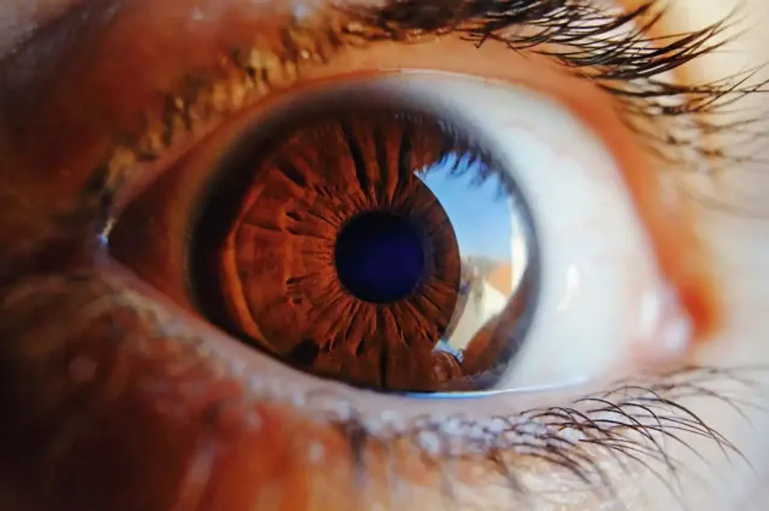
 

Function of the retina

Function of the retina

The retina is the light-sensitive, transparent structure at the back of the eye. Light is focused on the retina by the cornea and lens. The macula, or core portion of the retina, has a high density of color-sensitive photoreceptor (light-sensing) cells. Cones are the cells that generate the clearest visual pictures and are in charge of center and color vision. Rod photoreceptor cells, which respond to lower light levels but are not color sensitive, are found in the peripheral portion of the retina, which surrounds the macula. The rods are in charge of peripheral vision as well as night vision.

The optic nerve transmits photoreceptor-generated impulses (cones and rods). A small nerve branch connects each photoreceptor to the optic nerve. The optic nerve is linked to nerve cells that provide information to the brain's vision center, where they are processed as visual pictures.

The optic nerve and retina contain a dense network of blood vessels that transport blood and oxygen. The choroid, which is the layer of blood vessels between the retina and the outer white layer of the eye called the sclera, contributes to this supply of blood vessels. The central retinal artery (the other primary blood supply to the retina) enters the retina near the optic nerve and branches off within the retina. Blood flows from the retina into the central retinal vein's branches. Within the optic nerve, the central retinal vein exits the eye.

A doctor dilates the pupil of a patient's eye when inspecting the retina. This permits ophthalmoscopy to observe the retina in considerably greater detail (shining a light through a magnifying lens and into the back of the eye).

Ophthalmologists often diagnose and treat retinal diseases. An ophthalmologist is a medical specialist who specializes in the diagnosis and treatment of various forms of eye problems (both surgical and nonsurgical). Treatment is usually provided by an ophthalmologist who specializes in retinal problems.


What is retinal disease?

retinal disease definition

Blindness prevalence declines and causes of blindness alter as countries get wealthy and per capita income rises. Cataract and corneal scar are the most common causes of blindness in underdeveloped African countries. Glaucoma and diabetic retinopathy will be the primary causes of blindness in a middle-income Latin American country. Because cataract surgery is more widely available, fewer individuals are blinded as a result of cataract. Glaucoma and cataract will remain prevalent and significant disorders in an affluent country, but retinal disease will cause the majority of blindness.

Retinal disorders are already the leading cause of pediatric blindness across the world. Some of these children are blinded by hereditary retinal disorders, such as retinitis pigmentosa, which cannot currently be treated or avoided. Many of them, however, develop retinopathy of prematurity, which may be avoided and treated in its early stages. Adult blindness in India was caused by retinal disorders far more frequently than previously considered.

Diabetes is becoming more prevalent in underdeveloped countries. Diabetes is estimated to affect 8–10% of the Indian population, and the incidence is rising. Although population-based studies indicate that diabetic retinopathy is not now a leading cause of blindness in India,4 this is expected to alter in the future.

Our own efforts will contribute to a rise in the prevalence of retinal problems. At the moment, around 10 million cataract procedures are performed each year. It is hoped that by 2020, this figure would have risen to more than 30 million. Almost bulk of this expansion will occur in poorer, developing countries. Greater cataract surgery will result in more posterior segment cataract surgery consequences, such as retinal detachment and retained lens debris. These issues are very manageable if you have access to a qualified and well-equipped vitreo-retinal surgeon.

Given these tendencies, retinal disorders are projected to be a substantial and growing concern in every area of the world.


Most common retinal diseases

common retinal diseases

1. Retinal tear

When the vitreous gel that fills the eye begins to shrink, it pulls on the retina with enough force to tear the retinal tissue. The vitreous tends to separate from the retina as we age, resulting in a posterior vitreous detachment (PVD).

A posterior vitreous detachment affects more than 60 percent of people over age 70.

In most circumstances, a PVD is not associated with any difficulties. Some people, however, are born with a more sticky vitreous, causing it to peel away from the retina in an unnatural manner, resulting in a retinal tear.

A retinal tear can occur as a result of an eye injury or damage to the retina in rare situations. A retinal tear can produce unexpected visual alterations such as black patches, floaters, and flashing lights. Retinal tears increase the likelihood of retinal detachment and significant vision loss.

Laser surgery or freezing therapies known as cryotherapy are commonly used to repair retinal tears.


2. Retinal detachment

When fluid travels through a retinal tear, it causes the retina to separate from the surrounding tissues in the back of the eye, cutting off its blood supply and ability to operate correctly.

  • Retinal detachments affect 5 in 100,000 people, annually.
  • Middle-aged and elderly populations are affected more frequently, with approximately 20 in 100,000, annually.

The symptoms of a retinal detachment vary depending on the amount of the separation and can range from no symptoms to seeing floaters, flashing lights, and a shadow that limits peripheral vision and, in rare cases, central vision.

Three main causes and forms of retinal detachment include:

  • Rhegmatogenous. This is the most prevalent kind as a result of a retinal tear. It might be caused by age, an eye injury, surgery, or nearsightedness.
  • Tractional. This is most commonly seen in diabetics, when blood vessels at the rear of the eye get damaged and produce scar tissue. This scar tissue strains on the retina, resulting in retinal tissue detachment.
  • Exudative. This happens when fluid accumulates behind your retina and pulls it away from the tissue linked to it from behind. It is most commonly caused by age-related macular degeneration, injury, or retinal edema.


To heal any tears in the retina and reconnect the retina to the back of the eye, laser treatments, surgery, or freezing treatments (cryotherapy) may be used to cure a retinal detachment.


3. Diabetic retinal disease

Diabetic retinopathy is a serious sight-threatening complication of diabetes that can lead to blindness.

High amounts of sugar in the circulation induce this condition, which causes gradual retinal degeneration.

Diabetes wreaks havoc on the body's microscopic blood vessels, particularly the retina's tiny capillaries. These small blood vessels eventually leak blood and other fluids into the eye, causing the retinal tissue to enlarge and create foggy or impaired vision. New aberrant blood vessels are formed as these blood vessels are damaged. These new vessels are more fragile and prone to leaking and bleeding fluid into the eye.

  • Diabetic retinopathy affects 1 in 3 people with diabetes.
  • 95% of people with diabetic retinopathy can avoid severe vision loss with early treatment. 


There are three different types of diabetic retinopathy:

  • The initial stage of the illness is Non-Proliferative Retinopathy (NPR), also known as Background Retinopathy. At this stage, excessive blood sugar levels in the retina induce microaneurysms, which cause damage to the walls of the small retinal capillaries. Microaneurysms ultimately burst and bleed, and the resulting hemorrhages are known as "dot-and-blot" hemorrhages due to their form.
  • The most severe type of the illness is Proliferative Retinopathy (PR). Abnormal blood vessels begin to form in the retina at this stage. If these new blood vessels rupture, they can cause bleeding into the vitreous, the gelatinous stuff that fills the eye, resulting in retinal scarring, severe visual impairments, and even blindness.
  • Diabetic macular edema (DME) is a diabetic retinopathy consequence. DME arises when damaged blood vessels leak fluid into the macula, which is important for crisp center vision and the ability to perceive minute details.
  1. Diabetic macular edema can produce hazy or wavy vision, blind patches, and poor colors. These symptoms have the potential to impair reading, writing, driving, and face recognition.

Treatment of diabetic retinopathy includes anti-VEGF injections and/or laser surgery.


4. Epiretinal membrane

Cellophane maculopathy, commonly called as macular puckers, is another name for epiretinal membranes (ERMs). Epiretinal membranes are semi-translucent, fibrocellular, and avascular, with few or no blood vessels.

epiretinal membranes (ERMs) originate on the inner surface of the retina and cause little symptoms; but, if they damage the macula, they can cause painless visual abnormalities and vision loss.

An ERM is made up of glial cells that develop and collect on the retina's surface. The membrane, which resembles a wrinkled piece of cellophane, continues to compress and tug on the retina over time, causing visual aberrations such as hazy or crooked vision, as well as vision loss.

Research has shown that ERM affects up to 2 percent of patients over age 50 and 20 percent over age 75.


5. Retinitis pigmentosa

Retinitis pigmentosa

Retinitis pigmentosa (RP) is a rare, genetic, ocular disease that causes retinal damage and vision loss.

  • RP is one of the most frequently occurring forms of inherited retinal degeneration.
  • RP affects approximately 1 in 4,000 people worldwide.

In its early stages, retinal cells in the portion of the retina responsible for mid-peripheral vision begin to degenerate, resulting in impaired night vision (nyctalopia), mid-peripheral visual field loss, and trouble seeing in low light. The disease's fast development continues to damage cells in the central visual field, resulting in tunnel vision, decreased visual acuity, and loss of color vision.

The progression of RP causes symptoms in both eyes at a similar rate.

Later stages of RP involve sensitivity to bright lights owing to the emergence of an extreme glare (photophobia) as well as the appearance of flashing, shimmering, or whirling lights in the visual field (photopsia).

Because RP can be caused by a variety of gene mutations, its course varies from person to person. In some situations, central vision is not impacted until the individual reaches the age of 50, but in others, people have considerable vision loss in early adulthood.

Most people with RP will eventually lose a major portion of their vision. Currently, there is no cure for RP. Many patients learn to utilize low vision aids, which are designed to amplify current center vision in order to broaden the field of view and minimize glare.


6. Central vein occlusion

Central retinal vein occlusion (CRVO) is a disorder in which the central vein responsible for draining blood from the retina becomes obstructed, commonly as a consequence of a blood clot. Diabetes or glaucoma may potentially contribute to this problem.

Central retinal vein occlusion generally presents in only one eye.


There are two different forms of CRVO:

  • Non-ischemic CRVO— This is a milder type that produces retinal cell leakage and macular edema. This variety is normally asymptomatic, however some patients complain of impaired or distorted vision. This kind may evolve to a more severe variety.
  • Ischemic CRVO— This is a more severe variant that causes the retinal cells to shut, resulting in discomfort, redness, irritation, and vision loss with a lower probability of vision restoration.

Anti-VEGF injections are commonly used to minimize new abnormal blood vessel development and retinal edema in patients with central retinal vein blockage. Bevacizumab (Avastin®), ranibizumab (Lucentis®), and aflibercept (Eylea®) are examples of anti-VEGF medications.

Laser surgery may also be recommended for a more permanent treatment.


7. Branch retinal vein occlusion

Branch retinal vein occlusion (BRVO) occurs when a branch of the retinal vein becomes blocked, as opposed to central retinal vein occlusion (CRVO), which happens when the central retinal vein becomes blocked. Branch retinal vein occlusion is mainly caused by a blood clot that prevents blood drainage. The major cause of visual loss linked with BRVO is macular edema.

Occlusion of a branch retinal vein generally results in a rapid loss of vision. BRVO, on the other hand, can go undetected if it does not impact the center of the retina. Anti-VEGF injections and/or laser surgery are commonly used to treat branch retinal vein blockage.

Retinal neovascularization is a potentially fatal consequence of BRVO. This disorder causes ischemia, or a lack of blood flow to the retina, which results in the formation of aberrant new blood vessels. These aberrant blood cells have the potential to induce a vitreous hemorrhage and subsequent vision loss.

Scatter laser photocoagulation treatment is performed to halt aberrant blood vessel development if this issue arises.


When to see an eye doctor?

If you've observed any changes in your vision, make an appointment for a full eye exam as soon as possible. Your eye doctor can identify any indications of ocular illness, even in its early stages, using a battery of tests.


Retinal disease treatment

Retinal disease treatment

The second reason for retinal disease's low priority is the idea that nothing can be done to address these disorders. True, there are several retinal degenerations for which there is no therapy. Patients, on the other hand, can benefit enormously from a precise diagnosis, a full explanation, and a clear prognosis; the provision of low vision assistance; and genetic counseling.

Even if the condition is incurable in one eye, prevention in the other eye may be viable. The Age Related Eye Disease Study, for example, found that daily vitamin and zinc supplementation lowered the incidence of macular degeneration in the other eye in individuals with age-related macular degeneration in one eye.

Recent research has demonstrated that surgery for retinal detachment can be quite beneficial in India and East Africa. Retinas were successfully reattached in 70–80% of patients, and even with 'macula-off' detachments, more than 60% of eyes could see 6/60 or better. A quarter of all retinal detachment surgeries restored sight to a blind person.


Equipment

The third concern is the equipment's availability and dependability. Newer technology provides substantial advancements. Traditional fundus cameras, for example, use film to record the pictures. This is pricey, as is the development of the films. Digital fundus cameras are more expensive to purchase, but they record the picture straight to a computer and do not require film, which significantly decreases operating expenses.

The early Argon lasers were large, costly, and delicate. Newer diode lasers, on the other hand, are durable and portable. While they are not inexpensive, their operating expenses are quite low. Assuming a $40,000 cost and a five-year life expectancy (which is conservative), the cost per treatment is $20 if 400 treatments are performed each year. Volume is the key to cost effectiveness; if the number of treatments is reduced, the cost per treatment rises.

Vitrectomy equipment is considerably more costly and might be more difficult to keep in good working order. It is important, however, not only for retinal surgery, but also for controlling congenital cataract, trauma, and complex cataract surgery. Because the capital cost of lasers and vitrectomy devices is so costly, they should be employed only in centers with a large enough patient volume to justify the investment.


Conclusion 

To summarize, retinal illness is projected to grow increasingly prevalent in emerging countries. Treatment of retinal disorders is becoming more successful and may be cost effective, even in a poor country eye clinic. Because of technological developments, retinal disease treatment equipment, while still expensive, is now considerably more acceptable for usage in a poor nation.

However, a fundamental constraint is the scarcity of skilled workers. Ophthalmic education should equip eye care practitioners not just for today's difficulties, but also for future advancements. This means that we need more ophthalmologists from poor countries with sub-specialty training in retinal disease to instruct future generations of eye care professionals.