Overview
Septoplasty is a surgical procedure designed to correct a deviated septum, which can obstruct airflow and lead to various breathing difficulties. The surgery involves straightening or repositioning the nasal septum, which is the cartilage and bone that divides the nasal passages. For individuals suffering from chronic nasal congestion, sinus infections, or difficulty breathing through their nose, septoplasty surgery can significantly improve their quality of life.
This procedure is not performed for cosmetic reasons, unlike rhinoplasty, which is aimed at altering the external appearance of the nose. Instead, septoplasty focuses on restoring proper nasal function, allowing individuals to breathe more freely and effectively. If you’ve ever wondered how septoplasty surgery works or if it could be the solution to your breathing issues, this guide will provide a detailed explanation of the procedure and its benefits.
What is Septoplasty?
Septoplasty is a medical procedure that addresses a deviated septum, which occurs when the nasal septum (the cartilage and bone that divides the nasal cavity) is crooked or displaced, often causing difficulty breathing. A deviated septum can also lead to problems such as frequent sinus infections, snoring, and other complications related to airflow.
During the septoplasty procedure, the surgeon works to straighten or reposition the nasal septum to restore proper airflow. Unlike rhinoplasty, which is typically performed for aesthetic reasons to change the shape of the nose, septoplasty focuses solely on functional improvements. Patients often experience immediate relief from symptoms such as nasal congestion, improved breathing, and fewer sinus issues post-surgery.
The procedure is typically performed under local or general anesthesia, depending on the complexity of the surgery and the patient’s preferences. The recovery time is generally shorter than rhinoplasty since the surgery is focused on the internal nasal structure rather than altering the external appearance.
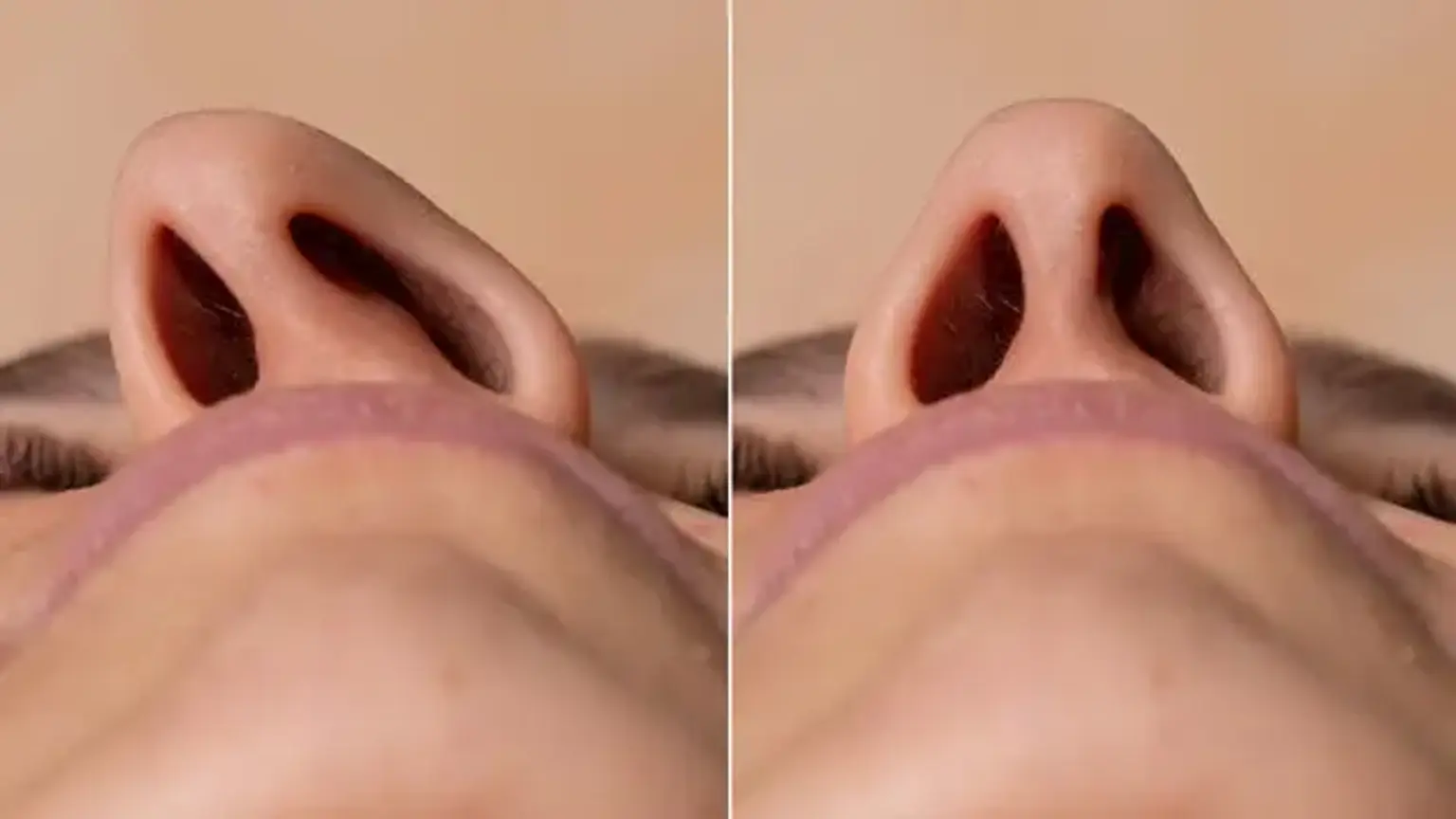
Understanding the Nasal Septum
The nasal septum is the structure inside the nose that separates the two nostrils. It consists of both bone and cartilage and is essential for maintaining proper airflow through the nasal passages. Ideally, the septum is straight, allowing air to flow evenly through both sides of the nose.
However, in many individuals, the septum may become deviated, or displaced, often due to injury or natural development. A deviated septum can cause a variety of issues, including:
Nasal Obstruction: A deviated septum can block airflow through one or both nostrils, leading to difficulty breathing through the nose.
Chronic Sinus Infections: Misalignment of the septum can hinder proper drainage of the sinuses, increasing the risk of infections.
Snoring: A blocked nasal passage can contribute to snoring and disturbed sleep.
Facial Discomfort: In some cases, a deviated septum can cause pressure in the face, especially around the sinuses.
Septoplasty aims to correct these problems by straightening the septum, which improves airflow and alleviates the symptoms caused by nasal obstruction. The procedure is highly effective in treating conditions related to nasal obstruction and chronic congestion.
The Septoplasty Procedure: Step by Step
The septoplasty procedure is typically performed on an outpatient basis, meaning patients can usually go home the same day. The surgery generally takes about 30 minutes to an hour, depending on the complexity of the septal deviation. Here’s a breakdown of what to expect during the procedure:
Anesthesia: Patients are given anesthesia to ensure they are comfortable and pain-free during the procedure. Depending on the severity of the deviation and the patient’s preference, the surgeon may use local anesthesia (numbing the area) or general anesthesia (putting the patient to sleep).
Incision: The surgeon makes a small incision inside the nose, typically along the mucous membrane that covers the nasal septum. This incision is usually hidden from view, so there are no visible scars on the exterior of the nose.
Correction of the Septum: Once the incision is made, the surgeon carefully lifts the mucous membrane and accesses the deviated septum. The surgeon will then straighten the septum by removing or repositioning the cartilage and bone as necessary. In some cases, excess cartilage or bone may be removed to improve airflow.
Reshaping and Reinsertion: After the septum has been corrected, the mucous membrane is repositioned, and any incisions are closed with dissolvable stitches.
Post-Surgery Care: Once the surgery is completed, patients are monitored for a short time to ensure there are no immediate complications. The patient may be advised to rest and avoid strenuous activities for several days. After the procedure, there may be some swelling, bruising, or discomfort, but these effects usually subside within a week or two.
Septoplasty surgery is designed to provide long-term relief for individuals suffering from a deviated septum and associated symptoms. With minimal downtime and effective results, this procedure has helped millions of patients breathe easier and improve their overall nasal function.
Septoplasty vs Rhinoplasty: Understanding the Difference
While septoplasty and rhinoplasty are both nasal surgeries, their purposes and procedures differ significantly. Septoplasty is performed to correct a deviated septum and improve nasal function, specifically by straightening the internal structure of the nose. Its main goal is to enhance breathing by restoring proper airflow.
On the other hand, rhinoplasty is primarily a cosmetic surgery aimed at changing the external appearance of the nose, such as reshaping the bridge, tip, or nostrils. While rhinoplasty can sometimes address functional issues like a deviated septum, its primary focus is aesthetics.
Some patients opt to combine both procedures to improve both the appearance and function of their nose. When performed together, these surgeries can offer a comprehensive solution, enhancing both the look and breathing capability of the nose.
Septoplasty for Deviated Septum
A deviated septum occurs when the cartilage and bone in the nasal septum are displaced, causing one side of the nose to be smaller than the other. This can lead to symptoms like nasal obstruction, snoring, and difficulty breathing through one or both nostrils.
Septoplasty is the recommended treatment for this condition. The procedure straightens the septum, alleviating the blockages and improving airflow. For many individuals, it offers immediate relief from persistent nasal congestion, sinus infections, and other related issues. This procedure is particularly effective for patients who have difficulty breathing due to a deviated septum and want to avoid long-term reliance on medication or nasal sprays.
Risks and Complications of Septoplasty
Like any surgery, septoplasty carries some risks, though they are generally rare. Possible complications include:
Infection: As with any surgery, there's a risk of infection at the incision site.
Bleeding: While bleeding is common right after the procedure, excessive bleeding may occur in some cases.
Scarring: Though rare, scarring can occur inside the nose if healing is not optimal.
Persistent symptoms: In some cases, patients may not experience complete relief from breathing problems.
Most of these risks can be minimized with proper care, choosing an experienced surgeon, and following post-operative instructions. The procedure itself is safe when performed by a qualified professional, and the benefits generally outweigh the risks for those suffering from a deviated septum or related conditions.
Benefits of Septoplasty Surgery
The main benefit of septoplasty surgery is improved breathing. When the nasal septum is deviated, airflow is obstructed, leading to difficulty breathing through the nose. After septoplasty, patients often experience:
Improved airflow and better overall breathing.
Reduced snoring, leading to better sleep quality.
Fewer sinus infections due to better drainage.
Increased comfort and relief from chronic nasal congestion.
Patients often report feeling a significant improvement in their quality of life, as better breathing can lead to better sleep, more energy, and a decrease in daily discomfort.
Septoplasty Recovery: What to Expect
Recovery from septoplasty surgery is relatively straightforward, though it does require some patience. Immediately after surgery, patients can expect some swelling, bruising, and mild discomfort. Here’s what you should know about the septoplasty recovery process:
First Few Days: Rest is essential. You may experience some pain, which can usually be managed with prescribed medications. Swelling and bruising around the nose and eyes are common.
Nasal Packing: In some cases, the surgeon may use nasal packing to prevent bleeding. This will typically be removed within 24-48 hours.
Post-Surgery Care: Patients should avoid blowing their nose for at least a week to prevent complications. Keeping the head elevated helps reduce swelling.
Full Recovery: Most people can return to normal activities within 1-2 weeks, although strenuous exercise and heavy lifting should be avoided for 3-4 weeks.
Following your surgeon’s post-operative care instructions will help ensure a smooth recovery and optimal results.
Septoplasty Surgery Cost: Factors to Consider
The cost of septoplasty surgery can vary widely depending on factors such as location, surgeon expertise, and whether the procedure is combined with other treatments. On average, the cost of septoplasty in the United States can range from $3,000 to $8,000, while in Korea, the price may be significantly lower, with costs ranging between $1,500 and $3,500.
Factors that influence cost include:
Surgeon’s experience: More experienced surgeons may charge higher fees.
Medical facility: High-end clinics and hospitals with advanced technology may be more expensive.
Location: Medical tourism in places like Korea can offer competitive prices while maintaining high standards of care.
If the procedure is medically necessary (for example, to correct a deviated septum causing breathing problems), some health insurance plans may cover part or all of the costs. It’s essential to check with your insurer to understand what’s covered.
Is Septoplasty Covered by Insurance?
Many insurance companies will cover septoplasty surgery if it is deemed medically necessary. If you have chronic nasal congestion, frequent sinus infections, or difficulty breathing due to a deviated septum, your insurance may provide coverage for the procedure.
However, if the surgery is considered cosmetic (such as when combined with rhinoplasty for aesthetic purposes), insurance may not cover the costs. It’s important to work closely with your healthcare provider to ensure that the surgery is properly documented as medically necessary to increase the chances of insurance coverage.
Patients should always confirm insurance details with their provider and consider obtaining pre-authorization before undergoing the procedure.
Recovery and Post-Operative Care in Korea
After septoplasty, the recovery process typically lasts around 1-2 weeks, with full recovery taking a few months. In Korea, medical facilities are equipped to ensure a smooth recovery:
Post-Surgery Care: You’ll be monitored for a few hours after surgery. If nasal packing is used, it will be removed within 48 hours.
Swelling and Bruising: Mild swelling and bruising, especially around the eyes, are normal. Ice packs and head elevation can help reduce these effects.
Avoiding Strain: It’s important to avoid strenuous activities and nose-blowing for at least a week. Gentle nasal care will be advised.
Korean clinics provide excellent post-operative support, including follow-up consultations to monitor healing and address any concerns during recovery.
Why Septoplasty is Popular in Korea
Korea is renowned for its advanced cosmetic and functional surgeries, including septoplasty. Known as a hub for medical tourism, Korea offers high-quality, affordable care, making it an attractive destination for patients seeking nasal surgery.
The reasons septoplasty is popular in Korea include:
Skilled Surgeons: Korean surgeons are highly experienced in nasal surgeries, combining technical expertise with precision.
Advanced Technology: Korea is known for its cutting-edge medical technology, which ensures high success rates and minimal complications.
Affordable Costs: Compared to Western countries, the cost of septoplasty in Korea is often more affordable, without compromising on quality.
Many international patients travel to Korea for nasal surgeries, benefiting from world-class care at a fraction of the price they might pay elsewhere. Additionally, Korea’s reputation for excellence in cosmetic surgery draws patients who may wish to combine septoplasty with other aesthetic procedures.
Septoplasty and Rhinoplasty Combined in Korea
In some cases, patients opt to combine septoplasty and rhinoplasty to address both functional and aesthetic concerns. While septoplasty is performed to correct a deviated septum and improve breathing, rhinoplasty focuses on altering the appearance of the nose. Combining these two surgeries allows patients to enjoy both enhanced nasal function and a more aesthetically pleasing nose.
The benefits of combining these procedures include:
Improved breathing and aesthetic appearance in one surgery.
Single recovery period, minimizing the total time away from regular activities.
Cost savings since undergoing both surgeries at once may be more affordable than having them separately.
In Korea, this combination of septoplasty and rhinoplasty is particularly popular due to the country’s expertise in both functional and cosmetic nasal surgeries. Patients from around the world often travel to Korea to have both procedures done by highly skilled surgeons, ensuring excellent results in both appearance and function.
Best Septoplasty Surgeons in Korea
When considering septoplasty surgery in Korea, it’s essential to choose a qualified and experienced surgeon. Korea is home to some of the best surgeons in the field of nasal surgery, offering world-class medical expertise. Here’s what to look for when selecting a surgeon:
Experience and Specialization: Look for surgeons who specialize in septoplasty and have years of experience in performing this procedure. Many surgeons in Korea are also skilled in combining septoplasty with rhinoplasty, which is a bonus if you’re considering both procedures.
Patient Reviews and Before/After Photos: Research patient testimonials and look at before-and-after photos to assess the surgeon’s skill and the outcomes of their surgeries.
Reputation of the Clinic: Choose a clinic or hospital with a reputation for quality care and successful outcomes. Internationally recognized medical institutions are a good sign of expertise and patient satisfaction.
Consultation: A thorough consultation is key. The best surgeons will take the time to assess your nasal issues, explain the procedure in detail, and answer any questions you have, ensuring you are fully informed before making a decision.
By selecting the right surgeon, you increase the chances of achieving the best results and a smooth recovery from septoplasty surgery.
Non-Cosmetic Septoplasty: Focusing on Function
While many people associate septoplasty with cosmetic surgery, it’s important to note that septoplasty is primarily a functional procedure. The main goal is to improve nasal airflow by correcting a deviated septum. Many patients undergo septoplasty not to change the appearance of their nose but to resolve issues like chronic nasal congestion, sinus infections, or difficulty breathing.
Non-cosmetic septoplasty is essential for individuals who experience:
Chronic nasal blockage or difficulty breathing through the nose.
Frequent sinus infections due to blocked sinuses.
Snoring caused by a deviated septum.
Sleep apnea symptoms related to nasal obstruction.
The surgery is not performed to alter the shape of the nose but to restore natural breathing. By focusing solely on functional improvements, non-cosmetic septoplasty can significantly improve the quality of life for many patients, helping them breathe easier and reducing related health issues.
Global Popularity of Septoplasty Surgery in Korea
Korea has become a global leader in medical tourism, particularly for nasal surgeries like septoplasty. The country’s renowned expertise in cosmetic surgery, combined with its advanced medical technology and relatively affordable prices, makes it a popular destination for patients seeking septoplasty.
The reasons for Korea’s popularity in this field include:
Expert Surgeons: Korean surgeons are highly skilled in performing complex nasal surgeries, including both septoplasty and rhinoplasty, offering patients excellent outcomes.
State-of-the-Art Facilities: Clinics and hospitals in Korea are equipped with the latest medical technology, ensuring the safest and most efficient procedures.
Affordable Costs: Compared to Western countries, septoplasty and other nasal surgeries in Korea are often more affordable, attracting medical tourists from all over the world.
Patients from countries like the United States, Canada, and many parts of Europe travel to Korea to undergo septoplasty, benefitting from high-quality care at a fraction of the cost. The global popularity of septoplasty surgery in Korea continues to grow, making it a leading destination for those seeking functional and aesthetic improvements to their noses.
Pre-Surgical Assessments and Safety Protocols
Before undergoing septoplasty, a thorough pre-surgical assessment is necessary. This ensures you’re in good health and that the procedure is right for you. Key steps include:
Health Evaluation: The surgeon will review your medical history, any existing conditions, and current medications. A physical examination of your nasal passages will also be conducted.
Imaging: A nasal endoscopy or CT scan may be done to get a detailed view of the nasal structures and the severity of the deviation.
Blood Tests: Some surgeons may require blood tests to check for conditions that could affect healing or anesthesia.
Safety protocols during surgery include ensuring a sterile environment to minimize infection risks. Anesthesia will be administered by trained professionals, and the surgery will be performed in a fully equipped medical facility. Post-surgery, clear aftercare instructions will be provided to ensure safe recovery.
FAQs about Septoplasty Surgery
How does septoplasty improve breathing?
Septoplasty corrects a deviated septum, opening up blocked nasal passages and allowing air to flow freely, making breathing easier.
How long does the recovery take?
Most people can return to normal activities within 1-2 weeks, although it can take up to 3-4 weeks to fully recover from septoplasty.
Is septoplasty covered by insurance?
If septoplasty is deemed medically necessary, such as for correcting breathing problems, it may be covered by insurance. However, cosmetic procedures are usually not covered.
Can I combine septoplasty with rhinoplasty?
Yes, many patients combine septoplasty and rhinoplasty for both functional and cosmetic improvements. This combined surgery can save time and costs.
What are the risks of septoplasty?
Risks include infection, bleeding, and scarring, but these are rare. Choosing an experienced surgeon minimizes these risks.
Conclusion
Septoplasty surgery offers life-changing benefits, especially for individuals with a deviated septum that causes chronic nasal obstruction and breathing difficulties. With improvements in airflow, snoring, and sinus health, many patients find significant relief. In addition to the functional benefits, septoplasty can enhance overall well-being and quality of life.
For those considering this procedure, particularly in places like Korea, the combination of expert surgeons, advanced technology, and affordable costs makes it an attractive option. If you're experiencing nasal congestion or related symptoms, septoplasty could be the solution you've been seeking to breathe easier and enjoy better health.