Single Ventricle Defects Surgery
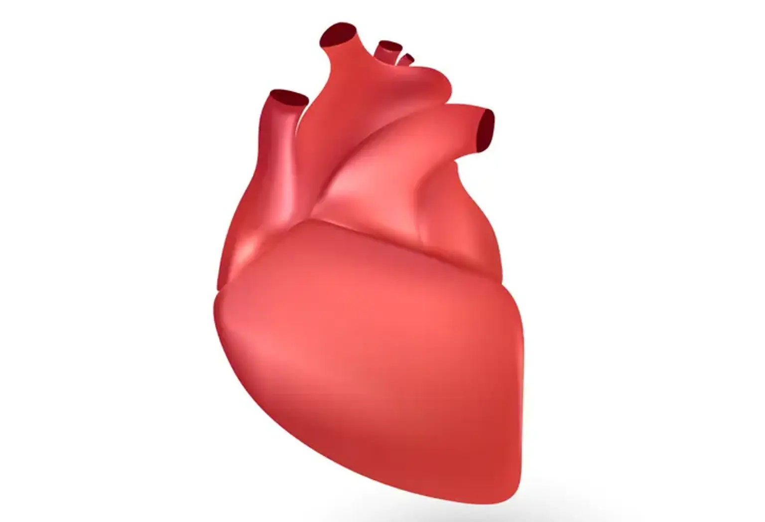
Congenital heart disease (CHD), also known as a congenital heart defect, is an issue with the heart's or blood vessels' architecture that develops before birth. When the walls of the heart, the valves of the heart, and the arteries and veins near the heart do not grow normally before birth, abnormalities occur. These flaws may or may not cause problems with a person's circulatory system. If they do, blood flow may be impeded, shifted in the wrong direction or to the wrong location, or completely blocked. Some of these defects are minor and may not cause issues, while others are more serious and may result in life-threatening problems.
Because of advancements in diagnosis and treatment, most babies born with congenital heart disease survive into adulthood. Adults, including those who received treatment as kids, can develop signs and symptoms of the illness later in life. It's also possible that cardiac abnormalities that weren't significant enough to treat as a child have deteriorated and now necessitate treatment. As a result, it is essential to consult your physician.
What is Norwood Procedure?
Hypoplastic left heart syndrome (HLHS) is a severe congenital cardiac abnormality (existing at birth) characterized by an underdeveloped left side of the heart. The left half of a typical heart is responsible for pumping oxygenated blood into the aorta (the large artery that transfers blood to the body). There are several issues in a child with HLHS: the mitral valve is too tiny or totally closed, the left ventricle is too small, and the aortic valve is too tiny or completely closed. As a result, the left side of the heart becomes completely incapable of supporting the circulation required for the body's organs. HLHS is deadly without treatment, frequently during the first hours or days of life.
Three types of open-heart procedures (involving splitting the breast bone and general anesthesia) are required to treat HLHS. The Norwood treatment (within a few days of birth), the Glenn procedure (within six months of birth), and the Fontan procedure must all be done in a certain order (approximately at 2 to 4 years of age).
Before Norwood Procedure
Your doctor may take a variety of tests before the surgery, including:
- Diagnostic techniques and tests
- Ultrasound of the heart (echocardiography)
- Computed tomography (CT scan)
- Magnetic resonance imaging (MRI)
- X-rays
- Electrocardiogram (ECG)
Norwood Procedure
This treatment takes about 5 to 6 hours on average, although preparation and recovery time can add up quickly. In most cases, the procedure is carried out in a cardiothoracic operating room (OR). Generally speaking, throughout this procedure:
- Your kid will be anesthetized before the doctor inserts a breathing tube into their lungs and connects it to a ventilator through their throat. This will allow your baby to breathe during the operation. A general anesthetic will be administered by your doctor (will make your kid feel sleepy).
- The surgery begins with the doctor dividing the breastbone (sternum) in half to expose your baby's heart. After that, your doctor spreads both halves apart to gain access to your baby's heart (open-heart surgery).
- The heart must be quiet for this type of surgery. Your doctor will first insert tubes into your baby's heart so that blood may be pumped through his or her body using a heart-lung machine. This equipment replaces the heart's pumping motion and the lungs' oxygenation by supplying oxygen to the blood.
- After the blood has been transferred to the bypass machine for pumping, your surgeon will use a cold solution to arrest the heart.
- Your doctor will begin the surgery once the heart has been stopped. The Norwood operation goals are to ensure that blood reaches the lungs and to better control blood flow to avoid lung and heart damage.
- The pulmonary artery, which leaves the right side of the heart, is separated first. Sewn shut at the far end (the end nearest to the lungs). The near end (nearest to the heart is sewn into the aorta). To make the new aorta bigger and stronger, a patch is sewn in this location. All blood leaving the heart now passes through the pulmonary valve and out the body through the new aorta on the right side.
- Hollow tube (shunt) is sewn into place that joins the new aorta to the pulmonary artery. Some of the blood delivered to the body is redirected to the lungs via the shunt.
- The septal wall that separates the two upper chambers of the heart (atria) is removed. This allows red blood to circulate from the left upper chamber to the right upper chamber after returning from the lungs. After that, the blood travels to the right lower chamber before exiting the body.
- After the procedure, the doctor will double-check everything to ensure it is working correctly. The doctor will then let the blood circulate through the bypass machine back into your baby's heart after it has been tested.
- The machine will be switched off once the procedure is completed. The tubes will be taken out, and the sternum will be stitched together with stitches or surgical staples.
What Happens after Norwood Procedure?
Your infant will be transported to the cardiothoracic intensive care unit (CTICU) for a few days after the surgery. Your infant will be in the care unit for several days. The cardiac team will do the following throughout this time:
- Keep an eye on your baby's vital signs, such as heart rate and respiration.
- Give your baby pain medication to ensure he or she is not in any discomfort.
- Make sure your newborn's medications are taken care of.
- Make note of the tubes (drains) that were inserted into your baby's chest during surgery.
- Take better care of the sternal incision (chest wound) and any additional incision sites.
- Confirm that your newborn can feed well by mouth.
- Will provide you with instructions to follow during the recovery of your kid.
What is Bidirectional Glenn Shunt?
The bidirectional Glenn (BDG) shunt, also referred to as a bidirectional cavopulmonary anastomosis, is a surgical procedure used in pediatric cardiac surgery to enhance blood oxygenation for babies with one functional ventricle due to a congenital heart defect. The Fontan operation decreases the amount of blood volume that the heart must pump by creating a bidirectional shunt.
Bidirectional Glenn Shunt Uses
Univentricular heart defects are predicted to occur in 0.3 to 0.5 per 1,000 live births. These patients require an aortopulmonary shunt in the newborn period, which is supported medically with prostaglandin and later surgically with the first cardiac shunt surgery. When the patient's oxygen saturation starts to drop, they are considered for the Glenn bidirectional shunt since they will outgrow the shunt over time. For newborns born with congenital single ventricle defects, the Glenn surgery is routinely performed around 4 to 6 months of age. Following the Glenn bidirectional operation, these patients usually require a Fontan procedure between the ages of 18 and 36 months.
Hypoplastic left heart syndrome, tricuspid atresia, double-inlet left ventricle, and double-outlet right ventricle are some of the congenital cardiac defects for which this treatment may be used. Congenital univentricular cardiac defects have a natural history of cyanotic heart failure at an early age. These patients have been able to survive into adulthood thanks to the bidirectional Glenn shunt and Fontan operation.
Bidirectional Glenn Shunt Contraindications
To support pulmonary blood flow, a patient's circulation following bidirectional Glenn shunt insertion requires an adequate systemic venous return. High pressures or valvular blockages, on the other hand, prevent pulmonary blood flow and consequently oxygenation.
The bidirectional Glenn is contraindicated in patients with moderate to severe pulmonary hypertension. The reason for this is that pulmonary vascular resistance is too high to allow adequate oxygenation. High pulmonary vascular resistance, rigid ventricle tissue, and atrioventricular valve dysfunction are examples of contraindicated physiologic parameters. Blockage of blood flow at the atrial septum should be evaluated and treated in patients with hypoplastic left heart syndrome.
Bidirectional Glenn Shunt Procedure
The surgical goal of the bidirectional Glenn shunt surgery is to manage pulmonary blood flow and heart volume load. This must be balanced with sufficient oxygenation and oxygenated blood supply throughout the body. Detaching the superior vena cava from the right atrium and linking the cranial segment of the SVC to the pulmonary arteries creates this modified circulatory system (shunt). A surgical anastomosis looks like this. As a result, venous blood from the upper body reaches the SVC and circulates through pulmonary circulation in a low-pressure circuit, comparable to a two-ventricle system. The remaining systemic venous blood, on the other hand, flows through the inferior vena cava, mixing with oxygenated blood coming from the pulmonary circulation.
Bidirectional Glenn Shunt Risks
Many of the dangers and consequences associated with the Glenn bidirectional shunt failure are thought to be caused by thrombosis. The key factors that enhance the risk of complications and failure are right-side dominant circulation, higher pulmonary vascular resistance, and prolonged surgical and recovery times.
Approximately 35% of individuals develop problems after surgery. Emergency cardiac catheterization, new neurologic impairment, reoperation, cardiac arrest, and feeding issues requiring procedural intervention were the most prevalent complications, according to data published by the National Pediatric Cardiology Quality Improvement Collaborative registry in the 2010s. Longer cardiopulmonary bypass time, higher central venous pressure (CVP) or pulmonary arterial pressure, and specific malformations such as an unbalanced atrioventricular septal defect or a history of total anomalous pulmonary venous connection repair have all been identified as risk factors for unfavorable outcomes.
Failure of the procedure after the bidirectional Glenn shunt can be classified as procedural failure, cardiac dysfunction related to surgery, or cardiac dysfunction leading to death before further surgical intervention. The procedure failed in 6.5 percent of patients, according to retrospective studies. The bidirectional Glenn procedure has a reported death rate of 0 percent to 2.5 percent, but up to 70% of patients may die if the bidirectional Glenn shunt fails, with many decompensating before further surgery can be done. Early additional intervention, according to current studies, is a promising route for improving future outcomes. Right ventricular dominance, extended pleural drainage, prolonged admission to the ICU/hospital, or the need for ECMO to support oxygenation are all predictors of a failed surgery.
What is Fontan Procedure?
The Fontan operation is the last in a series of surgeries aimed at improving heart function in children born with severe single-ventricle diseases including hypoplastic left heart syndrome (HLHS). The operation does not correct the congenital cardiac abnormality. In the absence of a second ventricle, it permits the heart to work more efficiently.
Treatment for kids born with only one ventricle has improved, allowing more children to enjoy full lives. Before surgeons discovered the three-surgery series, HLHS was thought to be inoperable and fatal.
Newly oxygenated blood is reduced by mixing with deoxygenated blood in only one ventricle, completing its route throughout the body.
The Fontan operation rebuilds the veins to deliver blood directly to the lungs from the lower body. The route skips the heart, preventing deoxygenated and oxygenated blood from mixing.
Cardiothoracic surgeons at any children's heart center are experts at rebuilding the heart and redirecting blood flow so that the heart isn't strained and more oxygen-rich blood is transported throughout the body.
While the Fontan technique and other therapies have saved lives, children with this abnormal blood circulation pattern are at risk for issues in other parts of the body, such as the digestive tract, as they grow older.
Fontan clinics usually follow these infants as they grow and catch growing issues. A patient can see cardiologists, gastroenterologists, and other specialists in one visit. The multidisciplinary practice provides a natural environment for doctors to compare notes and share perspectives on a patient's treatment plan, in addition to convenience.
Fontan Procedure
The Fontan operation involves detaching the inferior vena cava (the vein that returns blood from the lower body) from the heart and reconnecting it to the pulmonary artery.
Between the conduit and the right atrium, a small opening called a fenestration is frequently built. This lets some blood return to the heart directly. It serves as a relief valve as the lungs adapt to the increased flow from the lower body. A minimally invasive cardiac catheterization treatment can be used to close the fenestration later in life.
With all of the connections in place, blood from the lower body now flows directly to the pulmonary artery, then to the lungs, bypassing the heart. The patient's single ventricle is now responsible for pumping oxygen-rich blood from the lungs to the body and back.
Because the abnormality prevents high- and low-oxygen blood from mixing in the heart, more oxygen reaches the body.
Fontan surgery patients often spend one to two weeks in the hospital recovering. They also receive medications to support their heart and blood flow. Your hospital's physicians and staff will provide your child with 24-hour care and supervision.
Fontan Procedure Complications
Exercise intolerance, heart failure, right atrial dilatation and arrhythmia, systemic and hepatic venous hypertension, portal hypertension, coagulation disorders, pulmonary arteriovenous malformation, venovenous shunts, and lymphatic dysfunction (e.g., ascites, edema, effusion, protein-losing enteropathy, and plastic bronchitis) are all complications of Fontan circulation. Postoperative assessment of Fontan surgery patients is best done with magnetic resonance imaging, and heart transplantation is still the only curative therapy for those with failed Fontan circulation.
Biventricular Repair and Conversion
For certain children born with a borderline (tiny) right or left heart, complex biventricular repair and biventricular conversion are therapeutic options. One of the heart's two lower chambers, called ventricles, isn't large or healthy enough to function properly in certain conditions. The majority of individuals with borderline cardiac disease are treated with single ventricle palliation.
In a biventricular repair, two working ventricles are constructed in one surgery.
A biventricular conversion is a process of transforming a single ventricle palliated patient's heart into a heart with two functioning ventricles.
If a patient's ventricle is too tiny for biventricular repair or conversion right away, the small ventricle may need to be reconditioned through a series of treatments known as staged recruitment. The eventual aim of staged recruitment is biventricular conversion, which involves switching from a single ventricle heart to one with two functioning ventricles.
Each child's cardiac condition and unique heart architecture determine the type and number of surgeries and procedures required to complete this procedure.
Why Consider Biventricular Repair Instead of a Single Ventricle Palliation?
The Fontan operation is performed on most kids with a single ventricle heart defect after a sequence of three heart surgeries called single ventricle palliation. These procedures allow a single functional ventricle to perform the function of two ventricles.
While this technique works successfully for some infants, single-ventricle circulation causes long-term issues for others. Because one ventricle is pumping blood to the body as well as the lungs, it is more likely to become fatigued. Many children who have only one ventricle experience long-term liver and lung problems and may require a heart transplant in the future.
Because of these considerations, surgeons attempt to achieve a two-ventricle (biventricular) heart repair or conversion whenever possible. Doctors have discovered that this strategy improves children's long-term quality of life. Many patients have benefited from these techniques thus far.
Not all patients are eligible for biventricular repair or conversion, and there are dangers associated with this procedure, which is why it's vital to have a dedicated team examining each patient and closely monitoring the results. Your team of specialists works directly with your family throughout the process to ensure that you make the best decisions for your child.
Conclusion
Hypoplastic left heart syndrome, tricuspid atresia, single ventricle, imbalanced AV septal defect, mitral atresia, and other cardiac abnormalities have a functionally single ventricle. The physiology of a single ventricle differs from that of a biventricular heart, and the delicate balance of blood flows between the pulmonary and systemic circulations must be preserved. These babies are typically managed in three stages via staged total cavopulmonary connection. Inter-stage (between stages) mortality is pretty prevalent (5-15 percent). Between Stages one and two, it occurs more frequently than between Stages two and three. Restricted atrial communication, blockage of the aortic arch, stenosis of the pulmonary arteries, AV valve incompetence, shunt obstruction, and intercurrent diseases are all linked to inter-stage mortality. Clinical evaluations and echocardiographic or other imaging examinations should be performed on a regular basis to detect these problems, which should be addressed immediately once discovered. Even mild coexisting diseases must be aggressively managed.