Skull Base Surgery
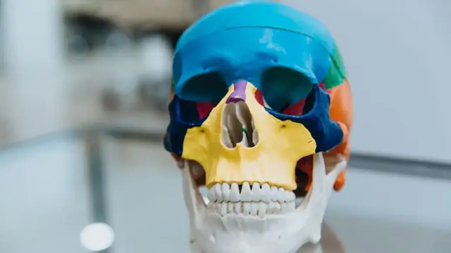
The face and the cranium, which enclose the brain, are made up of bones and cartilage that make up the skull. On top of the skull, you can touch the craniums' bones. The eye socket, the roof of the nasal passage, some sinuses, and the bones that encircle the inner ear are all formed by the five bones that make up the bottom, or base, of the cranium. The base of the skull is a dense and complex location with numerous openings through which the spinal cord, numerous blood vessels, and nerves all travel.
Noncancerous and cancerous growths, as well as anomalies on the underside of the brain, the skull base, or the top few vertebrae of the spinal column, may be removed during skull base surgery. Skull base surgery may be performed using a minimally invasive endoscopic approach because this is such a challenging place to view and reach. The surgeon introduces instruments into the skull through natural openings such as the nose or mouth, or a small opening just above the brow. ENT surgeons, maxillofacial surgeons, neurosurgeons, and radiologists are among the professionals needed for this procedure.
The only option to remove growths in this part of the body before endoscopic skull base surgery was to make an incision in the skull. This type of surgery may be required in some circumstances.
What is Skull Base Surgery?
Skull base surgery is a highly specialized, minimally invasive surgical method for assessing, diagnosing, and removing benign or malignant growths on the underside of the brain, the base of the skull, and the upper spinal vertebrae. It may also be beneficial in the treatment of congenital abnormalities and deformities.
Rather than reaching the brain through a craniotomy, skull base surgeons employ specific devices introduced through the skull's natural holes (e.g., nose, mouth, and above the eyes). In order to obtain access to these locations before the invention of skull base surgery, large sections of the skull and/or facial musculature had to be removed. Skull base surgery has several advantages, including a lower chance of infection and injury to cerebral structures and nerves, as well as a reduced risk of deformity and a faster recovery period.
Anatomy of the Skull Base
At the base of the skull, many critical structures, such as major blood vessels and vital nerves, leave and enter. The brain is separated from the eyes, nose, ears, and other facial structures by the base of the skull. It begins above the brows and descends to the back of the neck, where the foramen magnum can be found.
The front cranial fossa, the middle cranial fossa (focused around the pituitary), and the posterior cranial fossa, which comprises the brainstem and cerebellum, are split into three parts from front to back.
The brainstem connects the brain and the spinal cord, and it contains nerves that control respiration, blood pressure, eye movements, and swallowing. The foramen magnum links the brainstem to the spinal cord. Coordination and balance are controlled by the cerebellum, which is located behind the foramen magnum.
The fact that each of the skull's sections is at a distinct level adds to the skull's complexity. The anterior cranial fossa is highest and the posterior cranial fossa is lowest when a person is standing and gazing ahead.
Skull Base Surgery Approaches
Historically, the skull base has been difficult to reach, making brain abnormalities in this area difficult to manage. However, surgical breakthroughs and advances in neuroimaging have made skull base disorders approachable and surgically treatable.
The world-class neurosurgeons use the most up-to-date procedures and technology to make this treatment as minimally invasive as possible. Either of the two approaches to skull base surgery can be used to achieve the maximum levels of safety and effectiveness. Physicians adopt a technique in each situation based on the location of problems and what is best for the patient overall.
- Pterional approach. It's a flexible anterolateral neurosurgical method that gives you access to the cranial fossae many structures. It is critical for neurosurgeons to master and be well-versed in the multilayer architecture of the brain.
- Frontolateral approach. It is a straightforward, reliable, and efficient operation that provides enough space for intracranial manipulation while protecting the brain and other intracranial structures to the maximum extent possible. For the treatment of paraclinoid aneurysms, the frontolateral approach could be a useful alternative to the pterional method.
- Craniotomy. This is an open surgery method in which pieces of the skull are removed temporarily to allow the neurosurgeon to access the brain. This procedure can now be performed utilizing minimally invasive procedures for some brain disorders, reducing the size of incisions and the opening in the skull.
- Transsphenoidal surgery. This is a minimally invasive treatment in which an endoscope is used to execute the procedure through the nostrils or the inside of the mouth. Only absolutely tiny incisions are created, and they are usually unnoticeable.
- Suboccipital approach. A section of the bone behind the ear is removed in the suboccipital approach. To treat the acoustic neuroma, the cerebellum is carefully pushed back.
Skull Base Surgery Indications
When a brain abnormality is discovered around the skull base, skull base surgery is performed. The neurosurgeons at the Skull base surgery center always investigate conservative treatments first, such as medication, however, surgery is frequently the primary treatment for the condition.
Skull base surgery is used to treat a variety of brain disorders, including skull base tumors, aneurysms, trigeminal neuralgia, brain abscesses, hematomas, arteriovenous malformations, and hydrocephalus.
Pituitary Tumor
These are growths in the pituitary gland that are dysfunctional. The pituitary gland is in charge of creating hormones that regulate a variety of bodily activities. Changes in the levels of these hormones can be caused by tumors. The majority of tumors do not produce hormones. It's possible that the tumor will grow large enough to press upon optic nerves, causing vision loss.
The majority of pituitary tumors can be excised through the nose with the help of an endoscope and tiny devices. To establish a passage to the tumor, the sinuses are exposed. The tumor is resected after the bone between the sinuses and the tumor is excised.
Craniopharyngioma
This is a non-malignant tumor that forms near the pituitary gland and is extremely rare. Hormone levels may be affected, and vision may be lost as a result of these tumors. This is more common among children and the elderly.
Many craniopharyngiomas can be excised through the nose with the help of an endoscope and tiny devices. To make a path to the tumor, the sinuses are opened. The tumor is resected after the bone between the sinuses and the tumor is resected. At the termination of the surgery, tissue from within the nose is used to close the gap. Radiation is frequently used to stop any remaining tumor from growing.
Meningioma
This is a tumor that arises from the brain's surrounding tissue. These tumors can develop everywhere there is a lining around the brain or spinal cord. These tumors aren't malignant, but they can put pressure on the brain's structures. When this form of tumor develops on the bottom of the brain, near the sinuses, an endoscopic method may be a suitable option.
Endoscopic method for surgical resection of this sort of tumor on the bottom of the brain in an area that contacts the sinuses may be a possibility. The bone that divides the brain from the sinuses is cut in this case, enabling the tumor to be excised. At the completion of the surgery, tissue from within the nose is used to close the gap.
Rheumatoid Arthritis of the Cervical Spine
Rheumatoid arthritis is a chronic inflammatory disease. Occasionally, this can result in a mass that compresses the spinal cord due to a process in the fluid between the bones at the top of the spine. Patients with this condition may have gradual weakening in their arms or legs.
Through the nose, you can reach the top of the spine. The tumor pressing on the spinal cord can be removed with endoscopes and tiny devices. Patients can recover muscle strength in their arms and legs by eliminating this pressure.
Clival Tumor
This is an uncommon type of cancer that develops near the base of the skull, which is located on top of the spine. When malignancies are discovered in this location, they can typically be removed using endoscopes and devices that work through the nose.
An endoscopic method can often be used to remove clival tumors at the base of the skull. Depending on the size and location of the tumor, a combination of endoscopic and open surgery may be required. After the defect has been removed, tissue from within the nose is used to close it. Following surgical excision, radiation therapy is frequently required.
Petrous Apex Cholesterol Granuloma
These are benign cysts that can develop deep into the skull and are extremely rare. These cysts can develop and press against vital ear nerves and tissues. These tumors are frequently related to ear structures, however, due to their location, they can easily be accessed through the back of the sinuses.
An endoscopic technique can be used to open the cyst if the tumor reaches the sphenoid sinus wall. This method avoids hearing loss and increases the chances of the cyst staying open.
Skull Base Surgery Preparation
When doing skull base surgery, the neurosurgeons form a multidisciplinary team to achieve the best possible outcome. In addition to the neurosurgeon, the surgical team may include any of the following doctors:
- Head and neck surgeon
- Ophthalmologist
- Plastic surgeon
- Otolaryngologist
- Oral and a maxillofacial surgeon
Skull base surgery begins with general anesthesia, so the patient does not experience discomfort during the procedure, whether it is performed via craniotomy or transsphenoidal procedures.
Skull Base Surgery Types
Craniotomy
The neurosurgeon makes an incision in the scalp, usually at the region of the lesion at the base of the skull, and then carefully reflects the skin and muscle to show the skull. By temporarily removing a bone flap while the skin and muscle are reflected, the neurosurgeon can clear the path in the skull. Towards the end of the procedure, the bone flap will be replaced. Bone removal is a sensitive treatment that necessitates advanced technical abilities to avoid injuring structures along the base of the skull. The removal of bone is intended to enable access to the tumor while avoiding brain manipulation. The opening is as small as feasible; its size is determined by the amount of treatment required for the specific problem.
The dura mater may be seen through the hole. The neurosurgeon makes an incision in the dura mater with surgical scissors, exposing the brain.
The neurosurgeon can begin efficiently maneuvering to the lesion after the brain has been exposed. The skull is filled with delicate structures like brain tissue, nerves, and blood vessels, leaving little place for equipment. To provide a clear image of the numerous structures and accurately navigate to the brain problem, the neurosurgeon employs an operating microscope and other specialized equipment. The neurosurgeon also uses stereotactic techniques to help plan the optimal path to the abnormality, employing technologies such as magnetic resonance imaging (MRI) and computed tomography (CT) scans, as well as computer technology, to build three-dimensional views of the brain.
Following that, more treatments are conducted to address the problem, depending on the brain pathology.
After the problem has been treated, the neurosurgeon stitches the dura mater incision and replaces the bone flap with titanium plates and screws. Finally, the neurosurgeon sutures up the scalp incision. The procedure is now complete.
Transsphenoidal Surgery
Transsphenoidal surgery, unlike craniotomy, usually does not necessitate noticeable incisions, and if it does, they are very small. In addition, the surgeon performs the surgery through the nose or mouth.
The procedure begins with the tip of an endoscope being inserted into one of the nostrils and the sphenoid sinus at the rear of the nasal cavity being opened.
An endoscope or microscope gives the surgeon a better view of the surgical region deep inside the nose by providing light and magnification. Instruments are inserted through the nose to fix or eliminate the brain abnormalities, and the neurosurgeon obtains the greatest level of precision by employing stereotactic techniques to create a three-dimensional map of the patient's nerves, blood vessels, and other brain structures. Any incisions produced during the removal or correction of the brain abnormality are stitched. The operation is finished.
How is Skull Base Surgery Performed?
Talk to the doctor about any medications or supplements you're taking, including herbal or vitamins. It is essential that the doctor be aware of them since certain medications or supplements, such as warfarin and aspirin, might cause excessive bleeding during surgery, and the doctor will need to assess if the medication plan has to be altered.
Also, let the doctor know if you have any dietary or pharmaceutical sensitivities.
You will be instructed to cease eating and drinking at midnight last night before the surgery because general anesthesia will be administered. If the doctor has instructed you to take specific drugs on the day before surgery, take them with a little sip of water.
Wear loose, comfortable attire on the day of the procedure. You will be told not to wear any cosmetics, jewelry, or nail polish. Wear glasses rather than contact lenses.
Make every effort to prepare light for the hospital stay. Toiletries, dentures, and supplementary clothing for when they are discharged from the hospital are some of the essentials that patients want to carry.
You will not be allowed to drive yourself home following surgery, so make arrangements for transportation home from the hospital.
How Long Will I Stay in the Hospital?
Patients who have transsphenoidal surgery usually stay in the hospital for one or two days before being discharged. Those who get a craniotomy frequently stay for a week or more.
Will I Need to Take Any Special Medications?
Patients who have transsphenoidal surgery are given pain and nausea medications, and those who have a craniotomy may be given additional medications to avoid brain swelling and seizures.
Skull Base Surgery Follow-up
Depending on the handled brain disorder and whether any new symptoms arise after skull base surgery, such as vision impairment, a patient will be arranged for several follow-up visits with the neurosurgeon and possibly other specialist doctors, such as an endocrinologist, neurologist, otolaryngologist, or ophthalmologist.
During these visits, the neurosurgeon will assess the patient's recovery in extensive detail. The neurosurgeon may arrange imaging examinations to see the brain or do other investigations, depending on the type of brain problem being treated.
In order to have the greatest potential for recovery and long-term success, patients are highly advised to attend all follow-up visits. Patients should always notify the doctors of any new or deteriorating symptoms while recovering, even if the changes appear mild.
When Can I Resume Exercise?
Patients who undergo transsphenoidal surgery should anticipate beginning light exercises like swimming or running two weeks following surgery, and any other exercise four weeks later. The next day after surgery, patients are frequently walking, but they may realize that they exhaust easily and need to rest frequently. This is very normal.
Patients who have had a craniotomy are advised to get up and walk every day if they have the energy, but they should wait until their neurosurgeon gives them permission to do so. Because skull base surgery is more invasive than transsphenoidal surgery, recovery time will be longer. Patients can begin with modest activity and gradually progress to more vigorous exercise once the neurosurgeon has given his or her approval. However, until the patient has made a full recovery, any type of exercise should be done with a partner or under observation.
Skull Base Surgery Rehabilitation
The extent to which rehabilitation or physical therapy is required is primarily determined by the type of brain disorder being treated. In most cases, skull base surgery does not necessitate rehabilitation or physical therapy.
Skull Base Surgery Long-term Limitations
Any long-term limitations are mostly determined by the brain condition being addressed. Skull base surgery does not frequently result in long-term restrictions.
Conclusion
Skull base surgery is a field that has had a lot of growth in the last few years and is still being described in terms of its scope. Endoscopic techniques can address a wide range of anatomic locations and diseases, and questions about their efficacy remain unsolved for the time being. Surgical treatment for benign disease has mostly acquired acceptability; nevertheless, endoscopic care for the malignant disease remains contentious, and more data is needed to address these issues. This is an issue that can only be addressed with more patients and longer follow-ups, which will hopefully be seen in the coming years. It's vital to remember that a minimally invasive method should never be used as an excuse for avoiding surgery. The extent of resection should be equivalent to or better than the extent of resection achieved with an open method, and if the surgeon believes that the endoscopic approach will impair the surgery's completeness, another approach should be used. Finally, while deciding which strategy to take, the one that traverses the fewest neurovascular systems to reach the lesion is usually the best. This will be the endoscopic technique from below for the great majority of midline skull base lesions.