Small Bowel Obstruction
Overview
Small bowel obstruction is a frequent surgical emergency caused by mechanical bowel blockage. Though it can be caused by a variety of pathologic diseases, intra-abdominal adhesions are the most common cause in the industrialized world.
What is Small Bowel Obstruction (SBO)?
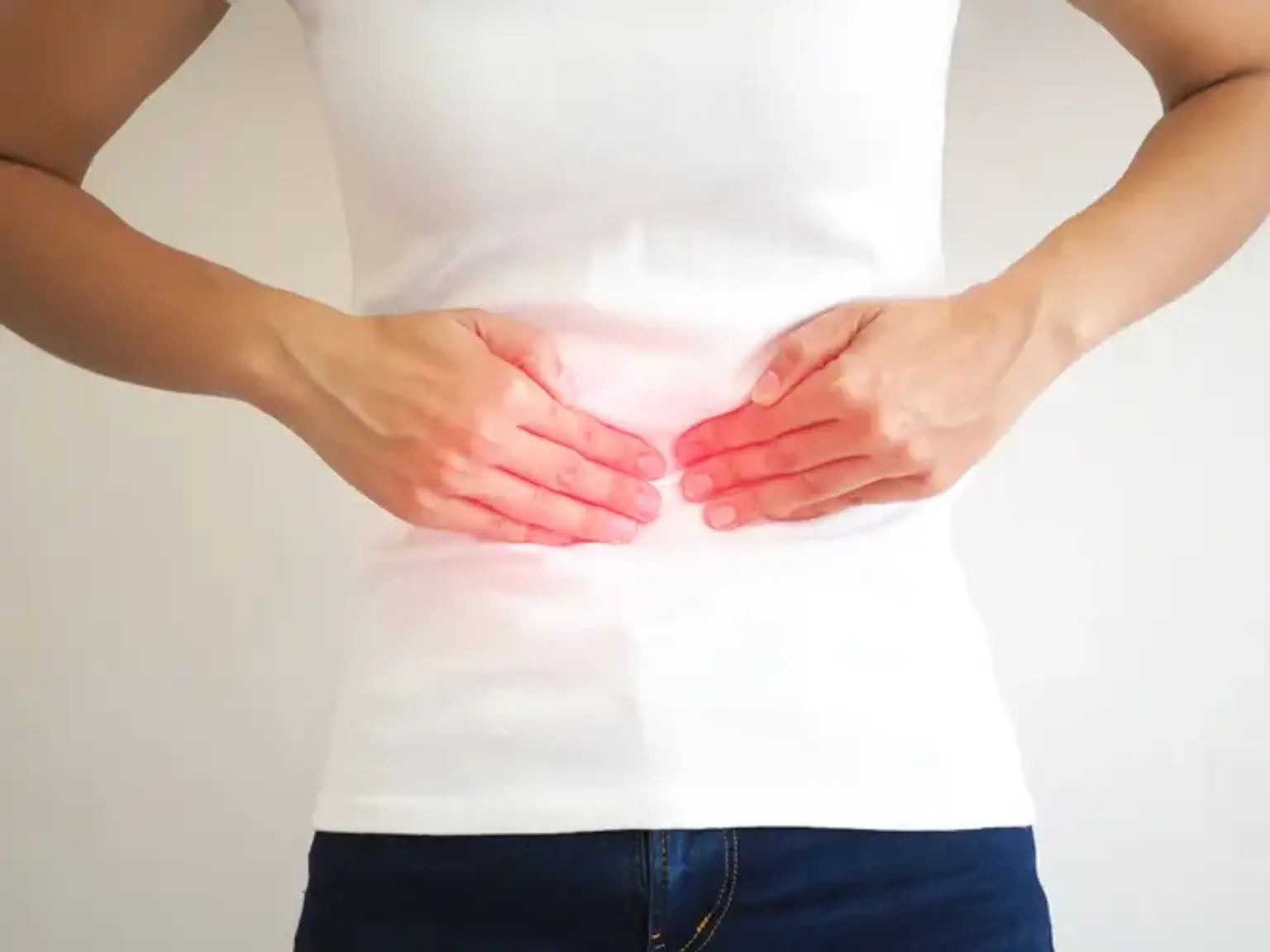
Small Bowel Obstruction (SBO) is a mechanical disturbance in the gastrointestinal tract's patency, resulting in emesis (which may be bilious), extreme constipation, and abdominal discomfort.
Scar tissue, hernias, or cancer are the most common causes of small bowel blockages. Most blockages in the United States are the result of previous procedures. After being touched during an operation, the bowel frequently produces scar bands (called adhesions). Scars are more prone to occur when bowel procedures are performed often. A minor intestinal blockage can occur if the colon becomes stuck in adhesions. In extreme situations, the blood supply may be damaged, and gut tissues may die. This is a potentially fatal circumstance.
There are two types of small bowel obstruction:
- Functional — there is no physical blockage, however, the bowels are not moving food through the digestive tract
- Mechanical — there is a blockage preventing the movement of food.
The gut twisting causes proximal bowel distention and distal bowel decompression. Peristalsis may initially increase, resulting in frequent bowel motions. Vomiting can occur as a result of proximal bowel distention. The twisted gut will initially cut off venous blood flow, causing edema and inflammation of the colon wall. The third fluid spacing is also common.
Ischemia and bacterial translocation are possible due to the thicker and inflamed gut wall. Bacterial translocation, most usually caused by Escherichia coli, can result in peritonitis and bacteremia. As the colon twists further, the arterial supply is cut off, resulting in intestinal ischemia and, if left untreated, perforation, peritonitis, and death.
Causes of Small Bowel Obstruction
At some point in life, the patient may have had prior abdominal operations, inflammatory bowel disease, cancer, or a hernia. Complaints of stomach discomfort, distention, nausea, and vomiting are the most prevalent. The abdominal discomfort might be either progressive or intermittent. It may be linked with constipation, obstipation, flatus, or even loose bowel motions.
Bowel noises may be low-pitched and high-pitched. On physical examination, abdominal discomfort may be widespread or localized, with or without distention. Rebound, guarding, and stiffness may be symptoms of peritonitis and indicate late findings that may be present depending on the time of presentation. Examination for hernias, surgical scars, lumps, especially those in the rectum, and fecal impactions may reveal the origin. There might also be signs and symptoms of dehydration and sepsis.
Pathophysiology of Intestinal Obstruction
Blockage occurs without vascular compromise in simple mechanical obstruction. Above the blockage, ingested liquids and food, digestive secretions, and gas gather. The distal portion collapses when the proximal bowel distends. Normal mucosal secretory and absorptive processes are impaired, and the gut wall becomes edematous and congested. Severe intestinal distention is self-perpetuating and progressive, exacerbating peristaltic and secretory dysfunctions and raising the risks of dehydration and strangulating blockage.
Strangulating blockage develops in roughly 25% of individuals with small-bowel obstruction due to reduced blood flow. It is frequently linked to hernia, volvulus, and intussusception. In as little as 6 hours, a strangulating blockage can lead to infarction and gangrene. Venous blockage occurs initially, followed by arterial occlusion, leading in gut wall ischemia. The ischemic gut edematizes and infarcts, resulting in gangrene and perforation. Strangulation is uncommon in large-bowel blockage (except with volvulus).
Perforation can occur in an ischemic section (usually the small bowel) or when there is significant dilatation. The danger is increased if the cecum is dilated to a diameter of 13 cm. Perforation of a tumor or diverticulum may also develop at the blockage location.
Types of bowel obstructions
Bowel obstructions can vary depending on the severity of the blockage.
1. Complete obstructions
A severe intestinal obstruction can completely block a section of the intestine. This may obstruct the passage of all solids, liquids, and gases via the digestive tract. Passing a stool or gas will be difficult, if not impossible, for someone who has a total blockage.
2. Partial obstructions
A partial bowel blockage is usually milder. Some of the intestine is blocked by these blockages, but not all of it. This will slow but not completely block the passage of solids, liquids, and gases through the digestive system. A partial intestinal blockage can result in pain, bloating, and diarrhea.
3. Pseudo-obstruction
Intestinal pseudo-obstruction is an uncommon illness that induces intestinal obstruction symptoms without the existence of a blockage. It happens when muscle or nerve problems prohibit food, liquids, and gas from passing normally through the intestines.
Symptoms of Small Bowel Obstruction
SBO symptoms can be classified as partial or total vs simple or strangulated. The traditional SBO symptoms of nausea, vomiting, stomach discomfort, and constipation are not always present. SBO-related abdominal discomfort is frequently described as crampy and intermittent. Without therapy, stomach discomfort might worsen due to intestinal perforation and ischemia. As a result, having a clinical suspicion for the illness is critical for early detection and care.
Furthermore, the clinical presentation of the individuals varies, and no one clinical sign may reliably identify the majority of SBO patients. According to several research, the lack of flatus and/or feces and vomiting are the most prevalent presenting symptoms, with abdominal discomfort/distention being the most common physical examination findings. Other investigations have revealed that stomach discomfort is prevalent in the majority of SBO patients.
Some signs and symptoms associated with SBO include the following:
- Nausea/vomiting (60-80%): The vomitus can often be bilious in nature
- Constipation/absence of flatus (80-90%): Typically a later finding of SBO
- Abdominal distention (60%)
- Fever and tachycardia: Late findings and may be associated with strangulation
Diagnosis of Small Bowel Obstruction
A physical examination may be sufficient to detect small intestinal blockage, but further diagnostics are frequently necessary for surgical assessment and therapy. While a physical examination was previously used to detect small intestinal obstruction, the development of computed tomography (CT) has greatly enhanced the accuracy and characterization of this condition. Although radiographs are frequently employed as a supplemental imaging modality, ultrasonography is more sensitive and specific than radiographs. Furthermore, ultrasonography does not expose patients to radiation and allows for speedy and repeated exams.
Plain radiography has a low sensitivity of 50% to 80%. It can be used to screen for evident air-fluid levels and free intra-abdominal air, but it cannot be used to rule out small intestinal blockage. Obstruction occurs when the small bowel diameter exceeds 6 millimeters, the large bowel exceeds 12 centimeters, and the cecum exceeds 15 cm.
The gold standard imaging modality is an abdominal computed tomography scan. If the patient's renal function is normal and there are no contraindications, intravenous (IV) contrast should be administered. A non-contrast study may be obtained if the patient has subnormal renal function. A radiology professional should be consulted to determine which research should be conducted. Oral contrast is unnecessary in the evaluation of small intestinal obstruction since it might result in a delayed diagnosis and consequences. Magnetic resonance imaging (MRI) may be helpful for young individuals who have previously undergone repeated computed tomography scans.
Point-of-Care Ultrasound
The following steps may be taken while performing a point of care ultrasound:
- With the patient supine, the highest frequency transducer feasible should be used to give enough depth in the patient. This is often a linear high-frequency transducer of 5 MHz to 10 MHz in pediatric patients, and a curvilinear transducer of 3 MHz to 5 MHz in adult patients.
- Begin in the transverse plane from the right lower quadrant of the abdomen. Serial compressions should be applied every 3 cm along all four abdominal quadrants, terminating in the left bottom quadrant.
- The transducer should then be used in a longitudinal or sagittal position to compress the bowel in all abdominal quadrants, concluding with the right lower quadrant.
- A dilated small bowel measuring more than 3 cm indicates an obstruction or ileus. More than 3 mm of edematous gut wall indicates an obstruction or other intestinal inflammatory etiology. Bowel and free fluid noncompressibility implies blockage. For blockage, anterograde-retrograde peristalsis is used. Finally, the depiction of a transition point is tailored to the blockage. On ultrasonography, a transition point is represented by a dilated, thick, noncompressible intestine adjacent to a tiny, decompressed gut.
Ultrasound is not a substitute for a computed tomography scan and should not be used to postpone surgical consultation. It is beneficial in circumstances where it can help with diagnosis and rule out alternative possibilities.
Routine laboratory tests are also required to rule out concurrent conditions such as intestinal ischemia, inflammation, and the degree of dehydration. A complete blood count (CBC), lactic acid, a complete metabolic profile (CMP), urine tests, and coagulation investigations may be performed.
Treatment of Small Bowel Obstruction
Patients who have a suspected intestinal blockage should be admitted to the hospital. The treatment of acute intestinal blockage must occur concurrently with the diagnosis. A surgeon should always be present.
Supportive therapy for small- and large-bowel obstructions is the same: nasogastric suction, intravenous fluids (0.9% saline or lactated Ringer's solution for intravascular volume repletion), and a urinary catheter to monitor fluid output. Test findings should guide electrolyte supplementation, however in situations of frequent vomiting, serum sodium and potassium are likely to be reduced. If intestinal ischemia or infarction is suspected, antibiotics (e.g., a 3rd-generation cephalosporin, such as cefotetan 2 g IV) should be administered prior to operational investigation.
Specific measures
Adults with duodenal obstruction are treated with resection or, if the lesion cannot be removed, with palliative gastrojejunostomy.
Complete small intestinal blockage is treated with an early laparotomy, however surgery might be postponed for 2 or 3 hours to enhance fluid status and urine output in a critically unwell, dehydrated patient. When feasible, the problematic lesion is excised. If a gallstone is the source of the blockage, it is removed with an enterotomy, and a cholecystectomy is not required.
Repair of hernias, removal of foreign substances, and lysis of the problematic adhesions should all be done to prevent recurrence. In the absence of peritoneal symptoms, simple intubation with a long intestinal tube (many physicians believe a regular nasogastric tube to be equally successful) rather than surgery may be considered in certain patients with early postoperative blockage or recurring obstruction caused by adhesions.
In adult patients with gastrointestinal tract cancer, disseminated intraperitoneal carcinoma clogging the small bowel is a leading cause of mortality. Bypassing the blockage, either surgically or with endoscopically implanted stents, symptoms may be temporarily relieved.
A single-stage resection and anastomosis, with or without a temporary colostomy or ileostomy, can sometimes be used to treat obstructing colon cancer. If this operation is not possible, the tumor may be removed and a colostomy or ileostomy made; the stoma may be closed subsequently. A diverting colostomy with delayed resection is occasionally necessary.
It is debatable whether an endoscopic stent should be used to temporarily ease the blockage. Although stenting can help with the palliation of a left-sided obstructing malignancy in patients who cannot endure surgery, there is a risk of perforation, and some studies have revealed a worse survival probability when a stent is used to bridge a possibly curable obstructing disease.
Perforation is common when diverticulitis causes blockage. Removal of the affected region may be challenging, but it is recommended if there is perforation and widespread peritonitis. Resection and colostomy are performed, but anastomosis is delayed.
Fecal impaction is most commonly seen in the rectum and can be removed manually or with enemas. A fecal concretion, whether alone or in combination with barium or antacids, that causes full blockage (typically in the sigmoid) necessitates laparotomy.
In the elderly, cecal volvulus is treated by excision and anastomosis of the affected segment or by cecostomy, which fixes the cecum in its usual position. An endoscope or a long rectal tube may often decompress the loop in sigmoid volvulus, and resection and anastomosis can be postponed for a few days. Recurrence is almost unavoidable in the absence of a resection.
Complications of Small Bowel Obstruction
A bowel obstruction can lead to other issues, such as:
- Dehydration
- Tissue death in the bowels
- Abscess within the abdomen
- Kidney failure
- Intestinal tears
- Pulmonary aspiration
- Sepsis
People who have had surgery for obstructions are also at risk of other complications, including:
- Abdominal adhesions
- Bowel paralysis
- Nerve damage
- Short bowel syndrome
- Wound reopening
At worst, it can result in multiple organ failure and death. That is why it is critical to address intestinal blockages as soon as possible.
How to Prevention Bowel Obstruction?
A healthy lifestyle is an excellent approach to reduce the risk of intestinal blockage. Even little exercise might help keep the bowels healthy.
Dietary and lifestyle changes
Simple dietary and lifestyle adjustments can help people digest food more smoothly and reduce the impact of intestinal blockages. Dietary adjustments that may benefit someone with intestinal blockages include:
- Eating smaller portions more often
- Avoiding large amounts of high fiber foods, such as whole grain cereals and nuts
- Focusing on eating soft or liquid meals
- Limiting the intake of caffeine, which can irritate the bowels
- Avoiding tough or stringy foods, such as celery or dried meat
Exercising regularly and staying hydrated can also aid regular digestive function.
Conclusion
Small bowel obstruction (SBO) is a medical emergency that necessitates prompt diagnosis and treatment. Typically manifests as stomach discomfort, bloating, vomiting, and inability to pass flatus or feces per rectum. Clinical characteristics are used to make a diagnosis, which is then verified using computed tomography. Treatment consists of nasogastric decompression and intravenous fluids. Because surgery may be necessary, the diagnosis necessitates a prompt surgical evaluation.