Spinal Compression Fracture
Overview
As we age, our bones shrink and our bone strength declines. Osteoporosis is a condition that causes bones to become exceedingly weak and brittle. It frequently grows slowly over time, with no signs or discomfort until a bone fractures.
What is Spinal compression fracture?
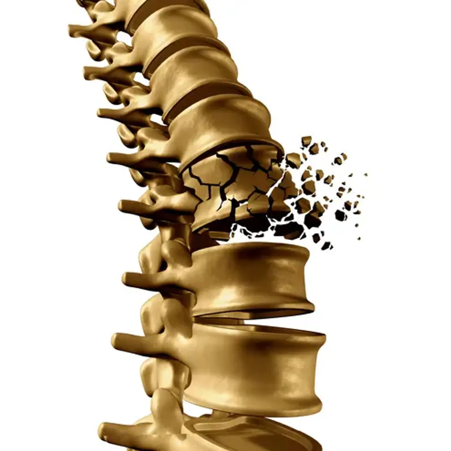
Spinal compression fractures, also known as vertebral compression fractures, are cracks or breaks in the bones of the spine produced by excessive pressure. Healthy bones can usually endure a lot of stress. Bones that have been weakened by a tumor or osteoporosis, on the other hand, can break with even minor force.
Osteoporosis-related fractures most commonly occur in the spine. Spinal fractures, also known as spinal compression fractures, are predicted to occur 1.5 million times every year in the United States. They are nearly twice as prevalent as other osteoporosis-related fractures, such as fractured hips and wrists.
Osteoporosis does not cause all spinal compression fractures. However, when the illness is present, a fracture is frequently the patient's first indication of a weaker skeleton due to osteoporosis.
Vertebral compression fractures (VCFs) of the spinal column develop as a result of an axial/compressive (and, to a lesser degree, flexion) pressure on the bone, resulting in a fracture. VCFs, by definition, jeopardize the anterior column of the spine, putting the anterior half of the vertebral body (VB) and the anterior longitudinal ligament (ALL) at risk. This results in the distinctive wedge-shaped malformation.
VCFs do not affect the VB's posterior half, nor do they involve the osseous components or the ligamentous complex (PLC). The former differentiates between a compression fracture and a burst fracture. The consequences of these compression fractures are connected to the structural stability and propensity for deformity development. Compression fractures are often regarded as stable and do not need surgical intervention.
VCFs are the most often described fragility fracture in the literature. Each year, around 1 to 1.5 million VCFs occur in the United States (US). Based on age and gender-adjusted incidence, it is predicted that 25% of women aged 50 and over have at least one VCF. Furthermore, it is believed that 40% to 50% of people over the age of 80 have experienced a VCF, either acutely or incidentally during clinical workup for another disease.
Anatomy
The spine is made up of 24 bones called vertebrae that are placed on top of each other. These bones join together to form a canal that protects the spinal cord. The spinal cord and nerves are also components of your spine. These "electrical wires" run through your spinal canal, conveying information from your brain to your muscles. Nerve roots emerge from the spinal cord via holes in the vertebrae.
Flexible intervertebral disks are located between your vertebrae. The thickness of these flat, spherical disks is roughly a half-inch. When you walk or run, they cushion the vertebrae and function as shock absorbers.
What causes a compression fracture?
VCFs are the most prevalent fragility fracture due to the most common cause, osteoporosis. Compression fractures, on the other hand, have a bimodal distribution, with younger individuals suffering these injuries as a result of high energy processes (fall from a height, MVA, etc)
Because of the ligamentous and structural changes that occur when one moves from the thoracic to the lumbar level, inherent regions of instability make this a common location of injury. Traditional teaching states that the spinal column may be split into three sections:
- The anterior column (also known as the anterior longitudinal ligament, the anterior annulus, and the anterior section of the vertebral body)
- The middle column (the posterior vertebral body, the posterior annulus, and the posterior longitudinal ligament), as well as
- The posterior column (ligamentum flavum, neural arch, facets, posterior ligamentous complex).
If two of these three columns are damaged, the injury is deemed unstable, and the patient may require surgery. Compression fractures, by definition, only require anterior column damage. As a result, VCFs are regarded as "stable" fracture patterns. When a fracture pattern involves the central column, it is classed as a burst fracture, which lacks the stability of a VCF.
Pathophysiology
The spinal column will revolve around a center of axis upon a fall or trauma. Because of the flexion/extension of the spine, an axial force is also applied. An axial force greater than the forces acceptable by the vertebral body causes a compression fracture, with greater forces culminating in a burst fracture.
The compression fracture's subsequent kyphotic (forward flexion of the spine) deformity may affect spine biomechanics, putting additional strain on other spine levels. The changed biomechanics increase the likelihood of further fractures and progressive deformity. The presence of an osteoporotic compression fracture raises the possibility of another compression fracture.
What are the symptoms of a Spinal compression fracture?
Once the patient has been stabilized, the initial evaluation of spine fractures includes an assessment of the neurologic function of the arms, legs, bladder, and intestines. Organization and patience are essential for a complete examination. It should be noted that many high-energy compression fractures are coupled with abdominal, brain, and extremities injuries, all of which should be investigated. Strength, as well as sensitivity and reflexes, should be evaluated. It is also critical to examine the skin along the back and record the existence of soreness on palpation. Documentation is critical since these first findings will very certainly be utilized as a baseline for all subsequent studies.
How is a Spinal compression fracture diagnosed?
Anterior-posterior (AP) and lateral radiographs of the affected region are taken in individuals with suspected back injuries. In the trauma scenario these initially, should be acquired supine with spine precautions until cleared by the spine team or bracing has been administered. Standing radiographs with the brace can assist guide therapy at some time since a supine posture can artificially minimize a displaced fracture.
In all trauma situations, a CT scan should be acquired. An MRI will show disruption of the posterior ligamentous complex if a suspected posterior column injury cannot be validated on CT. Radiographs with 30 degrees of traumatic kyphosis (forward flexion of the spine) and 50% vertebral body height loss have traditionally been considered unstable fractures, however recent research is altering this assumption. In addition, any neurologic impairment demands an MRI for further examination. An MRI is unlikely to be required for elderly individuals with low energy compression fractures. Serial standing lateral radiographs acquired in the clinic will aid in the course and healing of the fracture.
Bone Density Testing
When you suffer a vertebral compression fracture, you should be evaluated to see if you have osteoporosis and, if so, how serious the disease is. X-rays frequently reveal bone thinning along the spine, a disease known as osteopenia, or low bone mass. Osteopenia is a precursor of osteoporosis, a condition in which the bone becomes increasingly weak and prone to fracture.
Dual-Energy X-ray Absorptiometry (DEXA), a form of bone mineral density scan, can be used to evaluate the amount of bone loss. The DEXA results will assist your doctor in estimating your risk of more fractures in your spine and other sections of your body. Your doctor will use the DEXA results to determine whether to treat your bone density decrease with drugs.
How is a Spinal compression fracture treated?
Determining the necessity for surgery might be difficult at times. In 2005, a categorization system was implemented to improve management consistency and give easy treatment suggestions. The Thoracolumbar Injury Classification and Severity (TLICS) Scale uses the integrity of the PLC, injury morphology, and the patient's neurological status to provide a score (one to ten) that can guide intervention: a score less than four suggests non-surgical treatment, a score greater than four suggests surgical treatment, and a score of four is managed either surgically or nonsurgically.
Of course, these are basic principles for trauma victims, and each case should be thoroughly reviewed. Interestingly, recent research has revealed that historical factors such as loss of vertebral body height, segmental kyphosis, and canal impairment do not predict the need for surgery in neurologically intact individuals. There have yet been no randomized trials comparing surgery to brace therapy in "unstable" compression fractures.
Orthosis/bracing techniques provide cautious care for four to twelve weeks. When there is radiographic evidence of healing and the patient is no longer painful over the fracture site, the bracing can be removed. While a thoracolumbosacral orthosis (TLSO) can treat midthoracic and upper lumbar VCFs, lower lumbar VCFs may require a lumbosacral corset for effective immobilization.
Bracing is not a benign procedure and can be challenging in patients with barrel chests, pulmonary impairment, or obesity. These elements must be taken into account. Analgesic medicines and bracing might be difficult for some individuals to handle. If bracing is ineffective or unacceptably painful, the doctor may pursue percutaneous fracture stabilization techniques.
The surgical alternatives are heavily influenced by the fracture characteristics and brain damage. Compression fractures seldom need instrumented stabilization. The most typical surgical considerations for these individuals are cement augmentation in the form of vertebroplasty or kyphoplasty. Vertebroplasty, which was originally designed for spinal hemangiomas, is a minimally invasive technique in which cement is injected into the vertebral body through the pedicle. Supine stance with extension improves spinal alignment throughout the surgery; the vertebroplasty itself is not intended to restore alignment.
Cement augmentation should be explored for individuals who have failed a trial of conservative therapy or are hospitalized owing to discomfort and reduced function caused by a VCF. Recent studies have indicated that kyphoplasties can result in considerably faster improvements in quality of life, function, discomfort, and mobility.
What is kyphoplasty?
Kyphoplasty is a minimally invasive surgical treatment that uses a unique form of bone cement to mend a broken vertebra. We use a tiny needle to put a balloon into the fracture to assist restore the height of the collapsed bone, then inject cement into the space produced by the balloon, guided by real-time X-ray pictures. The cement hardens in a matter of minutes, reinforcing and stabilizing the bone and preventing it from collapsing again.
Kyphoplasty is typically safe, although it does involve dangers, just like any other form of surgery. These include issues with bleeding, infection, and anesthesia. Although uncommon, bone cement can seep out of the bone and cause nerve irritation or damage. Discuss the risks and advantages of kyphoplasty with your doctor to see if it is the appropriate therapy for you.
The balloon kyphoplasty, can be done under local or total anesthesia. You will be conscious but not in pain if local anesthetic is administered, but you will be sleeping if general anesthesia is used.
Living with a compression fracture
Osteoporosis-related compression fractures generally become less painful with medication and rest. They normally recover after three months. However, some can have long-term consequences. Medicines for osteoporosis can help prevent future fractures, but they cannot repair an existing fracture. If you have osteoporosis, you should get it treated before you get compression fractures.
Compression fractures induced by traumas usually heal in around 8 weeks. However, if surgery is required, it may take longer. Cancer-related compression fractures have variable results depending on the kind of cancer and how well it responds to therapy.
There is an increase in mortality in older individuals with osteoporotic compression fractures as compared to age-matched controls. Survival rates have been reported to be 53.9% after three years, 30.9% after five years, and 10.5% after seven years.
Complications
Nonoperative treatment of these fractures may result in ongoing back discomfort and the advancement of a kyphotic deformity. There is a substantial possibility that patients may have vertebral body collapse as well as further fractures in the future.
Several problems have been found using cement augmentation. There have been instances of neurologic harm during the surgery, however this is an uncommon event. The increased stiffness of a cement-filled vertebral body produces additional stress on the neighboring levels, which might result in subsequent fractures.
However, as previously stated, individuals in this category are frequently at risk for this regardless of surgical therapy. Most patients will have cement extravasation, although this does not appear to be clinically significant. Embolization of the cement is uncommon, but it can result in life-threatening consequences such as pulmonary embolism or stroke.
What can I do to prevent a compression fracture?
The greatest strategy to help avoid compression fractures is to prevent osteoporosis or treat it if you already have it. Discuss a bone density test with your healthcare practitioner to see if you are at risk for osteoporosis and what you can do about it. Don't smoke and restrict your alcohol use to lower your chance of osteoporosis and certain forms of cancer. Weight-bearing activities to strengthen your muscles and bones, as well as balancing exercises to lower your chance of falling, are both essential.
If you have osteoporosis and wish to begin an activity regimen, first consult with your osteoporosis expert. Exercise is essential for good health. However, you may need to avoid or modify some workouts. This is determined by the severity of your osteoporosis.
Conclusion
A spine compression fracture occurs when excessive tension is applied to one or more vertebrae, causing them to collapse. Spine compression fractures are dangerous, causing painful or incapacitating symptoms that impair your quality of life. Fortunately, therapy can assist in repairing the damage and alleviating symptoms.