Spine trauma
Last updated date: 06-Jun-2023
Originally Written in English
Spine Trauma
Overview
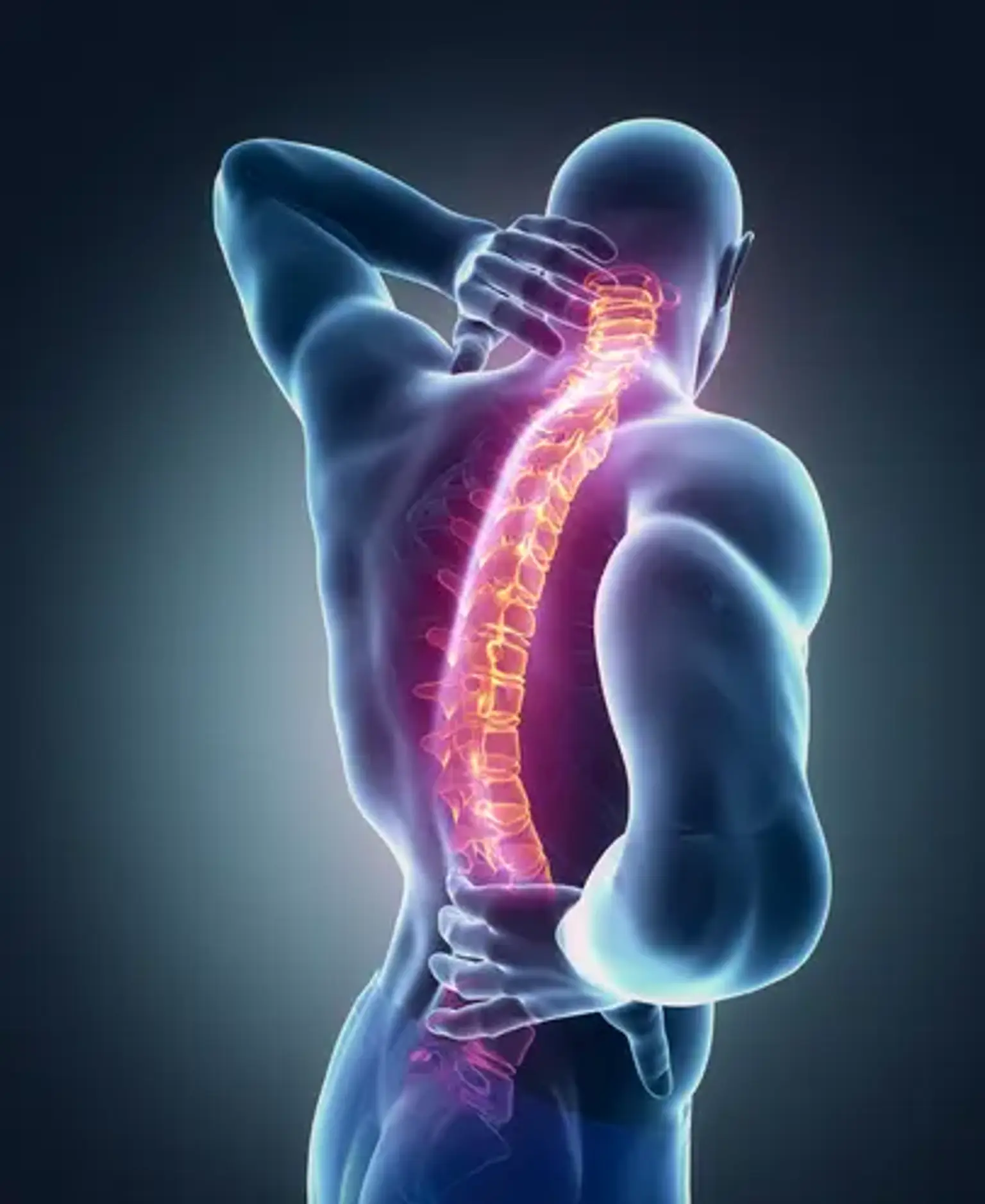
Spine trauma is caused by a sudden injury to the spinal cord or the bones that surround it. This condition most commonly affects the cervical spine (in the neck) or the thoracic spine (in the upper back). Fractures, crushing or compressing, and bone dislocation are all examples of trauma. Following the initial trauma, additional injury may occur as a result of swelling and inflammation, as well as internal bleeding and accumulation.
What is Spinal Trauma?
An injury to the spinal cord and spinal column, or the bones surrounding the spinal cord, causes spinal trauma. Damage to the spinal cord, which contains nerves that carry messages between the brain and the body, can result in permanent changes in bodily functions.
A traumatic blow to the spine that fractures, dislocates, crushes, or compresses one or more vertebrae can result in a spinal cord injury (vertebrae). It could also be caused by a penetrating injury to the spinal cord, such as a gunshot wound.
Minor falls or accidents can sometimes result in a significant spinal cord injury. This can occur if there is underlying weakness, dislocation, or instability caused by an untreated condition known as degenerative spine disease.
The Central Nervous System
Your central nervous system is a brain extension. It connects your brain to the rest of your body as a soft tissue column that travels down your spine. It is normally protected within the vertebral column, branching out into peripheral nerves and nerve tracts that extend into your extremities.
Motor and sensory tracts transport data from other parts of the body to the brain. Transmitting critical feedback about touch, weight, temperature, and pain.
Sustained spine trauma along the vertebral column will have an immediate impact on and disrupt the connection to the brain. The area that is impacted grows as the damaged area of the spine rises higher up the spinal column, which is why neck and upper spinal injuries pose the greatest threat.
Who gets spinal trauma?
Spinal cord trauma can affect anyone and is most often the result of an accident. Men are more likely than women to sustain a traumatic spinal cord injury.
Common causes of traumatic spinal cord injuries are:
- Motor vehicle accidents
- Falls
- Acts of violence (primarily gunshot wounds)
- Sports and recreational injuries
Arthritis, osteoporosis, cancer, inflammation, infections, and degenerative disc disease can all cause non-traumatic spinal cord injuries.
Spinal Trauma Symptoms
SCI presentations have a bimodal distribution. Trauma cases in younger adults frequently involve higher velocity injury in a healthy spine. Injuries to the elderly are common in later life and may be associated with a lower velocity injury in a vulnerable spine. These could be the result of a pathological vertebral fracture, the first sign of cancer, or a seemingly insignificant injury presentation.
Damage to the spinal cord can result in irreversible injury and either temporary or permanent neurological deficit. The natural progression of SCI, particularly rising spinal cord edema, may cause symptoms to worsen in the hours following an accident. Early SCI care can have a significant impact on these patients' long-term outcomes, with safe and appropriate transport to definitive care facilities being critical. Emergent surgical fixation and stabilisation of the spinal injury, where indicated, may provide the best outcome for patients and is the first stage of recovery.
Spinal cord injuries are classified as either complete (no neurological connection between the cortex of the brain and the lowest sacral spinal cord segment) or incomplete (with some connection maintained). Documentation of incomplete and/or motor preservation, as well as identification of unilateral deficits, are required by the American Spinal Injury Association (ASIA) Standards for Classification of Neurological Injury.
Following a traumatic injury, the spinal cord becomes edematous, and because the vertebral column has a limited capacity for swelling, normal neurological function is quickly compromised. Above the level of initial injury, this may affect about two nerve exit levels of the spinal cord. As swelling resolves over time, there may be recovery at the level of SCI but not always in the long tracts below.
Importantly, SCI trauma patients may present with a combination of motor and sensory neurological deficits that can be unilateral or bilateral, affecting the upper and/or lower body. Patients who are conscious may describe sensations such as numbness, burning pain, or a lack of feeling or movement. The emerging and frequently ascending nature of spinal injury signs and symptoms suggests that the SCI trauma patient requires exacting and ongoing assessment, as well as monitoring and management. Neurogenic shock and spinal shock are two serious consequences of a SCI.
Neurogenic shock is seen in SCI affecting the sixth or higher thoracic vertebrae, usually occurring within 30 minutes of cord damage and lasting six to eight weeks. It is caused by a decrease in vasomotor and sympathetic nervous system tone or function. Its key symptoms are hypotension, bradycardia, and poikilothermia.
Spinal shock is a combination of reflex loss, decreased reflexes, and autonomic dysfunction that occurs as a result of SCI. As a result, skeletal and smooth muscles are flaccid for hours to weeks.
A rapid and systematic primary and secondary survey of all patients with spinal trauma is required. The primary goals are to ensure injury mechanism understanding and pattern optimum management in the emergency setting, including secondary insult prevention and activation of the retrieval network, with timely transfer to an appropriate trauma facility.
Types of Spinal Cord Injury
Because most spinal cord injuries are caused by trauma, there are various ways injuries occur and different types of spinal cord injury. Motor vehicle accidents, falls, gunshot wounds, sports injuries, and surgical complications are some of the most common causes of spinal cord injury. The majority of cases can be divided into two types of spinal cord injury: complete spinal cord injury and incomplete spinal cord injury.
- A complete spinal cord injury causes permanent damage to the affected area of the spinal cord. Complete spinal cord injuries result in paraplegia or tetraplegia.
- An incomplete spinal cord injury is defined as partial spinal cord damage. The ability to move and feel is determined by the area of the spine injured and the severity of the injury. The health and medical history of the patient influence the outcome.
Types of Incomplete SCI Syndrome
- The most common incomplete SCI syndrome is central cord syndrome, which accounts for 15-25% of traumatic SCIs. Central cord syndrome is most commonly diagnosed in elderly patients who have pre-existing cervical spondylosis and stenosis and present after a fall that causes cervical hyperextension. Central cord syndrome is distinguished by disproportionate upper-limb motor impairment rather than lower-limb motor impairment, as well as bladder dysfunction and varying degrees of sensory loss.
- Brown-Séquard syndrome is most commonly seen in people who have penetrating traumatic SCI from gunshot or knife wounds. Loss of motor function, light touch, proprioception, and vibration sensation ipsilateral to the injury, as well as loss of pain and temperature sensation contralateral to the injury, are all symptoms of Brown-Séquard syndrome.
- Anterior and posterior cord syndromes are uncommon in the context of traumatic SCI, but are more common in patients with non-traumatic SCI of vascular origin.
Levels of Spinal Cord Injury
The level of spinal cord injury is determined by four sections of the spinal cord: cervical, thoracic, lumbar, and sacral. Each section of the spine protects various nerve groups that control the body. The type and severity of spinal cord injuries can vary depending on which part of the spine is injured.
How are acute spinal cord injuries diagnosed?
Following any traumatic injury, first responders assess patients in the field and attempt resuscitation while transporting them to the hospital. During this time, advanced trauma life support protocols dictate that initial care include airway, breathing, and circulation support, as well as immobilization of the potentially injured and unstable spinal column with a rigid cervical collar and backboard.
Although individual hospital approaches vary, most trauma patients will have a gross neurological examination (which includes a voluntary motor and sensory examination of each limb as well as a rectal examination) and spinal imaging (such as X-ray or CT imaging) if a SCI is suspected. Clinical examination or early radiographic imaging concerns are followed by advanced imaging and detailed neurological examinations.
Imaging
When investigating spine and SCI damage, plain X-ray, CT, and MRI are the most commonly used radiological tools. In the trauma room, X-rays of the anterior-posterior (AP) and lateral cervical spines, as well as the AP chest and AP pelvis, are taken. Although X-rays are not particularly sensitive for detecting subtle fractures involving the cervical spine, they are useful for detecting gross fracture dislocation injuries that are frequently associated with SCI. It is critical to ensure that any X-rays with visualization of the rostral half of the T1 vertebrae are adequate.
CT Scan
CT has largely replaced X-ray in the diagnosis of bone injuries in trauma patients. Some authors currently perform and recommend a high-resolution fine-cut CT scan of the cervical to lumbar spine. In certain cervical injuries, CT angiography can also be used to evaluate the bilateral vertebral arteries.
Although CT is extremely sensitive for diagnosing a spine fracture or dislocation, it is less effective at evaluating the integrity of soft tissue structures such as intervertebral discs, ligaments, the spinal cord, and nerve roots, which MRI is better suited for. The T2-weighted short-tau inversion recovery (STIR) sequence, in particular, enables the identification of injury-related edema and tissue disruption when evaluating for ligament or vertebral disc injury. An MRI can detect spinal cord transection and assess the presence of edema and/or hemorrhage.
In the treatment of patients with SCI and cervical facet dislocation, the timing of MRI can be critical. MRI prior to closed reduction (correcting the dislocation with traction) allows for the detection of disc herniation, which, if present, can lead to a deterioration in neurological status, though this is debatable. However, depending on the institution, MRI can significantly delay time to spinal cord decompression and requires an additional transfer of a patient with a highly unstable spine.
The most recent iteration of the American Association of Neurological Surgeons and Congress of Neurological Surgeons guidelines for the management of cervical SCI recommends MRI before performing an open reduction (realignment of the broken bones following surgery to expose the bones) or a closed reduction in an unconscious or uncooperative patient; if a disc herniation is identified, the guidelines recommend an anterior apex reduction.
MRI
Advanced microstructural techniques that can quantify physiological changes at a cellular level and assess axon integrity (for example, diffusion tensor imaging), myelination (for example, myelin water transfer), and the presence of key metabolites related to ischaemia, cell loss, or gliosis (for example, magnetic resonance spectroscopy) are likely to see increased integration in the care of SCI patients.
Electrophysiology
Numerous electrophysiological studies have been conducted to predict outcome as well as track and monitor recovery after traumatic SCI. Because it does not require the patient to be conscious or communicative, electrophysiology is an appealing tool. Several parameters, such as conduction time to motor neurons, cortical and spinal inhibition, spinal cord excitability (such as the H-reflex), and sensory impairment, have been studied and can be used to derive measures of physiological and anatomical function.
Electrophysiological measurements, while intriguing as a research tool, have not consistently demonstrated added value in predicting outcome in awake and alert SCI patients. However, electrophysiological measurements may provide insight into the mechanisms underlying the patient's functional recovery (for example, regeneration, plasticity, or adaptation), which may be useful as the field develops, for example, in patient selection for clinical trials.
Spinal Trauma Treatments
Recovery from spine trauma necessitates a dedicated team of spinal cord injury specialists (SCIs). To develop personalized treatment plans, our neurologists collaborate with neurological surgeons and rehabilitation specialists. Your treatment is determined by the severity of your spinal injury and your symptoms.
Unfortunately, full function following spine trauma may not be possible. Our spinal cord injury treatment program, on the other hand, helps you maintain as much independence as possible.
To maximize your capabilities at home and in your community, you will receive ongoing support and education. Rehabilitation programs can also help you improve your quality of life, allowing you to stay physically, emotionally, and socially healthy.
Immobilization
The most important thing to do immediately following a traumatic spine injury is to prevent further damage. It is possible to sustain a catastrophic injury while still being able to move. Any further movement after the injury can crush and splinter the bone, as well as permanently and irreversibly damage the spinal cord. To avoid this, first responders will retrain and immobilize the neck/spine.
Surgery
Traumatic spine surgery is divided into two stages. First, surgeons will remove any bone fragments, foreign materials, or substances, and attempt to realign any misplaced discs or vertebrae. The second phase of surgery aims to reinforce and strengthen the spine, restoring the structural integrity of the spinal column and preventing future injury.
Complications After Traumatic Spinal Cord Injury
Local Complications
Syringomyelia
Syringomyelia, which occurs when a fluid-filled cyst, or syrinx, forms within the spinal cord, affects about 3% of people with SCI. The cyst expands over time, causing spinal cord compression and progressive myelopathy years after the SCI.
Some people with syringomyelia have no symptoms and only require periodic monitoring. Patients with severe symptoms, on the other hand, may require a surgical decompression procedure.
Spasticity
Spasticity is a disorder characterized by long-term muscle contraction, resulting in stiff or rigid muscles. This can make any type of movement difficult, such as walking or talking. It can also have an impact on sleep.
Spasticity affects 65 to 78% of chronic SCI patients. Physical therapy, muscle relaxants, intrathecal drug therapy, botulinum toxin injections, and surgery are all common spasticity treatments.
Spinal Cord Injury—Systemic Complications
- Cardiovascular
A common SCI complication is abnormally low blood pressure, especially in people who have been injured in their cervical (neck) or thoracic (mid-back) spine. Approximately 60% of people have symptomatic orthostatic hypotension, which causes dizziness, weakness, and a brief loss of consciousness when rising from a sitting or lying position.
Wearing compression stockings or abdominal binding, as well as medication therapy, are common treatments.
- Autonomic dysreflexia
A damaging event below the level of injury, such as bowel impaction, bladder distension, or pressure sores, causes autonomic dysreflexia. This causes autonomic nervous system dysfunction, which prevents proper communication between the body and brain above the injury level. The autonomic nervous system is self-regulating in the sense that it operates without your conscious awareness. Bodily functions such as breathing, blood pressure, and heart rate become unregulated during autonomic dysreflexia.
Your doctor will prescribe drugs to help regulate your heart rate and relax your blood vessels if you have autonomic dysreflexia. This disorder can occur immediately following the SCI or years later, so long-term preventive health, including bowel and bladder care, is critical.
- Respiratory
Respiratory complications are the leading cause of death in chronic SCI patients. Cervical and thoracic SCIs can cause respiratory infections by weakening the chest and abdominal muscles. The common cold, bronchitis, and pneumonia are examples of common infections.
Patients may also develop fluid around the lungs and are more likely to develop sleep apnea (involuntary pauses in breathing) and respiratory failure. Your doctor may prescribe antibiotics to help clear an infection in your chest, but more serious respiratory issues may necessitate life-long ventilator dependency.
Other systemic complications of Traumatic spinal cord injury include:
- Secondary immunodeficiency: SCI can disrupt white blood cells, increasing your susceptibility to infections (including pneumonia, urinary tract infections, and wound infections).
- SCIs at or above L1-L2 (the first and second vertebrae in the low back) can cause bladder muscle dysfunction, resulting in problems emptying the bladder, urinary incontinence, and frequent urinary tract infections.
- Bowel dysfunction: According to 39% of people with SCI, bowel dysfunction has negatively impacted their quality of life. SCI can result in constipation and an increased risk of infection.
- Pressure sores are painful and most commonly appear on the buttocks, outer thighs, sacrum, feet, and ankles. If not treated promptly, these sores can become life-threatening.
- Neurogenic heterotopic ossification: Neurogenic heterotopic ossification occurs in up to 53% of people with chronic SCI and is defined as abnormal bone formation in connective tissue around joints. This is a common occurrence in the large joints of the hips, knees, elbows, and shoulders. It can cause discomfort, fever, and spasticity.
- Neuropathic pain: Up to 40% of patients with chronic SCI experience neuropathic pain. Neuropathic pain is complicated, and various treatments can help manage it. Prescription drugs, surgery, and alternative treatments such as acupuncture and cognitive behavioral therapy may be used in treatment.
Can Acute Spinal Cord Injuries be Prevented?
There is no sure way to prevent SCI, but there are many steps you can take that might lower your risk, including:
- Don’t drive when you are under the influence of alcohol or drugs.
- Always wear a seat belt while in a vehicle.
- Take precautions to avoid falls around your home.
- Keep firearms unloaded and locked away.
- Wear a helmet when riding a motorcycle or participating in any sports or activities that may result in head injury (bike riding, skiing, hockey, football, etc)
Living with an acute spinal cord injury
SCI recovery frequently necessitates long-term hospitalization and rehabilitation. Physical therapy will almost certainly be an important part of your recovery. Specialists will work with you to prevent muscle wasting and contractures, as well as to help you retrain other muscles to help with mobility and movement. Occupational therapy is another type of therapy that can help you learn new ways to do everyday tasks despite your new physical limitations.
A traumatic event that causes a SCI is devastating for you and your family. After hospitalization and rehabilitation, the healthcare team will help educate your family on how to care for you at home and understand the specific problems that require immediate medical attention.
Following hospitalization and rehabilitation, you will require frequent medical evaluations and testing to monitor your progress. It is critical to concentrate on improving your capabilities at home and in the community.
Following your injury, you may experience sadness or depression. If this happens to you or a loved one, your doctor may advise you to see a mental health professional. Both antidepressants and psychotherapy, also known as "talk therapy," are available to help treat depression.
Conclusion
SCI is defined as damage to the spinal cord that causes temporary or permanent changes in its function. SCI is classified as traumatic or non-traumatic1. Traumatic SCI occurs when an external physical impact (for example, a car accident, fall, sports-related injury, or violence) causes acute spinal cord damage, whereas non-traumatic SCI occurs when the primary injury is caused by an acute or chronic disease process, such as a tumor, infection, or degenerative disc disease.