Spondylopathy
Last updated date: 10-Jul-2023
Originally Written in English
Spondylopathy
Overview
Spondylopathies are vertebral disease. When there is inflammation, it is referred as spondylitis. Spondyloarthropathy, on the other hand, is a disorder involving the vertebral joints; nonetheless, many conditions involve both spondylopathy and spondyloarthropathy. Ankylosing spondylitis and spondylosis are two examples.
Clinically Relevant Anatomy
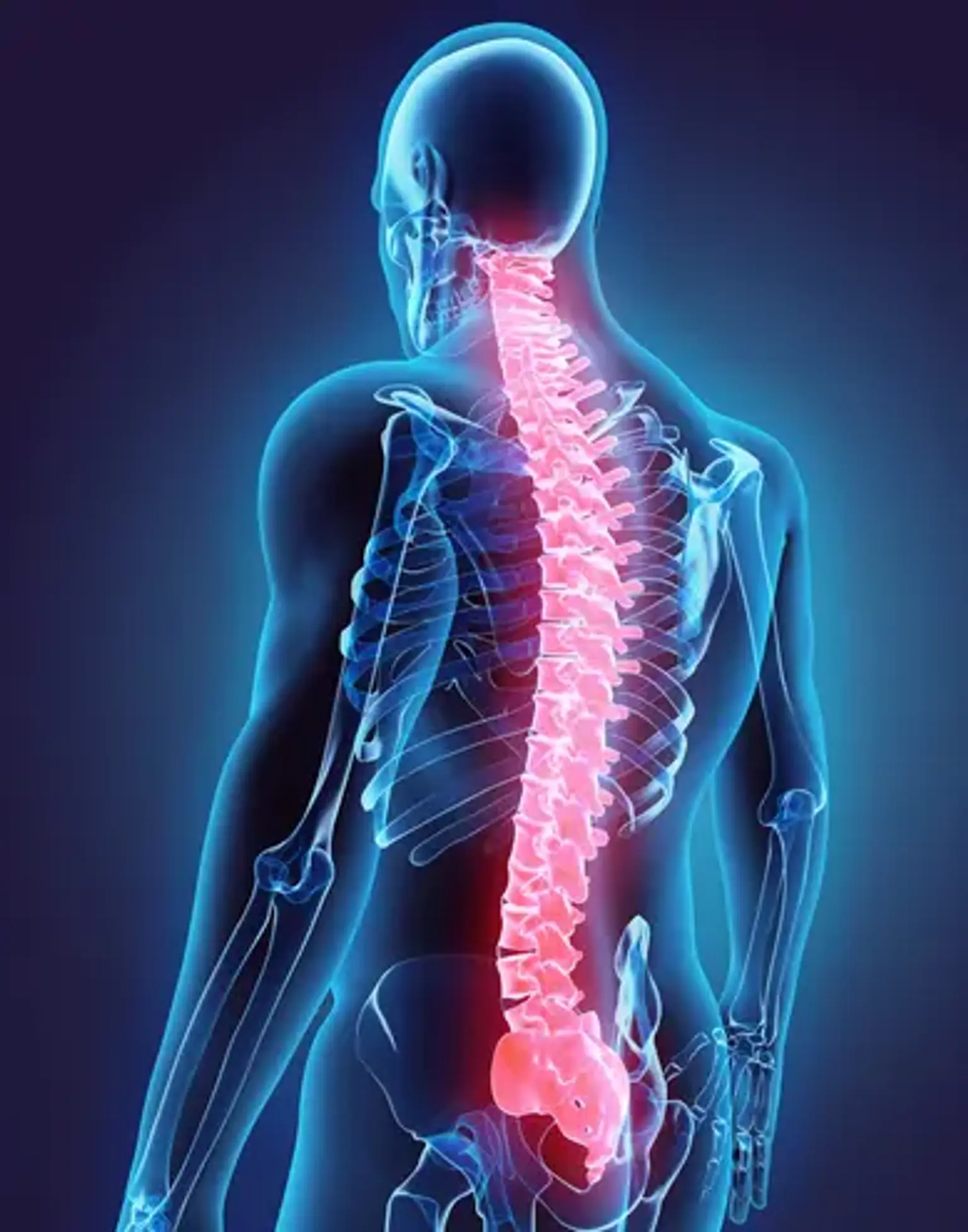
There are 24 vertebrae in the spinal column: seven cervical vertebrae, twelve thoracic vertebrae, and five lumbar vertebrae. Ligaments connect the vertebrae and intervertebral discs divide them. The discs are made up of an inner nucleus pulposus and an outer annulus fibrosis, both of which are made up of fibrocartilage rings.
Spondyloarthropathy patients are predisposed to inflammation at the areas where tendons, ligaments, and joint capsules adhere to bone. These are referred to as entheses.
The sacroiliac joint is made up of two compartments: a cartilaginous component and a fibrous (or ligamentous) compartment with very strong anterior and posterior sacroiliac ligaments. As a result, the SIJ is an amphiarthrosis with limited rotation and translation. Another distinguishing feature of sacroiliac joint (SIJs) is the presence of two distinct kinds of cartilage covering the two articular surfaces. While sacral cartilage is entirely hyaline, iliac cartilage is a combination of hyaline and fibrous cartilage. The sacroiliac joint is an articular enthesis because of its fibrocartilaginous components.
What tests are performed to confirm the diagnosis of spondylopathy?
Plain x-rays of the spine are frequently ordered in conjunction with certain basic blood tests such as hemoglobin, erythrocyte sedimentation rate [ESR], and CRP. HLA-B27, RA, ASLO, ANA, and other blood tests would be performed. You may be requested to undergo MRI scanning based on the results of these tests, or in the presence of any very serious indications or symptoms.
Plain x-rays show skeletal characteristics, but MRI shows the spinal cord, nerve roots, and the intervertebral disc between the two bones of the spine. Both of these tests are complementary in nature; doing one does not imply that the other is not required. A nuclear bone scan is seldom requested by a spine doctor or specialist.
Seronegative Spondyloarthropathies
Seronegative spondyloarthropathies are a family of joint disorders that classically include
- Ankylosing spondylitis (AS),
- Psoriatic arthritis (PsA),
- Inflammatory bowel disease (IBD) associated arthritis,
- Reactive arthritis (formerly Reiter syndrome; ReA), and
- Undifferentiated SpA.
Patients with seronegative spondyloarthropathy frequently appear with inflammatory joint pain that lasts longer than an hour and improves with exercise. NSAIDs may alleviate symptoms.
These illnesses have now been subdivided into three new categories: non-radiographic axial SpA, peripheral SpA, and juvenile-onset SpA. This group of illnesses has various clinical characteristics as well as genetic similarities.
Ankylosing Spondylitis
Ankylosing spondylitis (AS) is a chronic, inflammatory disease of the axial spine characterized by a variety of clinical signs and symptoms. The most prevalent symptoms of the condition are chronic back pain and growing spinal stiffness. The condition is characterized by involvement of the spine and sacroiliac (SI) joints, peripheral joints, fingers, and entheses. AS is linked with impaired spinal mobility, postural irregularities, buttock discomfort, hip pain, peripheral arthritis, enthesitis, and dactylitis ("sausage digits").
This condition may potentially damage extraskeletal organs. The most prevalent extraarticular symptoms of AS are inflammatory bowel disease (up to 50%), acute anterior uveitis (25–35%), and psoriasis (approximately 10 %). AS is also linked to a higher risk of cardiovascular disease.
The systemic inflammation seen in AS has been proposed as the source of this elevated risk. Since is also related with pulmonary problems, as reduced chest wall expansion and decreased spinal movement predispose patients to a restrictive pulmonary pattern.
Finally, AS increases the risk of spinal fragility fractures by at least a factor of two. These individuals are also more likely to suffer from atlantoaxial subluxation, spinal cord damage, and, in rare cases, cauda equina syndrome.
Ankylosing spondylitis Causes
The etiology of ankylosing spondylitis (AS) is mostly unknown, however there appears to be a link between the incidence of AS and the prevalence of human leukocyte antigen (HLA)-B27 in the same population. The prevalence of AS is roughly 5% to 6% among those who are HLA-B27 positive. HLA-B27 prevalence varies by ethnic group in the United States. HLA-B27 was present in 7.5 percent of non-Hispanic Whites, 4.6 percent of Mexican-Americans, and 1.1 percent of non-Hispanic Blacks.
Ankylosing spondylitis Symptoms
Because of the disease's pervasiveness and propensity to multi-organ system involvement, individuals suspected of having ankylosing spondylitis (AS) should undergo a complete, full-body assessment. Almost all patients will have back discomfort to some extent. The characteristic type of back pain in AS is "inflammatory" in nature.
Inflammatory back pain typically exhibits at least four of the five following characteristics:
- Age of onset less than 40 years,
- Insidious onset,
- Improvement with exercise,
- No improvement with rest, and
- Pain at night with an improvement upon arising.
Spinal rigidity, immobility, and postural abnormalities, particularly hyperkyphosis, are also frequent. As Ankylosing spondylitis can have numerous axial and peripheral musculoskeletal symptoms, as well as extra-articular characteristics, the history and physical exam should address all systems. A thorough medical history should be collected in order to rule out any linked illnesses (e.g., psoriasis, inflammatory bowel disease, uveitis, among others).
Ankylosing spondylitis Diagnosis
Although laboratory results in ankylosing spondylitis (AS) are often vague, they can aid in diagnosis. Approximately 50% to 70% of individuals with active AS exhibit higher acute phase reactants such as erythrocyte sedimentation rate (ESR) and C-reactive protein (CRP). A normal ESR and CRP, on the other hand, should not rule out the illness.
AS is characterized by a multitude of imaging abnormalities, particularly those involving the spine and sacroiliac joints. Plain radiographic changes in the SI joints might be quite mild in the early stages of AS, but they generally become more visible during the first decade. The most visible abnormalities are subchondral erosions, sclerosis, and joint fusion, and these radiographic alterations are usually symmetric.
A sequence of simple radiographic alterations typical of AS might occur gradually throughout the course of the disease. The "squaring" of the vertebral bodies, best observed on lateral x-ray, is an early symptom. This is characterized by a lack of normal concavity of the vertebral body's anterior and posterior borders as a result of inflammation and bone deposition. In the early stages, Romanus lesions, commonly known as the "shiny corner sign," can be detected on this radiographic picture.
These lesions are distinguished by tiny erosions and reactive sclerosis at the vertebral body corners. Ankylosis (fusion) of the facet joints of the spine, syndesmophytes, and calcification of the anterior longitudinal ligament, supraspinous ligaments, and interspinous ligaments are late-stage abnormalities.
The "bamboo spine sign," which refers to vertebral body fusion by syndesmophytes, is a hallmark radiographic finding in late-stage AS. The thoracolumbar or lumbosacral connections are commonly involved in a bamboo spine. This spinal fusion predisposes the patient to increasing back stiffness.
While conventional radiography is the most often used imaging modality in AS, MRI may be required to detect more subtle abnormalities such as lipid alterations or inflammatory changes. On short tau inversion recovery (STIR) and T2-weighted images with fat suppression, active SI joint inflammatory lesions present as bone marrow edema (BME). It should be mentioned that the presence of BME on MRI can be found in up to 23% of mechanical back pain patients and 7% of healthy persons.
Ankylosing spondylitis Management
Treatment objectives should include reducing pain and stiffness, preserving axial spine mobility and functional capacity, and avoiding spinal problems. Regular exercise, posture training, and physical therapy are examples of non-pharmacologic therapies. Long-term, daily nonsteroidal anti-inflammatory medications are used as first-line pharmaceutical treatment (NSAIDs). If NSAIDs are ineffective, they can be supplemented with or replaced with tumor necrosis factor inhibitors (TNF-Is) such as adalimumab, infliximab, or etanercept.
Response to NSAIDs should be evaluated four to six weeks after starting them, and TNF-Is should be evaluated twelve weeks after starting them. Although systemic glucocorticoids are not advised, local steroid injections may be tried. Referrals to specialists may be necessary based on the patient's clinical picture, probable consequences, and extra-articular symptoms of the illness. Rheumatologists can help with official diagnosis, therapy, and monitoring, while dermatologists, ophthalmologists, and gastroenterologists can help with non-musculoskeletal AS symptoms.
Cervical Spondylosis
Spondylosis is an umbrella word for several types of age-related spine deterioration. The vertebrae are the bones of the spine. There are three joints between each pair of vertebrae. An intervertebral disc is a joint located at the front of the spine.
Cervical spondylosis is a word that refers to a variety of progressive degenerative alterations that affect all of the cervical spine's components. It is a normal aging process that affects the majority of people during their fifth decade of life. Cervical spondylosis symptoms include neck discomfort and stiffness, which can be accompanied with radicular symptoms when neural structures are compressed.
Neck discomfort is a frequent complaint, ranking second only to low back pain in terms of prevalence. Given the high disease burden, severe disability, and economic expense, healthcare professionals must detect symptomatic cervical spondylosis and deliver evidence-based, cost-effective therapies.
Cervical Spondylosis causes
Age-related degradation of the intervertebral disc and cervical spinal components is the major risk factor and contributor to the prevalence of cervical spondylosis. Degenerative alterations in surrounding tissues, such as the uncovertebral joints, facets joints, posterior longitudinal ligament (PLL), and ligamentum flavum, all contribute to spinal canal and intervertebral foramina narrowing. As a result, the spinal cord, vasculature, and nerve roots can become compressed, leading to the three clinical syndromes associated with cervical spondylosis: axial neck pain, cervical myelopathy, and cervical radiculopathy.
Significant spinal trauma, a congenitally small vertebral canal, dystonic cerebral palsy affecting cervical muscles, and certain sports activities such as rugby, soccer, and horse riding can all contribute to an accelerated disease process and early-onset cervical spondylosis.
Pathophysiology of Cervical Spondylosis
Cervical spondylosis is caused by a degenerative cascade that results in biomechanical abnormalities in the cervical spine, which manifest as secondary compression of neuronal and vascular systems. When the keratin-chondroitin ratio rises, the proteoglycan matrix alters, resulting in a loss of water, protein, and mucopolysaccharides inside the intervertebral disc.
The nucleus pulposus loses flexibility as it shrinks and becomes more fibrous as a result of disc desiccation. As the nucleus pulposus loses its capacity to efficiently support weight-bearing loads, it begins to herniate through the fibers of the annulus fibrosus, contributing to disc height loss, ligamentous laxity, buckling, and cervical spine compression. The annular fibers become increasingly structurally damaged under compressive pressures as the disc dehydrates more, resulting in major changes in load distribution along the cervical spine.
As a result, the usual cervical lordosis is reversed. The annulus and Sharpey's fibers peel away from the vertebral body margins as the kyphosis progresses, resulting in reactive bone production. These bone spurs, also known as osteophytes, can grow along the ventral or dorsal borders of the cervical spine and can protrude into the spinal canal and intervertebral foramina.
Furthermore, disturbance in the load balance along the spinal column causes increased axial stresses on the uncovertebral and facet joints, causing hypertrophy or enlargement of the joints and hastening the production of bony spurs into the surrounding neural foramen. These degenerative alterations result in a loss of cervical lordosis and mobility, as well as a narrowing of the spinal canal.
Cervical Spondylosis symptoms
The timing of the pain, pain radiation, aggravating variables, and triggering events should all be addressed in the patient history. Symptomatic cervical spondylosis often manifests as one or more of the three basic clinical symptoms listed below:
Axial Neck Pain
- Patients frequently complain of stiffness and soreness in the cervical spine, which is worse in the upright position and is improved by bed rest when the strain on the neck is removed.
- Neck motion, particularly hyperextension and side-bending, is known to aggravate discomfort.
- Patients with upper and lower cervical spine disorders may have radiating pain into the back of the ear or occiput as opposed to radiating pain into the superior trapezius or periscapular muscles.
- Patients may occasionally present with unusual symptoms of cervical angina, such as jaw discomfort or chest pain.
Cervical Radiculopathy
- Depending on the nerve root(s) implicated, radicular symptoms might manifest as unilateral or bilateral neck discomfort, arm pain, scapular pain, paresthesia, and arm or hand weakness.
- Head tilt toward the afflicted side or hyperextension and side-bending toward the affected side aggravate pain.
Cervical Myelopathy
- It usually starts slowly, with or without neck discomfort (frequently absent)
- Hand weakness and clumsiness may first emerge, leading in an inability to accomplish tasks requiring fine motor coordination (e.g., buttoning a shirt, tying shoelaces, picking up small objects)
- There have been several instances of gait instability and inexplicable falls.
- Urinary symptoms (such as incontinence) are uncommon and usually emerge late in disease progression.
Due to growing axial neck discomfort with cervical spine movement, the patient may look immovable and stiff at the head and neck at first. Tender "trigger" areas in the superior trapezius muscles, cervical paraspinal muscles, and/or periscapular muscles are common.
A positive Lhermitte's sign is electric shock-like sensations spreading down the spine and into the limbs with cervical flexion, which is problematic for cervical spondylotic myelopathy (CSM). Hoffman's sign, which is elicited by flicking the patient's distal phalanx of the middle finger and detecting reflexive bending of the thumb and/or index finger, is a more specific sign for CSM.
Cervical Spondylosis Diagnosis
In the absence of "red flag" symptoms, plain radiographs are an adequate first imaging investigation for neck and upper extremity discomfort. However, imaging degenerative alterations frequently do not correspond with the prevalence of neck discomfort. Osteophyte development, disc space narrowing, endplate sclerosis, degenerative alterations of the uncovertebral and facet joints, and calcified/ossified soft tissues are all common radiographic findings.
The AP, lateral, and oblique views of the spine are sufficient to assess foraminal stenosis, sagittal alignment, and spinal canal size. If ligamentous instability is a problem, flexion and extension views should also be considered.
- Magnetic Resonance Imaging (MRI)
MRI is the preferred imaging method for evaluating brain structures and soft tissues. It enables for good vision of the whole cervical spine while avoiding the use of radiation on the patient. Sagittal and axial incisions can assist assess the level of nerve and cord compression and highlight the underlying alterations that are causing it (e.g., herniated discs, bony spurs, ligamenta flava hypertrophy, or facet joint arthropathy).
- Computed Tomography (CT)
CT is more sensitive than plain radiography in evaluating intervertebral foraminal stenosis in the presence of uncovertebral or facet hypertrophy because it offers a better delineation of bony structures. It is, however, less sensitive than MRI for evaluating soft tissues and nerve root compression.
- CT Myelogram
CT is particularly beneficial when paired with intrathecal contrast injection (myelography) to better assess the site and extent of neurological compression. It is more intrusive than an MRI, however it may be worth considering in individuals who are MRI contraindicated (e.g., pacemaker) or have a hardware artifact.
- Cervical Spondylosis Management
The severity of a patient's indications and symptoms determines the therapy method for cervical spondylosis. Treatment aims in the absence of "red flag" symptoms or substantial myelopathy are to ease pain, enhance functional capacity in daily tasks, and prevent lasting harm to neural systems. Symptomatic cervical spondylosis should be treated in stages, beginning with non-operative treatment.
- Non-surgical
A four- to six-week course of physical therapy, involving isometric and resistance exercises to strengthen the neck and upper back muscles, is the basis of non-surgical treatment.
For pain management, pharmacologic medicines such as nonsteroidal anti-inflammatory medications (NSAIDs), oral steroids, muscle relaxants, anticonvulsants, and antidepressants can be administered. For refractory axial neck pain, opioid analgesics can be used, although they are not advised as first-line or long-term treatment owing to probable side effects.
For symptomatic alleviation, durable medical devices should be considered. A soft cervical collar used for a short period of time might occasionally relieve acute neck discomfort and spasm. The usage of a cervical pillow at night may assist to decrease neck discomfort by maintaining normal cervical lordosis, which would enhance the distribution of biomechanical stresses between discs, enabling improved sleep quality.
Surgical
Patients with severe or worsening cervical myelopathy, as well as those with chronic axial neck discomfort or cervical radiculopathy after non-operative therapies have failed, should consider surgical surgery. These people must also have a pathological condition that is visible on neuroimaging examinations and corresponds to their clinical symptoms. The surgical strategy is determined by the clinical condition as well as the site(s) of pathology.
The anterior technique entails performing a cervical discectomy or corpectomy, followed by fusion using an autograft, allograft, or artificial intervertebral disc. Although anterior plates, metallic cages, and synthetic spacers can be utilized in combination with bone transplants to achieve equal fusion rates, the long-term effects are yet unknown.
An anterior approach is preferred in patients with radicular discomfort owing to central or bilateral disc herniation, but either an anterior or posterior approach is an option for a lateral disc lesion. Anterior cervical discectomy and fusion (ACDF) are used to treat patients with myelopathy and pathological compression up to three levels or when cervical lordosis is lost.
Partial discectomy, laminotomy-foraminotomy, laminoplasty, and laminectomy are all part of the posterior approach. In individuals with foraminal stenosis caused by bone spur development and/or lateral disc herniation, foraminotomy alone is sufficient. Laminectomy or laminoplasty is a clinical option for individuals who require decompression at four or more levels or who already have a fused anterior column.
A preserved cervical lordosis is necessary for a posterior approach because it permits the spinal cord to relocate dorsally after decompression. Patients with flexible cervical kyphosis will require more posterior cervical apparatus to assist restore normal lordosis and optimize spinal cord posterior shift.
Conclusion
Spondyloarthropathies are a category of inflammatory arthritides that share genetic predisposing factors as well as clinical characteristics. Ankylosing spondylitis, reactive arthritis (including Reiter's syndrome), psoriatic arthritis, inflammatory bowel disease-associated spondyloarthropathy, and undifferentiated spondyloarthropathy are the most common types of spondyloarthropathy. Spondylosis is a word used to characterize osteoarthritis of the spine, but it is also used to indicate any type of spinal degeneration.