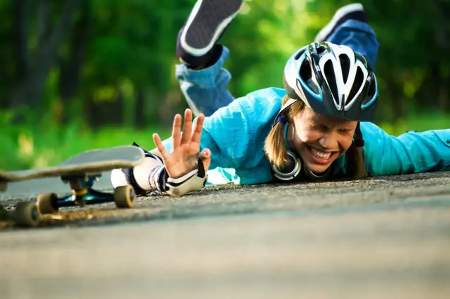
Sports Injury

Last updated date: 23-Mar-2023

Originally Written in English

Sports Injury

Overview

If you participate in sports, you are aware of the advantages of getting exercise while doing something you like. However, sports may also endanger your health. Every year, millions of individuals are sidelined due to sports injuries. While it is impossible to prevent all of these injuries, you may take steps to reduce your risk. Injuries caused by straining and overuse can be avoided by stretching before and after sports. Also, pay attention to your body's demands for rest.

Injuries are classified into two categories. Acute injuries occur unexpectedly. Chronic injuries accumulate over time. The following is a list of the most frequent sports injuries.


Achilles tendon injuries

Achilles tendon injuries

The Achilles tendon is the most often ruptured tendon in the lower leg. Adults in their third to fifth decade of life are the most likely to sustain the injury. Acute ruptures frequently appear with a quick onset of discomfort accompanied by a "snapping" or audible "pop" at the site of damage. Patients may experience feeling like they've been kicked in the lower leg. A ruptured Achilles tendon causes substantial pain and impairment.

Achilles tendon injuries are more common in those who are only active on a part-time basis (i.e., the "weekend warrior" athletes). According to reports, 20% to 25% of individuals have the injury misdiagnosed as an ankle sprain. Furthermore, people in their third to fifth decades of life are the most typically afflicted, with 10% reporting a history of prodromal symptoms. Risk factors include past intratendinous degeneration (i.e., tendinosis), fluoroquinolone usage, steroid injections, and inflammatory arthritides.


Etiology

Achilles tendon rupture can occur as a result of rapid forceful plantar flexion of the foot, direct trauma, or long-term tendinopathy or intratendinous degenerative diseases. Diving, tennis, basketball, and track & field are among sports that are frequently related with Achilles tendon rupture. Poor conditioning before exercise, extended use of corticosteroids, overexertion, and the use of quinolone antibiotics are all risk factors for Achilles tendon rupture.

Achilles tendon ruptures often occur two to four centimeters above the tendon's calcaneal insertion. The left Achilles tendon is more susceptible to rupture in right-handed people, and vice versa.

Achilles tendon injury appears to be complex in nature. Cyclists, runners, volleyball players, and gymnasts are the most susceptible to the ailment. Extreme pronation of the ankle puts significant strain on the tendon, resulting in damage. The combination of low seat height and severe dorsiflexion during pedaling may also play a role in overuse injuries in cyclists. 


Symptoms of Achilles tendon injury

Patients frequently complain with immediate, severe pain in the Achilles tendon area. Patients with Achilles tendon rupture are unable to stand on their toes or have extremely poor plantar flexion of the ankle on physical examination. Palpation may indicate a tendon rupture or bruises around the posterior ankle.

The Thompson test is used by the examiner to determine achilles tendon continuity in the presence of a suspected rupture. The patient is placed in the prone position, with the ipsilateral knee flexed to about 90 degrees. The foot/ankle is in a relaxed position. The examiner notices the presence and degree of plantarflexion at the foot/ankle when pressing the calf. This should be compared to the opposing side. A positive (abnormal) test result is highly linked to achilles rupture.


Achilles rupture Management


Operative Versus Nonoperative Management

Rest, elevation, pain control, and functional bracing are the first-line treatments for Achilles tendon rupture. The possible advantages vs hazards of surgical surgery are currently being debated. Both operational and nonoperative methods have shown high functional results and patient satisfaction in studies.

Healing rates with serial casting/functional bracing are comparable to surgical tendon anastomosis, however return to work may be somewhat delayed in those managed medically. Physical and orthotic treatment are required for all patients to help strengthen the muscles and increase ankle range of motion.

Rehabilitation is essential for recovering full ankle function. While the appropriate treatment technique is still being debated, the broad opinion includes the following:

  • Nonoperative treatment is frequently advised for those with major medical comorbidities or those who have relatively inactive lives.
  • The patient/surgeon dialogue should include a full review of the existing research demonstrating successful results with both treatment regimens, especially.

There are various methods for repairing the Achilles tendon, but all include reattaching the severed ends. The plantaris tendon or the gastrocsoleus aponeurosis may be used to strengthen the repair.

In general, the recovery rates of casting and surgical repair are comparable. The discussion about an early return to activities following surgery is increasingly being called into doubt. A cast should be worn for at least 6-12 weeks if one is used. The advantages of a non-surgical technique include no hospitalization charges, no wound issues, and no anaesthetic risk. The greatest downside is the chance of re-rupture, which is as high as 40%.


Anterior cruciate ligament (ACL) 

Anterior cruciate ligament (ACL) 

The anterior cruciate ligament (ACL) stabilizes and holds the knee joint together. A torn ACL is a sports injury that can occur while landing incorrectly, changing direction or stopping fast, or as a result of a direct impact to the knee. People who have an ACL tear may hear a pop and subsequently notice that their knee no longer operates. A torn ACL causes pain, edema, and loss of range of motion.

Walking may be challenging. A ruptured ACL must be surgically repaired, generally with a graft from another ligament in the patient's body. Significant rehabilitation is required following surgery to restore the strength and function of the knee joint. Some patients may choose not to have surgery based on their age, health state, and preferred degree of activity. Braces and physical therapy will not heal the issue in this scenario, although they may bring some alleviation.


Jumper’s knee

Jumper’s knee

"Jumper's knee," also known as patellar tendinopathy, is a painful knee ailment caused by tiny rips in the patellar tendon that mostly develops in sports demanding intense leaping and resulting in localized patellar tendon soreness. Tears are usually caused by a buildup of tension on the patellar or quadriceps tendon. As the name indicates, the ailment is widespread in athletes who compete in jumping sports that require a high level of leg extensor speed and power.

Volleyball, track (long and high jump), basketball, long-distance running, and skiing are examples of activities that frequently result in strong eccentric quadriceps loadings. The disorder is more frequent in teenagers and young adults, with a male predominance. A jumper's knee, contrary to popular assumption, does not include inflammation of the knee extensor tendons.


Etiology

Jumper's knee is an overuse injury to the knee extensor mechanism caused by repetitive mechanical stress from sports activity such as leaping, landing, acceleration, deceleration, and cutting. Micro-tearing of the knee extensor tendons can occur as a result of repeated repetition of these motions during a single workout session or from inadequate recovery between sessions.

The inferior pole of the patella, where the patellar tendon attaches, is the most probable component of the knee extensor mechanism to be impacted. Other less commonly implicated areas of the knee include the insertion of the quadriceps tendon to the superior pole of the patella and the insertion of the patellar tendon into the tibial tuberosity.

For the sake of simplicity, and because the majority of instances of jumper's knee are caused by a problem with the patellar tendon at its insertion in the inferior patella, we shall use the terms patellar tendinopathy and patellar tendinopathy interchangeably. It is important to note that patellar "tendinitis" is a misnomer because many physicians believe the illness is more tendinosis than tendinitis. Classic inflammatory cells are typically lacking in published investigations.


Jumper's knee Symptoms

Patellar tendinopathy is mostly diagnosed clinically by a thorough history and physical examination. Appropriate questions to prompt the diagnosis include: sport played, practice and competition schedule, position the athlete plays, and degree of performance. Typically, the patient may complain of well-localized discomfort and tenderness on the inferior point of the patella.

Patellar tendinopathy exhibits many of the same symptoms as other knee diseases, such as discomfort with extended sitting, squatting, and stair climbing. Patients may also report of pain during activities that require extended flexion of the knee, sometimes known as the "Movie Theatre sign." Load-related discomfort usually rises with stress on the knee extensors during activities that store and release energy in the patellar tendon. Loading causes sudden tendon discomfort, which normally disappears practically quickly as the weight is withdrawn. When a patient is resting, he or she rarely feels discomfort.

To discover important deficiencies at the hip, knee, and ankle/foot area, a comprehensive examination of the complete lower extremities is required. Excessive tension on the knee extensor tendons is often caused by misalignment of the foot, heel, or tibia, increasing the risk of tendinopathy.

There are several scales for assessing tendon overuse. The Victorian Institute Sports Tendon Assessment (VISA) score is a more accurate measure. The scale was especially designed to assess symptoms and functioning in patellar tendinopathy. The scale's inter- and intra-observer reliability and stability are high. It is a quick questionnaire that measures symptoms, simple functional tests, and the capacity to participate in sports.


Evaluation of Jumper's knee

There is currently no widely recognised gold standard diagnostic method. Ultrasound has various advantages, including the fact that it saves time and money, is non-invasive, consistent, and accurate, and provides a dynamic view of the knee structures. Ultrasound and magnetic resonance imaging (MRI) can both be used to diagnose patellar tendon problems. Imaging can also be utilized to help doctors determine the degree of a condition.

Anteroposterior, lateral, and skyline radiographs are advised to rule out any acute bone injuries or diseases. In chronic situations, radiographic alterations on the tendon may include elongation of the affected pole of the patella, calcification, an inferior traction spur known as an enthesophyte, and increased density within the patellar tendon matrix. These changes, however, are uncommon in the first six months of symptoms.

In chronic instances and for surgical planning, an MRI scan would be recommended. It can demonstrate patellar tendon thickening, which is more diagnostic than the presence of edema. Increased signal intensity foci have been found in both T1 and T2 imaging. MRI scans may detect the lack of the posterior edge of the fat pad in chronic instances.

The proximal section of the tendon is frequently damaged and thickened. Although it is not normally recommended, a bone scan can be performed in the early stages of the disease. This test will show increased blood pooling and localized tracer activity in the afflicted area.


Management

Sports Injury Management

There is no evidence-based therapy option for jumper's knee. Refractory response to treatment is also common for the ailment, leaving doctors and patients looking for other treatments.

In the early phases of the condition, most people with jumper's knee are handled medically and rehabilitatively. Early detection and diagnosis of jumper's knee are critical since it can develop.

Although nonsteroidal anti-inflammatory medicines have long been utilized, they have lately become less prudent as more physicians recognize that the condition is not inflammatory. As a result, NSAIDs may not give a long-term benefit in tendinopathy. Corticosteroid injections are not recommended since they increase the risk of patellar tendon rupture.

Treatment that is non-surgical and focuses on the following: To avoid tendon and muscle atrophy, use relative rest rather than immobility. Cryotherapy relieves pain and inhibits the neovascularization process, both of which lead to disease.

Modification of activities and sports training, including sufficient warm-ups and physiotherapy, can promote quadriceps and hamstring muscular flexibility. Eccentric training may play an important part in the recovery of a Jumper's knee.

Eccentric training has been demonstrated to have the same efficacy as surgical therapy. Before recommending surgical therapy, it is advised that eccentric training be done for twelve weeks.

Athletes must avoid actions that worsen the problem, such as excessive leaping or impact loading of the knee. The intensity of rehabilitative treatment and sport-specific exercise can be gradually raised when the discomfort subsides. Taping or infrapatellar strapping can assist reduce tension over the patellar tendon. It alleviates patellar tendon tension by changing the angle between the patella and the patellar tendon.

New approaches have lately evolved as a result of the resistant reaction to several first therapies. Dry-needling, sclerosing injections, platelet-rich plasma therapy, extracorporeal shock wave treatment, Aprotinin injection (a potent inhibitor of matrix metalloproteinases), and hyperthermia thermotherapy are examples of these treatments.

In situations of partial tendon rips and Blazina stage III pain that persists throughout rest and activity, surgical therapy is frequently necessary. Furthermore, it is still used as a last option in chronic refractory patients that have not responded to conservative therapy.

Traditionally, open debridement of the inferior pole of the patella, as well as debridement of the patellar tendon by excising the angiofibroblastic and degenerative regions, was the gold standard for surgical therapy of patellar tendinopathy.

Following that, the tendon is reattached using sutures or anchors as needed. Knee arthroscopy has recently acquired favor for tissue debridement and release. The arthroscopic surgery has been reported as being carried out via a straight inferior patellar portal.


Rotator cuff injuries

Rotator cuff injuries

Although the shoulder joint is a ball and socket joint, it compromises stability for mobility. The glenoid is a shallow rim that has been described as resembling a golf ball on a tee or a basketball on a dinner plate. The rotator cuff is made up of four muscles that originate from the scapula and insert on the superior humeral head to help with stability. The subscapularis attaches on the humerus's lesser tubercle and acts as an internal rotator.

For the first 30 degrees of abduction, the supraspinatus muscles attach onto the larger tubercle of the humerus and operate as an abductor. The infraspinatus attaches into the larger tubercle as well, although it is somewhat inferior to the supraspinatus and serves as an external rotator. The teres minor inserts inferior to the infraspinatus on the larger tuberosity and serves as an external rotator. Furthermore, they all serve as glenohumeral joint stabilizers. 

Rotator cuff damage can progress from injury to tendinopathy, then to partial tears, and lastly to total tears. Age is a crucial factor. Injuries varied from 9.7 percent in individuals aged 20 and younger to 62 percent in patients aged 80 and older (whether or not symptoms were present).

A tear in the rotator cuff of the contralateral shoulder is also possible with increasing age and unilateral discomfort. The average age for a patient with no cuff rupture in a research comparing individuals with unilateral shoulder discomfort was 48.7 years. Bilateral rips are 50% more likely beyond the age of 66. Furthermore, age was associated with the existence and type of tear but not with tear size.

Unfortunately, there is a paucity of strong information on the best way to manage tears in individuals under the age of 40. Tears are more traumatic and likely react better to surgery, although the function of non-surgical care has to be clarified.


Symptoms of rotator cuff injuries 

The origins of rotator cuff illness may all be traced back to discomfort. The pain might be sudden and severe as a result of a traumatic experience, or it can be gradual and mild at first, but slowly rising. In general, active people will present when they can no longer perform their sport, activity, or work without pain. To stay active, they frequently strive to adjust or change their biomechanics. They will only present when they can no longer adapt.

To maintain velocity, a baseball pitcher, for example, will begin to shift their action from an overhead movement to a more sidearm toss. Will they seek medical attention if the pain persists or the velocity decreases sufficiently? Depending on when the patient appears, the tendon may be tendinopathy, a partial tear, or a total tear.


Management of rotator cuff injuries 

Treatment is determined by the patient's age, functional demands, and the acuteness vs. chronicity of the tear. In the case of complete tears in individuals under the age of 40, surgical treatment is often advised, followed by suitable rehabilitation. These are often severe injuries that heal effectively after surgery. However, the data is limited and primarily focused on investigations of elderly people.

Some suggestions were created by the American Academy of Orthopedic Surgeons. They concluded that high-level evidence of surgical therapy for full-thickness tears was insufficient. Based on available evidence and expert judgment, the panel identified suitable usage criteria in the same article.

The authors examined five types of therapy. First and foremost, non-surgical management is always appropriate, as long as they result in increased function and decreased discomfort. Second, repairs may be necessary even if the patient responds to non-surgical treatment. Third, for healthy individuals who continue to be symptomatic, repair is the right therapy.

Fourth, debridement/partial repair and/or reconstruction may be indicated in situations of persistent, large rips. Finally, arthroplasty may be helpful for people who have painful pseudoparalysis due to an irreversible tear.


Ankle Sprain

Ankle Sprain

Acute ankle sprains are prevalent in both general care and emergency departments, and they can cause severe short-term morbidity, recurrent injuries, and functional instability. Appropriate initial examination and treatment can reduce the probability of these issues occurring.


Etiology

Ankle sprains are most usually caused by injuries to the anterior talofibular ligament (ATFL) and/or the calcaneofibular ligament (CFL) (CFL). Ankle sprains differ according to the cause of injury (high-energy vs low-energy traumas), foot posture, and rotational stress on the joint and supporting ligamentous structures.

Low-grade ankle sprains (grades I and II) cause straining or tiny rips of the stabilizing ligaments, whereas high-grade (grade III) ankle sprains impair the syndesmotic structures. In high-energy conditions, the process may potentially cause total tendon ruptures and ankle and foot fractures.


Symptoms of ankle injuries 

A practitioner should obtain a history of the mechanism of damage while evaluating an ankle injury. The practitioner should also look at whether the damage was caused by inversion, eversion, rotational stress, or direct impact, as well as a history of previous ankle injuries and the patient's ability to bear weight after the injury.

Inspection, palpation, and functional testing should all be part of the physical examination. Each of the three lateral ligaments should be palpated on the lateral malleoli's anterior (ATFL), inferior (CFL), and posterior (PTFL) sides. The medial ankle, as well as the whole fibula, should be palpated. The proximal fibula must be palpated to detect discomfort and/or crepitus, which may indicate a maisonneuve fracture pattern.

The latter is characterized by a high-grade ankle injury including an exerternal rotational force with an accompanying fibular fracture component dependent on the inversion or eversion position of the hindfoot. The maisonneuve fracture pattern damages the deltoid and syndesmotic ligaments, with the fracture force propagating proximally and exiting as a proximal fibula fracture.

To rule out a probable lisfranc injury, the foot should be palpated for concomitant injuries, notably around the base of the fifth metatarsal, the navicular, and the midfoot. Plantar-based ecchymosis is frequently detected during the inspection portion of the physical examination in the latter.


Management

The PRICE regimen is used in the first treatment of ankle sprains (protection, rest, ice, compression, and elevation). Resting the damaged ankle for 72 hours before gradually resuming activities as tolerated is a sensible method. Crutches might be used initially for comfort if necessary.

An elastic bandage, any lace-up ankle support, or a semi-rigid or inflated brace can be used to provide compression. Elevating the damaged ankle above the level of the heart as often as possible over the first 24 to 48 hours may help to reduce edema caused by the injury. When the discomfort and edema have subsided, range-of-motion exercises can be started. Analgesia can be achieved using nonsteroidal anti-inflammatory medications or acetaminophen.

To counteract recurring injuries, early functional rehabilitation programs should focus on regaining range of motion, followed by proprioception and neuromuscular training, and strength training (particularly the peroneal muscles). Once the edema and soreness have subsided and the patient has demonstrated complete range of motion, the strengthening phase should commence (active and passive). The functional braces should be used early in the strengthening phases and when the patient returns to exercise for the first time.


How to prevent or avoid sports injuries?

sports injuries

Stretching before and after sports or exercise is one of the greatest strategies to avoid injury. You should also never participate in a sport unless you are properly protected. In addition, your equipment should be in good working order. Drink lots of water before, during, and after sports to keep hydrated. Before you play or exercise, drink a couple glasses of water. During your activities, take a drink every 20 minutes. Drink additional water when you finish, depending on how much you sweated throughout the activity.


When to go to the doctor?

If your injury produces severe swelling, bruising, or an evident deformity, see a doctor very soon. Consult your doctor if the discomfort is severe or if the swelling or soreness does not subside within a few days. He or she may advise you to take medications in order to feel better. Ibuprofen and acetaminophen are examples of such medications (Brand name: Tylenol). These can assist with swelling and discomfort. Your doctor may also prescribe a device, such as a sling or splint, to aid in the healing of the wounded region.

Young athletes are more vulnerable to injury. Children who participate in the same sport all year are particularly vulnerable. These sports' repetitive motions can have an impact on their growth plates, which are the areas of long bones where growth happens. Some physicians advise participating in sports during a certain season. Play soccer in the spring, baseball in the summer, and basketball in the winter, for example, instead of all year. This variation can help to decrease the risk of overstressing the same joints all the time.


Conclusion 

Sports Injury

Physical activity is an important component of overall health maintenance. Certain measures, however, should be taken to reduce the risk of sports injuries. Sports injuries may be avoided by using the proper equipment and maintaining it.

Sports injuries are those that occur when participating in sports or exercising. Overtraining, a lack of fitness, and incorrect form or technique can all lead to sports injuries. Warming up properly reduces the chance of sports injuries. Sports injuries can cause bruises, strains, sprains, rips, and fractured bones. Muscles, ligaments, tendons, fascia, and bursae may all be impacted.

Wearing the right protective equipment can help protect the body from damage. Resting between workouts allows the body to recover and mend. Slowly beginning activity and gradually gaining strength, flexibility, and endurance allows muscles, bones, and other tissues to adapt to more challenging workouts, reducing the chance of injury. Finally, paying attention to the body and backing off at the first signals of pain, discomfort, tension, or overheating will help lessen the risk of sports injuries.