Stenting of COA

Last updated date: 12-Jun-2023

Originally Written in English

Stenting Of COA

Stenting Of COA

Overview

The Centers for Disease Control and Prevention (CDC) found that around 4 out of every 10,000 newborns born had coarctation of the aorta in a 2019 research that included data from 39 population-based birth defect monitoring programs from 2010 to 2014. This indicates that around 2,200 newborns are born in the United States each year with aortic coarctation. In other words, approximately 1 in every 1,800 newborns born in the United States each year is born with aortic coarctation.

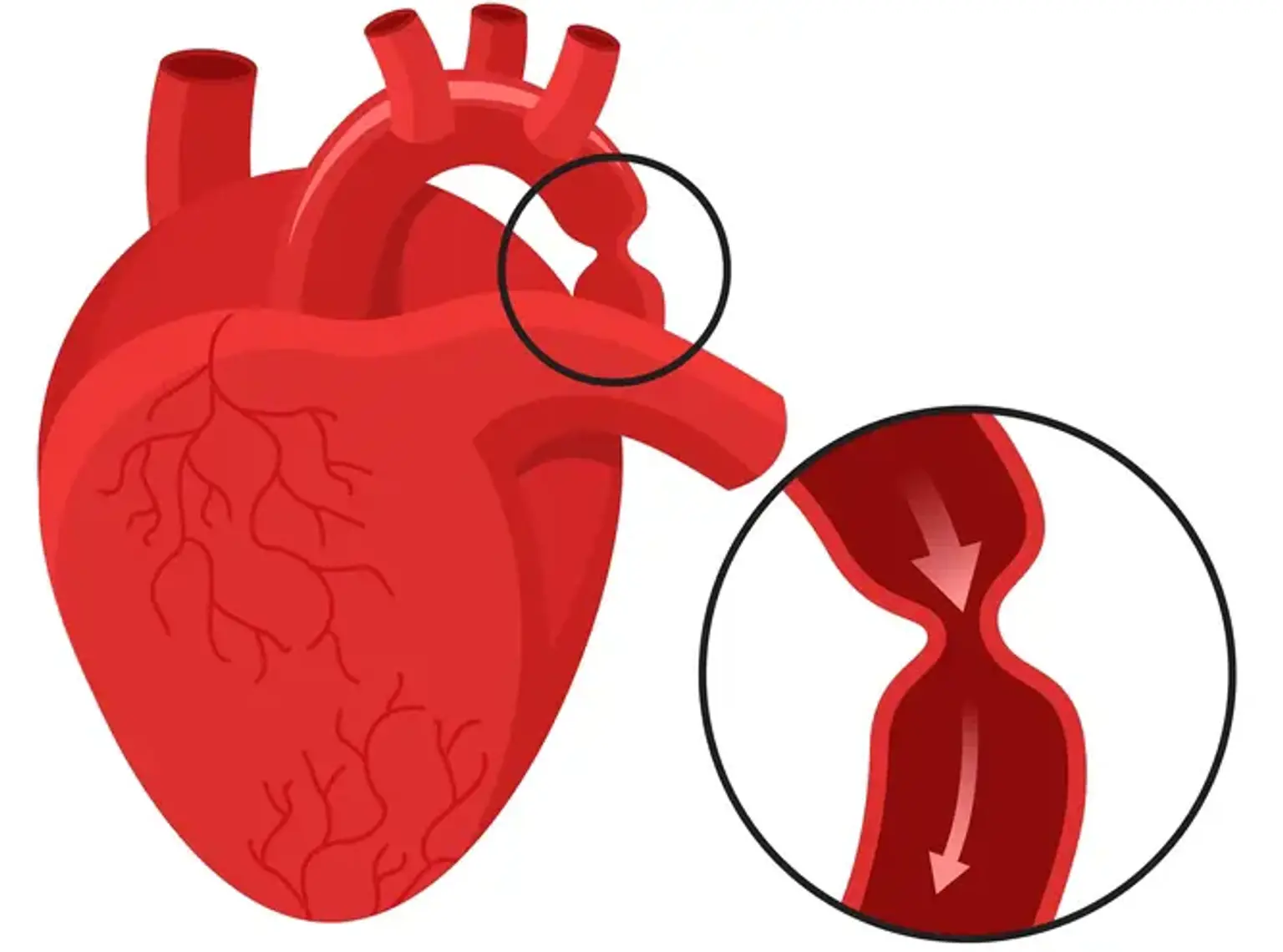
Coarctation of the aorta (COA) is a congenital cardiac defect. It indicates that the aorta is narrower than it should be. The aorta is a major artery that transports oxygen-rich blood from the left ventricle to the rest of the body. Because of this constriction, less oxygen-rich blood is delivered to the body.

The degree of constriction is variable. A young child with more aortic narrowing will have more symptoms. The symptoms will also appear at a young age. Coarctation can occur in infancy in some circumstances. Others may not see it till they are of school age or older. COA can be identified in infants, preschoolers, and teenagers. It is more common in men. If a family member has the condition, the chances of getting it are enhanced. It's also more common in some hereditary diseases, including Turner Syndrome. Aortic coarctation is frequently associated with other heart abnormalities. A bicuspid aortic valve is one of them.

Your child will be given medication to assist him or her relax throughout the COA Stenting procedure. A catheter is passed via a blood artery to the constricted section of the aorta by the healthcare practitioner. A balloon at the catheter's tip is inflated to stretch the region open. To maintain the aorta open, the provider may place a tiny metal mesh tube (stent) in the constricted location.


What Is Aortic Coarctation?

Aortic Coarctation

Aortic coarctation is a narrowing of the aorta. The aorta is the major blood vessel that transports oxygen-rich blood from the left ventricle of the heart to all of the body's organs.

Coarctation most commonly develops in a small section of the aorta just beyond where the arteries to the head and limbs branch off. The "juxtaductal" aorta is the name given to this section of the aorta.

The ductus arteriosus is a blood artery seen in most fetuses. Its wall has unique tissue that causes it to shut in the first hours or days of life. Extra ductal tissue may be the cause of coarctation.

The aortic arch may be narrow in newborns with coarctation (hypoplastic). Coarctation may also occur in conjunction with other cardiac abnormalities, most commonly effecting the left side of the heart. Coarctation is commonly associated with bicuspid aortic valve and ventricular septal defect. Coarctation can also be a symptom of more complex single ventricle heart abnormalities.

Aortic coarctation is frequent in people with hereditary diseases such as Turner's syndrome.

When there is a coarctation, the left ventricle must work harder to generate more pressure than usual in order to drive blood through the small section of the aorta to the lower half of the body.

If the narrowing is very severe, the ventricle may not be strong enough to do this extra work. This can lead to congestive heart failure or not enough blood flow to the organs of the body.


Diagnosis of Aortic Coarctation

Diagnosis of Aortic Coarctation

Aortic coarctation is present from birth. The age at which coarctation is discovered is determined on the degree of the narrowing.

The narrowing is significant enough in roughly 25% of instances of isolated coarctation to induce symptoms in the first days of infancy. When the ductus arteriosus constricts, the left ventricle is forced to pump against significantly greater resistance. This can result in cardiac arrest and shock. Symptoms occur soon because these neonates are healthy until the ductus arteriosus shuts. They are frequently severe.

Coarctation may not be discovered until the kid is many years old in people who do not exhibit heart failure as neonates. Coarctation is frequently suspected in these elderly adults as a result of a cardiac murmur or high blood pressure.

When a clinician is unable to feel pulses in a child's legs, coarctation is suspected. High blood pressure in the arms (but not in the legs) is possible. Typically, a cardiac murmur is present. It might be the loudest in the rear. This is the location of the aorta.

The diagnosis of coarctation is confirmed with echocardiography. This can look at the anatomy of the aorta. It can also evaluate for other cardiac anomalies that may be present. Occasionally other tests, such as a cardiac MRI or CT scan, may be used to look at the coarctation.

Diagnosis of coarctation of the aorta may require some or all of these tests:

  • Echocardiogram: sound waves create an image of the heart
  • Electrocardiogram (ECG): a record of the electrical activity of the heart
  • Chest X-ray
  • Pulse oximetry: a noninvasive way to monitor the oxygen content of the blood
  • Cardiac catheterization: a thin tube is inserted into the heart through a vein and/or artery in either the leg or through the umbilicus ("belly button")
  • Cardiac MRI: a three-dimensional image shows the heart's abnormalities


Signs & Symptoms of Aortic Coarctation

Symptoms of Aortic Coarctation

Coarctation of the aorta symptoms will occur during the first few days of life in severe instances. The symptoms will get more acute as the aorta narrows.

In infants where the coarctation of the aorta is severe or moderate, symptoms can include:

  • Labored or rapid breathing
  • Weak femoral artery pulse (taken in the groin area)
  • Heavy sweating
  • Poor growth
  • Pale or gray appearance
  • Heart murmur: extra heart sound heard when the doctor listens with a stethoscope

If the narrowing is mild, coarctation of the aorta symptoms may go unnoticed until the child is older or even an adult. In those cases, symptoms can include:

  • High blood pressure
  • Cold feet or legs
  • Difficulty exercising (gets out of breath quickly)
  • Dizziness
  • Fainting
  • Nosebleeds
  • Headaches
  • Leg cramps
  • Heart murmur


Indications for COA Stenting

Indications for COA Stenting

The most common indication for therapy is hypertension, however in many children under the age of 12, CoA is diagnosed during a workup for a heart murmur. In our experience, individuals over the age of 35 who arrive for treatment of CoA have been on antihypertensive drugs for an average of 6 years before their coarctation was discovered. Upper extremity hypertension is the most common reason for both the finding and subsequent treatment of a coarctation lesion. Other therapy indications include leg claudication, weariness, and headaches that are commonly linked with exercise or activities. Leg claudication affects roughly 2% to 5% of people, most of whom are over the age of 40.

The indication for stent placement in native and recurrent CoA was specified in a recent American Heart Association scientific statement. The selection between surgery, balloon angioplasty, and stent implantation for the treatment of coarctation remains controversial. In the CCISC's experience, the great majority of patients with late-onset CoA would choose balloon angioplasty or surgical repair of the coarctation segment (i.e., older than 4 years of age).  Once the patient weighs more over 25 kg, opinions on balloon angioplasty and transcatheter stent therapy of the coarctation section are evenly divided, with initial surgical treatment becoming less common.

In a recent study comparing coarctation stenting, surgery, and balloon angioplasty, surgical treatment utilizing resection and end-to-end anastomoses decreased significantly in children older than 8 years of age, reaching 50% at 12 years of age, with no patient older than 16 years undergoing the classic end-to-end resection of the coarctation segment. The aorta becomes less compliant and movable when the patient approaches early adulthood, and stent therapy becomes the treatment of choice.


Preparation Before COA Stenting Surgery

COA Stenting Surgery

  • Preparing for a hospital stay:

When your kid is prepared for COA stenting, providing emotional and physical support can help alleviate their concerns and worry. Packing age-appropriate toys and activities might help your youngster relax during their hospital stay.

These age-specific instructions might assist you in preparing your kid for a hospital stay.

  • Newborn to age 2:

Children of this age may detect your emotions and worry. Concentrate on preparing for the procedure. The more relaxed you are, the more relaxed your youngster will be.

Doctors will offer your child with infant bottles, diapers, and meals. You may want to bring your child's favorite pacifier, rattles, blankets, and washable toys.

  • Toddler:

At this age, children still don't understand enough to understand what's going on. Consider giving your child basic information about hospitalization a few days before admission.

Books, washable toys, and a soothing transitional object, such as a favorite stuffed animal or blanket, may provide comfort to your child while they are in the hospital.

  • Preschool:

Children of this age may misunderstand a hospital stay as punishment for misbehavior. Use brief explanations to reassure your child that their stay in the hospital will help them feel better.

To keep your child occupied while they're in the hospital, pack books, puzzles, toys, and a favorite stuffed animal or blanket.

  • School age:

At this age, your child will most likely comprehend why he or she is in the hospital. Allow lots of time for your child to ask questions and express any concerns regarding their medical condition.

Include movies, music, handheld video game systems, toys, and schoolwork while preparing for a hospital stay.

  • Tweens and teens:

Involve your child in the process from the start and encourage them to ask questions. You might even want to ask your kid what you can do to improve the experience.

Tweens and teens like watching movies, listening to music, and playing handheld video games. Don't forget to carry their homework so they can keep up with their studies.

  • Appointments before surgery:

Your child will undergo several preoperative testing, including a checkup, blood test, and a chest X-ray, a few days before surgery. Doctors will go through the surgery permission paperwork and go over what to expect before, during, and after your child's operation. This visit should last between two and three hours.

A few days before surgery, we’ll confirm admission time, preregister your child and provide additional instructions for the night before surgery.

  • If your child is scheduled for surgery on a Monday, we'll call between 1 and 7 p.m. on the previous Friday or 8 a.m. to noon the previous Saturday.
  • If your child’s surgery is scheduled on a Thursday, we'll call between 1 and 7 p.m. on the previous Wednesday.

Make sure your child avoids solid food (including chewing gum) after midnight the night before surgery. Doctors will provide specific instructions regarding liquid consumption and when to stop feeding infants.

  • The day of surgery:

On the day of your child's procedure, the surgery department will notify you of the time you should arrive at the hospital.

Our objective is to protect your child's health and avoid problems during and after surgery. If your kid exhibits signs of a respiratory infection or another disease, the operation may be postponed.

An anesthesiologist will answer any questions you have once your kid is hospitalized. Depending on their age, your kid may be given an oral sedative to help them calm. Children above the age of ten will be given an IV.

Your kid will then be brought to the operating room, where they will be given further anaesthetic via a breathing mask. Most children fall asleep in a short amount of time.

You can wait in the Surgery Family Waiting Room. A nurse will provide an update usually 60 to 90 minutes after your child was taken to the operating room and periodically throughout the surgery.


What Happens During COA Stenting?

General anesthesia

You will be given a sedative and either local or general anesthesia (be put to sleep). The procedure will be performed through tiny incisions in your groin made by your surgeon. Catheters are placed through the incisions to guide and deliver a stent-graft. Endovascular stent grafts are fabric tubes supported by metal wire stents (also known as scaffolding) that strengthen the aortic weak area.

The surgeon inserts the graft into the aneurysm using X-ray guidance. The transplant is then opened up inside the aorta and secured with metal hooks and stents instead of sutures (stitches).

The graft lets blood to pass through it without pulling on the COA by securely sealing the space with your artery above and below the COA.


What Happens After?

Cardiologist check-up

When your kid is discharged from the hospital, you will be given a time to meet with his or her cardiologist. You can make follow-up appointments with the cardiologist during your child's clinic visits in the future.

One week after your child leaves the hospital, you may have a follow-up appointment in the post-operative clinic. During this visit, your child may:

  • have their wound checked by the nurse practitioner.
  • have a chest X-ray, echocardiogram (heart ultrasound), or blood work.
  • see the dietitian or occupational therapist.

You and your kid will be able to ask questions and discuss any concerns at the follow-up consultation.

Please be on time and prepared for your appointments. Appointments may be delayed at times, or you may be need to wait for testing to be completed. Bring books, toys, food, diapers, and anything else you and your kid may require while waiting. Write down any questions you have before your visit and bring them with you to the clinic. If you are unable to attend an appointment, please contact the cardiology clinic as soon as possible to reschedule.


Risks & Complications

Risk of the treatment

This is a highly safe treatment in general, and specific steps are taken to reduce the possibility of problems. However, there are hazards, as with any surgical operation. Your doctor will go through the specific risks and advantages of the suggested operation with you.

Endovascular coarctation repair complications include blood seeping around the graft, the graft shifting away from its initial insertion, and the stent breaking. Paralysis, delayed rupture of the aneurysm, or infection are all rare but dangerous complications. Other dangers may exist.

When you visit with your doctor, please ask questions to ensure that you understand the procedure's risks, results, and why it is advised.


Conclusion

The aorta

The aorta (the major artery that transports blood from the heart to the body) is narrowed or constricted with this condition.

This narrowing affects blood flow as the arteries branch out to send blood to the upper and lower portions of the body through different vessels. High blood pressure or cardiac damage can be caused by CoA.

The reason is unknown in the majority of youngsters. Along with coarctation, some children may have additional cardiac abnormalities.

Symptoms are usually absent at delivery, although they might appear as early as the first week following birth. Congestive heart failure or high blood pressure can occur in a newborn.

If the blockage is minor, the heart will not be overworked, and symptoms may not appear. Coarctation is not always identified in children and adolescents until they have high blood pressure.

Cardiac catheter balloon dilation and stent insertion is also performed, most often in elderly patients and those with recurrent coarctation following surgery. In this situation, the balloon may extend the narrowing region while the stent offers strong support to prevent the artery from shrinking back to its original size.