Stereotactic Radiosurgery (SRS)

    Last updated date: 13-Mar-2023

    Originally Written in English

    Stereotactic Radiosurgery (SRS)

    Stereotactic Radiosurgery (SRS)

    Stereotactic radiosurgery (SRS) is non-surgical radiotherapy used as a treatment of functional disorders and tiny tumors of the brain. In comparison to conventional therapy, it can administer precisely focused radiation in fewer high-dose treatments, helping to maintain healthy tissue. Stereotactic body radiotherapy (SBRT) is the term used when SRS is utilized to treat body cancers.

    Typically, SRS and SBRT are done as outpatient procedures. Ask your doctor if you should arrange for a ride home after the procedure and whether you should skip meals, beverages, or medication several hours before the procedure. If you think you might be pregnant, if you're nursing a baby, or if you use oral diabetic medications or insulin, let your doctor know. Discuss any medical implants, claustrophobia, or contrast material sensitivities you may have.
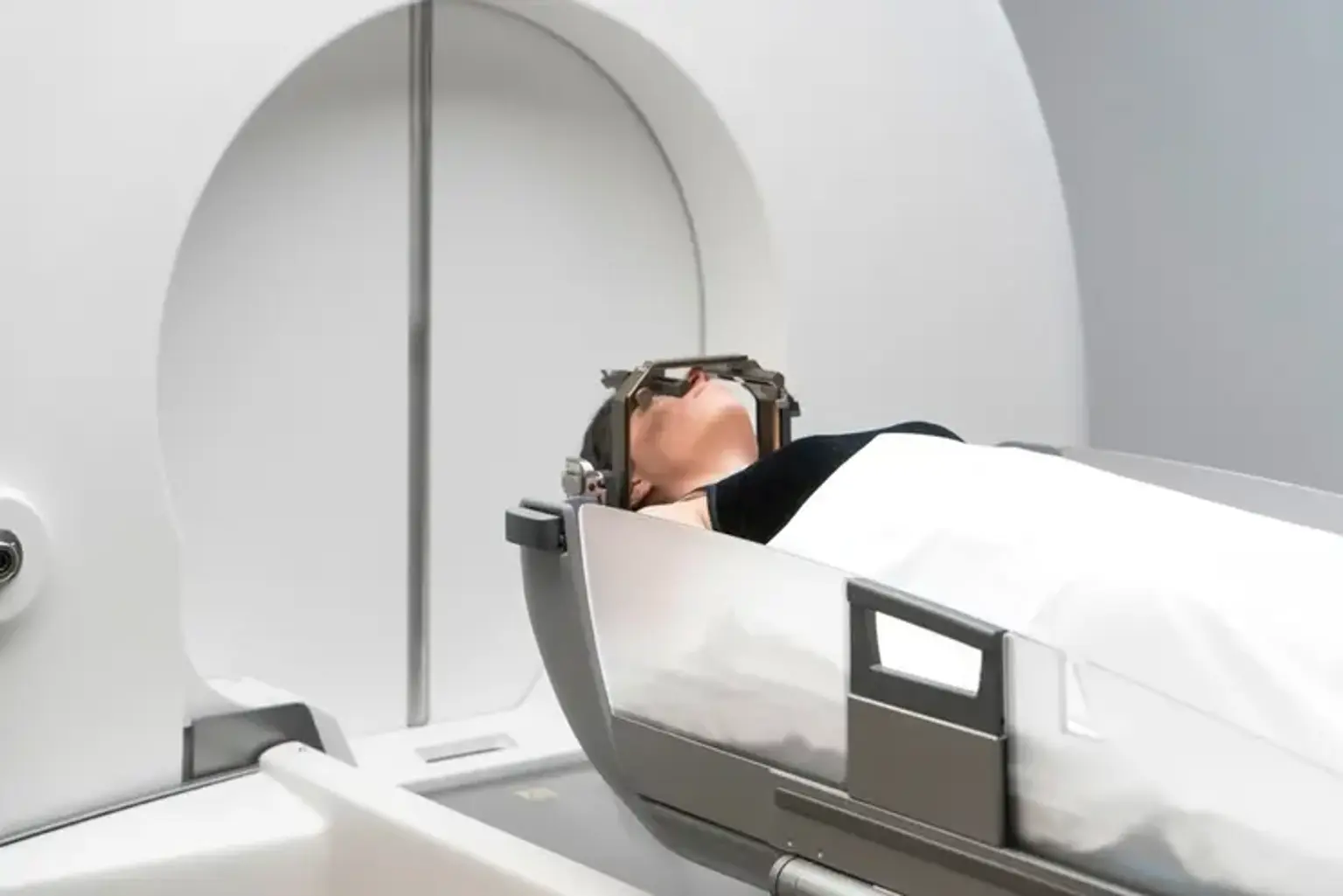
     

    What is Stereotactic Radiosurgery?

    Stereotactic Radiosurgery

    Small brain tumors and other functional disorders of the brain were the original target conditions for the development of stereotactic radiosurgery (SRS), a very precise radiation therapy technique. Stereotactic body radiotherapy is a technique used to treat body malignancies that uses the same concepts as cranial SRS, namely high precision radiation with delivery accuracy of one to two millimeters (SBRT).

    In contrast to conventional radiation therapy, SRS is a non-surgical process that provides precisely targeted radiation at substantially higher doses in just one or a few treatments. Only the development of incredibly advanced radiation technologies, which allow for maximum dose delivery to the target while limiting exposure to the surrounding healthy tissue, has made this treatment viable. Delivering doses that will completely eradicate the tumor and achieve long-term local control is the aim.


    Stereotactic Radiosurgery Indications

    Stereotactic Radiosurgery Indications

    SRS and SBRT are significant alternatives to invasive surgery, particularly for patients who are unable to have surgery and for tumors and abnormalities that are difficult to access, adjacent to key organs or anatomical areas, or that migrate within the body. SRS used to treat:

    • Many different kinds of brain tumors, including benign and malignant ones, primary and metastatic ones, single and multiple ones, tumor cells left over after surgery, intracranial tumors, and orbital and basal skull tumors.
    • AVMs are a tangle of dilated blood vessels that impair normal blood flow to the brain and occasionally bleed.
    • Other neurological diseases, such as tremors and trigeminal neuralgia (a nerve disorder in the face).

    The use of SBRT for the treatment of benign or malignant small-to-medium sized tumors in the body and common illness sites, such as the:

    • Lung
    • Liver
    • Abdomen
    • Spine
    • Prostate
    • Neck and head

    SRS functions basically in similar ways to other radiation therapies. The DNA of tumor cells is damaged rather than the tumor itself being removed. These cells can no longer reproduce as a result. After therapy, benign tumors often go smaller over the course of 18 months to 2 years. Tumors that are malignant or have spread to other organs may shrink more quickly, even within a few months. Arteriovenous malformations (AVMs) may start to thicken and slowly close off several years after receiving SRS treatment. Many cancers will stay intact and dormant for extended periods. This is considered a success as the goal is to stop tumor growth. Due to an inflammatory response within the tumor tissue, some tumors, particularly acoustic neuromas, may temporarily grow after SRS. Over time, however, this transient expansion may resolve or lead to a subsequent tumor regression known as pseudo-progression.


    Stereotactic Radiosurgery Tools

    Stereotactic Radiosurgery Tools

    There are three basic types of equipment, each of which makes use of various instruments and radiation sources:

    • The Gamma Knife uses 192 or 201 highly focused gamma-ray beams that are all directed at the target area. Small to medium-sized cerebral lesions respond well to treatment with the Gamma Knife.
    • High-energy x-rays, also known as photons, are delivered by linear accelerator (LINAC) devices, which are widely used. SRS, also known as fractionated stereotactic radiotherapy, can be applied to bigger tumors using the linear accelerator in a single session or over the course of several sessions. This kind of device is produced by numerous companies under the trade names Novalis Tx, XKnife, Axesse, and CyberKnife.
    • Although there are now far more facilities offering proton therapy than there were a few years ago, the utilization of proton beams or heavy-charged-particle radiosurgery is still rather uncommon in North America.


    How Does Stereotactic Radiosurgery Work?

    Stereotactic Radiosurgery Work

    SRS depends on the following technologies:

    • Procedures for localization and three-dimensional imaging that pinpoint the precise locations of the target within the body
    • Devices for keeping the patient immobilized, carefully positioned, and in the same position throughout therapy
    • Gamma or x-ray beams that are extremely focused and convers on a tumor or other abnormality
    • Image-guided radiation therapy (IGRT), which confirms the location of a tumor just before, and in certain cases, during the delivery of radiation, uses medical imaging. IGRT enhances the treatment's precision and accuracy.

    The tumor or anomaly is located within the body and its precise location, size, and shape are determined by three-dimensional imaging techniques like CT, MRI, and PET/CT. These images also aid in arranging the radiotherapy, which involves carefully placing the patient for treatment sessions and designing radiation beams to focus on the target area from various angles and planes.

    Although doctors may advise multiple stereo-tactically administered treatments, SRS is typically used to refer to a one-day procedure. This is significant for tumors larger than one inch in diameter because the volume of normal tissue treated increases according to the tumor size, and the adjacent normal tissue subjected to the single high radiation dose must be respected and limited. By spreading out the radiation delivery across several visits as opposed to one, you can increase safety and give healthy tissue time to recover in between treatments. As a result, fractionating the treatment maintains an acceptable safety profile while enabling the delivery of substantial dosages within the target. Usually referred to as fractionated stereotactic radiotherapy (SRT), this approach involves giving two to five focused radiation treatments, which are not generally delivered on consecutive days.


    Who is involved in Stereotactic Radiosurgery?

    A radiation oncologist, a medical radiation physicist, a radiologist, a dosimetrist, a radiation therapist, and a radiation therapy nurse often comprise the treatment team. They define the target(s) to be treated, identify any sensitive tissues or organs at risk, choose the proper radiation dose, approve the treatment plan, and interpret the outcomes of radiosurgical procedures. The radiation oncologist and, in some cases, a neurosurgeon lead the treatment team and oversee the treatment.

    • Imaging that identifies the targets in the brain or body that need to be treated is interpreted by a radiologist.
    • The precise radiation dose is delivered by a medical radiation physicist.
    • The physicist, or a dosimetrist working under their supervision, creates a treatment plan using specialized computer software; they calculate the exposures and beam configuration to conformally attack the target(s) to the prescribed dose.
    • The patient is placed on the treatment table by a highly skilled radiation therapist, who controls the equipment from a nearby, secure area. The radiation therapist can converse with the patient throughout the operation and can watch the patient through a window or on closed-circuit television. When using the Gamma Knife, the radiation oncologist may control the machine while the neurosurgeon and/or radiation oncologist may assist in positioning the patient.
    • The radiotherapy nurse evaluates the patient, informs the patient about the procedure, keeps an eye on the patient throughout the procedure, and assists with questions following the procedure.

    The interdisciplinary team that evaluates various treatment options for specific cases and assists in determining who may benefit from radiosurgery for brain lesions may include a neurologist or neuro-oncologist.


    Stereotactic Radiosurgery Preparation

    Stereotactic Radiosurgery Preparation

    Typically, SRS and SBRT are done as outpatient procedures. However, plan to stay in the facility for at least a half-day. If you require a driver to take you home afterward, they will let you know in advance. You might be instructed to abstain from food and liquids the evening before your treatment. Ask your doctor if you should take any prescriptions on the day of your treatment, then carry those medications with you. Additionally, if any of the following is true for you, you ought to let your doctor know:

    • To manage your diabetes, you use oral medicines or insulin.
    • You have an allergy to iodine or intravenous contrast substance.
    • You have a pacemaker, a prosthetic heart valve, a defibrillator, clips for brain aneurysms, chemotherapy ports or implanted pumps, neurostimulators, implants for your eyes or ears, stents, coils, or filters.
    • You are scared of small spaces.


    Stereotactic Radiosurgery Procedure

    Stereotactic Radiosurgery Procedure

    Stereotactic Radiosurgery Using the Gamma Knife

    Installation of the head frame, imaging of the tumor's site, computerized dosage planning, and radiation delivery are the four stages of gamma knife radiosurgery. Some institutions might substitute a plastic head mask for the head frame in certain situations.

    A tiny needle will be inserted into your hand or arm during the first stage to administer any necessary medication and contrast for imaging. Two locations on your forehead and two spots on the back of your head will be numbed with a local anesthetic by a neurosurgeon. Your head will be fitted with a box-shaped head frame, which will be secured to using specially made pins to prevent your head from moving until the treatment session is complete. The Gamma Knife beams are guided by this compact aluminum head frame to ensure that they are directed precisely where the therapy is required.

    After that, you will be brought to an imaging room for a magnetic resonance imaging (MRI) scan, which will precisely pinpoint the tumor's location in the head frame. A computed tomography (CT) scan may be used in place of or in addition to an MRI scan in some circumstances. You might also get an angiography if you're receiving therapy for an arteriovenous malformation. Gamma Knife software updates now enable pre-planning. As a result, to maximize the effectiveness of your therapy, you might receive your MR images for treatment planning ahead of time.

    While your treatment team locates the tumor(s) to be treated and creates a treatment plan using specialized computer software to best irradiate the tumor and reduce the dose to nearby normal tissues, you can unwind for an hour or two in the following phase.

    Before starting treatment, your head frame will be connected to the machine as you lay down on the Gamma Knife bed. With a pillow or wedge-shaped sponge under your knees and a blanket covering you, you will be made comfortable. The treatment team will then start your therapy by going to the control area outside the treatment room. A camera will enable the team to watch you at all times, and a microphone in the helmet will allow you to communicate with your doctor. The bed on which you are lying will recline into the Gamma Knife device. The procedure won't be noticeable to you, since the machine is extremely silent. Depending on the Gamma Knife model and the treatment strategy, the entire procedure may be carried out in one continuous session or it may be divided into several smaller sessions. The entire procedure could take up to four hours, or less. When the procedure is finished, a bell will ring, and the bed will move back to its original position. When the procedure is complete, you will sit up and the head frame will be taken off. You should typically be able to return home shortly afterward.


    Radiosurgery Using the Linear Accelerator

    The four stages of the Gamma Knife procedure, head frame setup, imaging, computerized dose planning, and radiation delivery are comparable to the linear accelerator (LINAC) SRS. LINAC technology is far more widespread and has been in use for a similar length of time as Gamma Knife technology. A portion of the LINAC machine, known as a gantry, moves around the patient while administering the radiation beams from various angles, in contrast to the Gamma Knife, which is stationary throughout the treatment. With LINAC-based SRS, getting the MRI first is also a more common pre-planning approach. Another frequently acquired CT has the frame attached.

    A robotic arm maneuvers the small LINAC around the patient while employing CyberKnife to deliver SRS. A simpler plastic head mask can be used to hold the head still during delivery with the CyberKnife, which can increase patient comfort. This method of SRS delivery does not require an invasive head frame. The incorporation of image guidance in treatment delivery makes frameless SRS development conceivable. Nowadays, frameless SRS is the most widely used LINAC-based SRS technique. To spare the patient the invasive frame placement operation, the Gamma Knife has also produced frameless technology. Additionally, frameless SRS permits fractionated SRS, or SBRT, which might be advantageous for large tumors or those in extremely serious sites.


    Stereotactic Body Radiotherapy (SBRT)

    Stereotactic Body Radiotherapy (SBRT)

    In most cases, stereotactic body radiotherapy (SBRT) entails one to five treatment sessions spread out over one to two weeks.

    You might be required to have a fiducial marker put in or close to your tumor for certain technologies, such as the CyberKnife, that base image guidance on an x-ray-based solution. This step is not necessary for the majority of CT-based image-guidance technology, though. Depending on where your tumor is, your radiation oncologist may collaborate with a pulmonologist, gastroenterologist, or radiologist to have one to four fiducial markers put close to the tumor if one is necessary. The placement of the fiducial marker is generally always performed without hospitalization.

    Following that, your radiation oncologist will decide how to simulate matching your body with the linear accelerator's beams. Patients are frequently accurately aligned and kept immobile throughout simulation and therapy using immobilization devices. You should let your doctor know if you have claustrophobia because some of these devices may hold you in a tight position. A CT scan is carried out over the treatment area once an immobilization device has been made for you. Your doctors might also perform a 4DCT, in which the CT scan collects information on the tumor's movement as you breathe. This happens frequently with liver or lung tumors. You'll be sent home after the scan is finished.

    Planning is the third component covered in the course. The radiation oncologist will coordinate the beam design that is most appropriate for your tumor with a radiation dosimetrist and a medical physicist. They might use MRI or PET/CT, for example, in addition to traditional imaging methods. The team will examine hundreds of possible beam combinations using specialist algorithms to determine which is ideal for your case.

    SBRT uses a linear accelerator for the delivery of radiation. Normal eating and drinking habits are not restricted, though some patients may take an anti-inflammatory, anti-nausea, or anti-anxiety medication before the procedure. You will be put into the immobilizer. Before you start radiotherapy, X-rays or CT scans will be used (depending on the type of image guidance unique to the LINAC technology) to align the radiation beams with the tumor. Based on these x-rays, the radiation oncologist will advise the radiation therapist on how to position you. The therapy will subsequently be given by the radiation therapist. During the course of the treatment, x-rays or a CT scan may be performed to follow the tumor's location. Up to an hour or more may be needed for treatment.


    Stereotactic Radiosurgery Risks

     Stereotactic Radiosurgery Risks

    Radiosurgery treatments resemble getting an x-ray. Generally, the x-rays won't be audible, visible, or tactile. One exception is that, even with their eyes closed, some patients receiving treatments for the brain may see light flashes while the machine is operating. There is no genuine treatment-related pain or discomfort.

    You should inform your doctor or nurse if you have pain for any other causes, such as back pain or discomfort from the head frame or immobilization device.

    There can be some mild bleeding from the pin locations that will be wrapped when the head frame is removed. You might have a headache; in which case you might ask for medicine to help you feel better.

    Patients who have undergone SBRT or radiosurgery can typically return to all of their regular activities in one to two days.

    In addition to issues brought on by the treatment itself, radiation damage to healthy cells in the treatment area can also have negative effects.

    The type and quantity of radiation you get as well as the area of your body being treated will all have an impact on how many and how severe side effects you experience. For your doctor and nurse to assist you in managing any adverse effects, you should discuss them with them.

    Early adverse effects from radiation therapy might occur during or right after treatment, although they usually fade away within a few weeks. The onset of late negative effects can be months or years away. Radiation therapy's early side effects frequently involve fatigue and skin issues. An increase in sensitivity, redness, irritation, or swelling of the skin in the treatment area is possible. Dryness, itching, peeling, and blistering are some more skin changes. Other early side effects may depend on the location being treated and may include:

    • Loss of hair in the treated area
    • Issues with the mouth and swallowing
    • Issues with digestion and eating
    • Diarrhea
    • Nausea and vomiting
    • Headaches
    • Swelling and pain in the treated area
    • Bladder and urine changes

    Rare late side effects can develop months or years after therapy and are frequently irreversible. They consist of:

    • Brain alterations
    • Spinal cord alterations
    • Lung problems
    • Kidneys changes
    • Changes in the colon and rectum
    • Infertility
    • Joints changes
    • Lymphedema
    • Mouth changes
    • Secondary cancer
    • Bones breaks

    Radiotherapy poses a negligible risk of acquiring cancer. Your radiation oncologist should continue to monitor you for recurrent and new cancers after your radiation therapy for cancer. The goal is to increase radiation therapy's capacity to eradicate cancer while reducing its effect on normal tissues to reduce treatment-related adverse effects. One approach used to do this is SBRT.


    Conclusion

    To treat tumors and other issues in the brain, neck, lungs, liver, spine, and other areas of the body, stereotactic radiosurgery (SRS) employs a large number of extremely accurately focused radiation beams. There is no incision, thus it is not surgery as we know it. Instead, stereotactic radiosurgery directs strong radiation doses to the affected area while having little effect on the healthy tissue around it. Stereotactic radiosurgery harms the targeted cells' DNA, much like other radiation treatments do. Tumors then shrink as the damaged cells lose their capacity to reproduce.