Supportive surgery
Last updated date: 03-Mar-2023
Originally Written in English
Supportive surgery
Overview
Cancer is a disease of the cells, which are the foundation of the human body. The body is continually producing new cells to help us in growing, replacing old cells, and healing damage. This procedure can go wrong, causing the cell to become atypical. The defective cell continues to divide, producing other abnormal cells, which might aggregate and form a mass known as a tumor. Tumors are classified into two types:
- Benign tumors are not cancer. They do not spread to other parts of the body.
- Malignant tumors are cancer. They can spread to other parts of the body.
Cancer may begin anywhere within the body since our bodies are made up of cells. Cancer is most commonly seen in the skin, intestines, breasts, prostate, and lungs. Primary cancer refers to the location where the cancer first appears. Doctors are sometimes unable to determine where the cancer began. This is referred to as cancer of unknown primary.
Cancer symptoms might vary greatly from person to person. However, there are a few factors that might signal early illness symptoms. Make an appointment with your doctor if you have any of the following symptoms:
- Unexplained weight loss.
- Chronic tiredness.
- Persistent pain.
- Fever that occurs mostly at night.
- Skin changes.
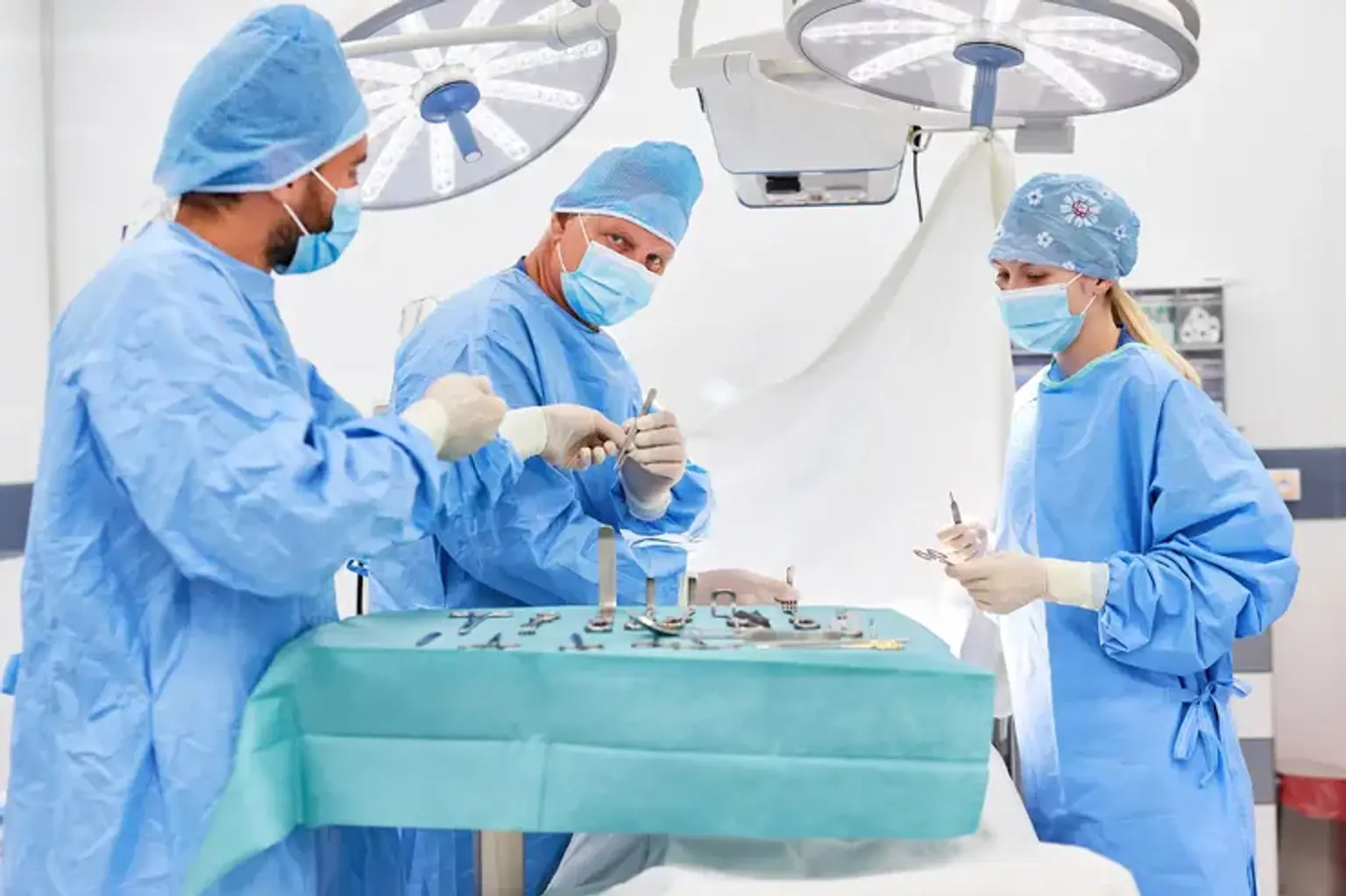
Surgery is used to treat several forms of cancer. Surgery is more effective for solid tumors that are localized. It is a local therapy, which means it just addresses the affected area of your body. It is not used to treat leukemia (blood cancer) or tumors that have spread. Surgery may be the sole option in some cases. However, you will almost always undergo additional cancer therapies as well.
How surgery is used for cancer?
Cancer is prevented, diagnosed, staged, and treated by surgery. Surgery can also alleviate (palliate) cancer-related discomfort or difficulties. Sometimes a single procedure can address more than one of these objectives. In other circumstances, various operations may be required over time.
Preventive surgery:
Preventive surgery is the removal of tissue that does not contain cancerous cells but has the potential to grow into a malignant tumor. Polyps in the colon, for example, may be considered precancerous tissue and removed by prophylactic surgery.
Preventive or prophylactic surgery is performed to remove body tissue that is likely to develop cancer, even if no evidence of cancer is present at the time of the operation. When a person has a disease that puts them at high risk for cancer, a whole organ may be removed. The operation is performed to lower cancer risk and assist prevent cancer, but it does not ensure cancer prevention.
Some women with a significant family history of breast cancer, for example, have an inherited mutation in a breast cancer gene (called BRCA1 or BRCA2). Because the risk of breast cancer is so great, it may be necessary to remove the breasts (prophylactic mastectomy). This implies that the breasts are removed before the malignancy is discovered.
Diagnostic surgery:
Diagnostic surgery aids in determining whether or not cells are malignant. Diagnostic surgery is performed to obtain a sample of tissue for testing and assessment (in a laboratory by a pathologist). Tissue samples aid in the confirmation of a diagnosis, identification of the kind of cancer, and determination of the cancer's stage.
Staging surgery:
Staging surgery is used to determine the degree of cancer or illness in the body. A surgical staging method is laparoscopy (in which a viewing tube with a lens or camera is placed through a tiny incision to study the inside of the body and take tissue samples).
Debulking surgery:
Debulking surgery eliminates a piece of a malignant tumor but not the entire tumor. It is utilized in cases where removing the entire tumor might cause harm to an organ or the body. Following debulking surgery, further cancer treatments, including as chemotherapy and radiation, may be employed.
Curative surgery:
Curative surgery is the removal of a malignant tumor or growth from the body. Curative surgery is used by surgeons when a malignant tumor is limited to a specific location of the body. This form of therapy is frequently regarded as the primary treatment. Other cancer therapies, such as radiation, may be utilized either before or after the operation.
Restorative surgery:
Restorative surgery is occasionally done as a follow-up procedure following curative or other procedures in order to improve or restore a person's look or the function of a bodily component. Women suffering breast cancer, for example, may require breast reconstruction surgery to restore the physical contour of the afflicted breast (s). Curative surgery for oral cancer can alter a person's mouth's shape and appearance. To remedy these consequences, restorative surgery may be undertaken.
Palliative surgery:
This operation is performed to treat complications induced by advanced cancer. Palliative surgery can be combined with other therapies to address an issue that is causing pain or incapacity. Some malignancies in the stomach (abdomen), for example, can develop large enough to block (obstruct) the intestine. If this occurs, surgery can be done to remove the obstruction. Palliative surgery may also be performed to manage pain when medication is ineffective. Palliative surgery relieves cancer-related symptoms and makes individuals feel better, but because the cancer is generally advanced, it is not used to treat or cure the malignancy.
Supportive surgery:
Because it does not cure cancer, supportive surgery is similar to palliative surgery. Instead, it aids the effectiveness of other cancer therapies. A catheter insertion to assist with chemotherapy is an example of supportive surgery.
Supportive surgery is performed to make it simpler for patients to receive other forms of therapy. A Port-A-Cath or Infusaport, for example, is a thin, flexible tube that may be surgically put into a big vein and attached to a little drum-like device implanted just beneath the skin. Instead of inserting needles into the hands and arms each time IV fluids, blood transfusions, or therapies are administered, a needle is inserted into the port's drum to administer treatments and take blood.
Tubes, Lines, Ports, and Catheters Used in Cancer Treatment
Tubes, lines, ports, and catheters may be used if you require surgery, chemotherapy, or other forms of treatment. The sort of equipment used is determined primarily by the purpose for its usage, but it will also be determined by your health and preferences, as well as the preferences of your cancer care team.
Tubes used to give medicine or nourishment:
Liquid medications or feedings may be administered via a tube inserted into the stomach or small intestine. They are frequently referred to as feeding tubes. If the feedings are only needed for a short period of time, a tube can be inserted via the nose and guided down to the stomach without requiring surgery. If liquid food is required for an extended length of time, surgery to place a feeding tube through the skin of the abdomen and straight into the stomach (a gastrostomy tube, or G tube) or the small intestine may be performed (a jejunostomy tube, or J tube).
Oxygen tubes:
If you require oxygen, it can be administered by a mask or tube (known as a nasal cannula) inserted just beneath your nose. The mask or cannula is connected to an oxygen tank or concentrator through tubing. Oxygen flows continually through the tubes on its own. It is critical to utilize oxygen carefully. Check that you understand how to maintain the tank and other equipment and that you follow all directions. Allow no smoking or the use of open flames, including candles, in locations where oxygen is being used.
Tubes used to drain fluids from the body:
Draining tubes may be used to help drain excess fluid that accumulates following surgery or a procedure, or as a result of a tumor obstruction. Draining tubes can be utilized in a variety of ways. A tube, for example, might be:
- A nasogastric (NG) tube, which is inserted via the nose and travels to the stomach, may be required if there is a blockage or obstruction. Alternatively, a tube can be inserted into the stomach (gastric tube) or rectum (rectal tube) to drain excess fluid or aid in the treatment of a blockage.
- Inserted between two ribs in the chest to drain excess fluid from the lungs or to assist maintain the lungs full with air.
- Inserted into the abdomen to remove excess fluid caused by some malignancies.
- Inserted into the bladder to drain urine following surgery or because to other issues that may arise.
- Put into a colostomy or the rectum to help drain intestinal waste if needed.
Intravenous (IV) lines, catheters, and ports
Intravenous (IV) lines are thin, flexible plastic hoses that connect a drug bottle or bag to a tiny needle or intravenous catheter (a small, flexible tube) inserted into a vein in your body. To ensure that no illnesses enter your bloodstream, IV lines must constantly be germ-free ("sterile"). IV supplies are never reused and are only used once. They are used to deliver medications, blood products, nutrients, or fluids directly into your bloodstream. They can also be used to extract blood for testing.
These catheters may deliver medications at various rates. The rate is determined by the type of therapy and the type of catheter utilized. Sometimes medications can be administered simply by inserting a syringe into the catheter and pumping the drug into it, while other times the medication is connected to a mechanical pump that ensures only a particular quantity of medication is delivered through the catheter each hour.
There are several types of IV lines and catheters. Which kind is utilized depends on the purpose of the IV, how frequently it is required, the sort of medicine administered through it, and the level of care required.
Peripheral IVs:
Regular IVs are inserted into a vein in your arm or hand and are only in situ for a brief time. These are known as peripheral IV lines. This is a one-inch-long plastic tube with a plastic hub. The catheter is inserted into a vein in your forearm or hand with a needle, and then the needle is withdrawn, leaving the catheter in the vein with the hub outside the skin. The hub is covered with an adhesive coating.
Central venous catheters (CVCs): Ports and catheters
CVCs are sometimes referred to as central venous access devices (CVADs), central catheters, or central lines. The catheter is a thin, flexible tube that is inserted into a big vein near your heart. Depending on the kind of CVC, the opposite end of the catheter, where medicine and fluid are administered, appears different. CVC catheters are larger and more flexible than peripheral IVs.
A CVC is not required for everyone receiving cancer therapy, although they can be useful in a variety of conditions. You may require a CVC if:
- You have fragile or difficult-to-find veins.
- One or both arms cannot be utilized to administer IVs.
- The therapy has harmed your veins.
- You must be administered a medicine that can be painful to the veins or cause skin injury if it spills outside of a peripheral IV.
- You will require hyperalimentation (TPN), a liquid kind of feeding administered through IV. This can be quite taxing on the veins and may be required for a lengthy period of time.
As long as a CVC is properly cared for and does not cause complications, it can remain in place for as long as you are receiving therapy. Some kinds can be left in place once therapy is completed. There are several types of CVCs.
Implanted port: This is a catheter that is put into a big vein near your heart, or occasionally into a vein in your arm or belly, via your chest. It features an access port at the catheter's tip, beneath your skin. Nothing protrudes from your skin, however there is a slight bulge where the port is. To utilize the port, your nurse will put a special needle into your skin near the port. The needle is then covered with a bandage and attached to a syringe or IV line to infuse or inject (deliver) your medicine or fluids. The majority of blood tests may also be obtained using a port.
Peripherally inserted central catheter (PICC line): This kind is placed into your arm and threaded via a vein to a larger vein near your heart. The catheter's tip protrudes through your skin and is covered by a dressing. When necessary, a nurse will attach it to an IV line to infuse or inject the necessary medicine or liquids. Blood can also be drawn in this manner at times.
Other types of CVCs: Some CVCs are threaded into a major vein near your heart after being put into your chest. The catheter's tip may have one, two, or three separate tubes protruding from the skin of your chest, each covered by a dressing. The tubes attach to an IV line, allowing drugs or fluids to be infused or injected, or the catheters can be used to take blood.
Before you agree to get a CVC, talk with your cancer care team about the type they recommend and why. They can help you decide if you need a CVC and the right type of CVC for you. Some of these devices can restrict certain activities, and safety can be a concern. Each type comes with its own specific care and possible problems and complications.
What are the problems that might happen with central venous catheters?
In CVCs, potential issues might arise. The type of catheter used determines the sort of complications that may occur. Possible complications with inserting a CVC:
- You may have discomfort where the catheter is inserted or where it rests beneath your skin.
- The needle or catheter used may cause harm to the vein or another blood vessel. This might result in bruising or bleeding at the puncture site, as well as infection.
- Before inserting the CVC, tests will be performed to ensure that your blood clots normally. Even when it clots normally, blood might seep out of the vein, causing bruising, strain on other blood arteries or organs, and other complications. In most situations, the bleeding is little and resolves on its own.
- When a CVC is put in the chest or neck, a condition known as a collapsed lung (pneumothorax) can occur. When a lung is ruptured, air accumulates in the chest outside the lung. This danger is considerably reduced by CVC implantation guided by ultrasonography or fluoroscopy.
- When the catheter is inserted, your regular cardiac rhythm may be disrupted. This is generally only transitory, and when the catheter is adjusted, the regular beat resumes. It seldom creates major issues.
- In rare situations, the catheter will enter the wrong location, such as an artery rather than a vein. If this occurs, the catheter must be removed. If no additional issues exist, the artery normally heals on its own.
Problems that could happen later with CVCs:
- A skin infection can begin where the catheter or port enters the body. More dangerous bloodstream infections are also possible. You (and anyone else who touches the catheter) can reduce the risk of infection by washing your hands before using it, changing the dressing properly, checking the skin each time the dressing is changed, and using cautious technique when using the catheter. If you observe any changes in the appearance of the skin surrounding the CVC, contact your cancer care team. Inform them if you get a fever or chills. These are symptoms of a blood infection.
- A hole or crack in the catheter might result in a fluid leak. This might happen if the catheter is frequently clamped in the same region, which can weaken that area. It might happen if you apply too much effort when flushing the catheter. If you observe any leakage, clamp the tube between your body and the source of the leak. Call your cancer care team as soon as possible to find out what to do next.
- The catheter in the vein may shift, move, or get kinked or twisted. If this occurs, it may need to be relocated or deleted.
- Clotted blood can obstruct any type of catheter. You can lessen this risk by flushing the catheter as directed. When a catheter gets occluded, it may sometimes be opened by administering specific medications, although it may need to be removed or replaced.
- Some CVCs can shift or be pulled out if they are not taped or sutured to the skin. If the CVC has been removed or the tubing protruding from your skin seems to be longer, contact your cancer care team straight once.
- Certain catheters must be clamped when not in use, and caps must be snugly screwed on to prevent air from entering. (A considerable amount of air in the catheter might trigger a medical emergency resulting in chest discomfort or shortness of breath.) Know how to clamp your catheter and keep a spare clamp on hand.
- A blood clot might develop around the catheter at times. This might result in arm, shoulder, neck, or head swelling. If you observe new swelling, contact your cancer care team straight away. Blood thinners may be used to treat the clot, but in certain situations, the CVC must be removed.
Conclusion
Surgery is used to prevent, diagnose, stage, and treat cancer. Surgery can also reduce cancer-related pain or complications. Sometimes, a single procedure can address more than one of these objectives. In other circumstances, different operations may be required throughout time.
Supportive surgery is similar to palliative surgery in that it does not cure cancer. Instead, it aids the efficacy of other cancer treatments. A catheter insertion to aid in chemotherapy is an example of supportive surgery. Learn everything you can about the tubes, lines, and equipment you have. Inquire about what to do and when to contact your cancer care team or another care team.
If you have a variety of tubes, you might wish to color identify them with colored tape. You might, for example, choose one color for IV lines, another for a feeding tube, and a third for oxygen tubing. Wrap tape around the catheter or tube around the site where medicine is inserted (if an IV) or where liquid food is connected (if a feeding tube), and write the kind of tube on the tape. Make a chart that indicates which color corresponds to which type of tube and put it in the location where you receive medicine or tube feedings.