Tachycardia
Overview
Tachycardia is a normal cardiac rhythm in which the heart beats faster than usual, causing an increase in cardiac output. While sinus tachycardia is common as a compensatory response to exercise or stress, it becomes concerning when it occurs at rest. Adults have a normal resting heart rate of 60 to 100 beats per minute, which varies depending on fitness level and the presence of medical comorbidities. It varies by age in children but is usually higher than the resting rate in adults (starting approximately between 100 and 150 beats per minute in infancy with a gradual reduction over the next six years).
The presence of tachycardia at rest could be the first sign of serious pathology. As a result, it is critical for the clinician to quickly identify the underlying cause of tachycardia and determine whether it requires urgent evaluation and/or treatment.
Sinus tachycardia can be caused by strenuous exercise, fever, fear, stress, anxiety, certain medications, and street drugs. It can also be caused by anemia, an overactive thyroid, or damage from a heart attack or heart failure. Supraventricular tachycardia is more common in people who smoke, drink too much alcohol, or consume a lot of caffeine. It's been linked to heart attacks in some cases. It is more common in women and children.
Treatments for ventricular tachycardia may include medication to reset the heart's electrical signals or ablation, which is a procedure that destroys the abnormal heart tissue that is causing the condition. A defibrillator may also be used by the doctor to interrupt rapid heart rhythms. A rapid heart rate does not always necessitate treatment, but it can be life-threatening in some cases.
Is Tachycardia so common?
Sinus tachycardia is a common, intermittent, and transient heart rhythm. Inappropriate sinus tachycardia is an exclusion diagnosis that occurs in patients who do not have an underlying cause for the sinus tachycardia. It is thought to be a rare condition that primarily affects young females and healthcare professionals. Postural orthostatic tachycardia syndrome is a common orthostatic disease seen after stress that is also commonly seen in young females (i.e., sepsis, pregnancy, surgery, or trauma).
The most common non-sinus tachydysrhythmia in young adults is atrioventricular nodal re-entrant tachycardia, which has a prevalence of 35 per 10,000 person-years or 2.29 per 1000 people. Women are two times more likely than men to develop paroxysmal SVT, and older people are five times more likely than younger people.
The most common symptomatic dysrhythmia in infants and children is supraventricular tachycardia (SVT). SVT is more common in children with congenital heart disease. The most common cause of SVT in children under the age of 12 is an accessory atrioventricular pathway causing re-entry tachycardia.
Types of Tachycardia
Tachycardia is a condition that makes the heart beat more than 100 times per minute. There are three types of it:
- Supraventricular: This happens when the electrical signals in the organ's upper chambers misfire and cause the heart rate to speed up. It beats so fast that it can’t fill with blood before it contracts. That reduces blood flow to the rest of your body.
- Ventricular: This is a rapid heart rate that starts in your heart's lower chambers. It happens when the electrical signals in these chambers fire the wrong way. Again, the heart beats so fast that it can’t fill with blood or pump it through the rest of your body.
- Sinus tachycardia: This happens when your heart’s natural pacemaker sends out electrical signals faster than normal. Your ticker beats fast, but it beats the way it should.
Causes of Tachycardia
Tachycardia can be caused by either physiological or pathological factors. It is commonly associated with catecholaminergic triggers such as exercise, stress, pain, and anxiety. Pathologically, there are two types of etiologies: cardiac and non-cardiac.
Cardiac etiologies:
- Supraventricular tachycardia: Supraventricular tachycardia is an arrhythmia originating above the ventricles as demonstrated by a narrow QRS complex on an electrocardiogram. If the rhythm is regular, atrial flutter, atrioventricular reentrant tachycardia (AVRT), atrioventricular nodal reentrant tachycardia (AVNRT), atrial tachycardia, or sinus tachycardia are the common etiologies. If the rhythm is irregular atrial fibrillation, atrial tachycardia with variable block, atrial flutter with variable block, multifocal atrial tachycardia, or frequent premature atrial contractions should merit consideration.
- Ventricular tachycardia: Ventricular tachycardia is an arrhythmia that starts in the ventricles and is characterized by a wide QRS on an electrocardiogram. It can be nonsustained (lasting less than 30 seconds) or sustained (lasting more than 30 seconds) (lasting greater than 30 seconds or with associated hemodynamic instability). Furthermore, if the QRS morphology is stable, it is classified as monomorphic, whereas if the PRS morphology is variable, it is classified as polymorphic.
- Torsades de pointes: It is a type of polymorphic ventricular tachycardia caused by either a congenital or acquired prolonged QT interval. Medication that prolongs the QT interval, female gender, hypokalemia, hypocalcemia, hypomagnesemia, ischemia, and structural heart disease are all risk factors for torsades de pointes.
- Myocarditis: It is an inflammatory process involving cardiac myocytes that usually occurs as a result of a viral infection. Other causes of myocarditis include bacterial and parasitic infection, drug or recreational substance ingestion, hypersensitivity reactions to medications or venoms, autoimmune disease, sarcoidosis, hypothermia, ischemia, radiation, and rejection of a transplanted heart.
- Cardiac tamponade: It is a set of symptoms caused by increased pressure in the pericardial space (typically hypotension, jugular venous distention, muffled heart sounds, pulses paradoxus, and interventricular septum bowing during inspiration). This pressure rise is caused by a pericardial effusion, which can be transudative, exudative, or sanguineous. Aortic dissection, intrathoracic neoplasm, recent thoracic procedures or instrumentation, radiation therapy, autoimmune disease, myocardial infarction, medication, uremia, or infection can all cause effusion.
- Acute coronary syndrome: It is a group of symptoms caused by ischemic heart disease. It can manifest as unstable angina, non-ST segment elevation myocardial infarction (NSTEMI), or ST-segment myocardial infarction (ST-segment myocardial infarction) (STEMI). Patients with obesity, hypercholesterolemia, hypertension, diabetes mellitus, age greater than 50 years, male gender, tobacco use, and a family history of heart disease are at increased risk for acute coronary syndrome.
Non-cardiac pathologic etiologies:
- Respiratory:
- Pulmonary Emboli: They are pulmonary vasculature disorders. They can be acute, chronic, or a combination of the two. Acute pulmonary emboli are more clinically significant and have higher morbidity and mortality rates. Acute pulmonary emboli are most commonly a fragment of a deep vein thrombus that has traveled through the circulation to the pulmonary vasculature, but fat emboli from an orthopedic injury, amniotic fluid emboli during pregnancy, and air emboli from central vasculature instrumentation are all possible causes.
- Hypoxia: it's a clinical state where tissues do not receive the necessary amount of oxygen to support their metabolic demand. Hypoxia can result from the inability to take in oxygen, transport the oxygen, or perform oxygen gas exchange.
- Gastrointestinal/Renal/Electrolyte:
- Hypoglycemia: It is defined as a condition in which the plasma glucose concentration falls below 70 mg/dL. It most commonly manifests as a drug-induced side effect of medical therapies. Critical illness, alcohol consumption, hormonal imbalance, bariatric surgery, inborn metabolic errors, insulinomas, and some pituitary and adrenal adenomas can also cause it.
- Dehydration: it results from an imbalance of the total body fluid intake and output. To maintain cardiac output in the setting of intravascular depletion, heart rate will increase.
- Hyperkalemia: It is defined as a potassium serum level that is higher than the standard accepted upper limit of normal (generally 5.0 to 5.5 mEq/L). The most common cause of hyperkalemia is falsely elevated potassium levels caused by sample hemolysis. Renal failure, rhabdomyolysis, excessive exercise, metabolic acidosis, insulin deficiency, tumor lysis syndrome, medication-induced, and increased potassium intake are some of the other etiologies.
- Hypomagnesemia: It is defined as a serum magnesium level less than 1.6 mg/dL. Hypomagnesemia can occur as a result of inadequate magnesium intake or absorption, as well as increased magnesium filtration or excretion. All of these conditions can be congenital or acquired. Prolonged nasogastric suction, malabsorption, acute pancreatitis, refeeding syndrome, late-term pregnancy, lactation, uncontrolled diabetes mellitus with glucosuria or diabetes mellitus complicated by diabetic nephropathy, post-kidney injury diuresis, proximal tubule injury, or medications that act on the proximal tubule, thick ascending loop of Henle, or descending loop.
- Hypocalcemia: It is a serum calcium level less than 8.5 mg/dL. Lack of sunlight exposure, nutritional deficiency, malabsorption, post-gastric bypass surgery, end-stage liver disease, chronic kidney disease, Vitamin-D dependent rickets, hypomagnesemia, hyperphosphatemia, medications, rapid transfusion of large volumes of citrate-containing blood, acute critical illness, osteoblastic metastases, acute pancreatitis, rhabdomyolysis, and mitochondrial gene defects can all cause it.
- Infectious Disease:
- Sepsis: It is a systemic inflammatory illness caused by an infection that leads to organ dysfunction and is a leading cause of death in the United States. It is a spectrum illness that can manifest as mild, severe, or intermediate disease. The high inflammatory burden in the early stages of sepsis reduces systemic vascular resistance. Patients (particularly pediatric patients) can be normotensive to hypertensive during this compensatory period because the body increases heart rate to compensate for decreased systemic vascular resistance.
- Vascular:
- Shock: It is a type of acute circulatory failure that results in insufficient tissue perfusion and hypoxia. Shock is classified into four types: distributive, hypovolemic, cardiogenic, and obstructive. Distributive shock is characterized by decreased systemic vascular resistance and is caused by sepsis, SIRS, anaphylaxis, neurologic trauma, or endocrinologic pathology.
Hypovolemic shock is characterized by intravascular volume depletion. It can be caused by hemorrhage or dehydration. Cardiogenic shock is distinguished by a decrease in cardiac output. Cardiogenic shock can occur as a result of an infarction, which causes large areas of ischemia in the ventricles, papillary muscle rupture, or ventricular septal rupture; infection of the myocardium; pericardial effusion, aortic dissection, acute arrhythmias; and cardiotoxic medications, especially in overdose.
Obstructive shock is distinguished by abnormal cardiac output despite normovolemia and normal cardiac function.
Common causes include tension pneumothorax, cardiac tamponade, restrictive cardiomyopathy, constrictive pericarditis, hemodynamically significant pulmonary emboli, coarctation of the aorta, or other cardiac outflow obstruction, restrictive cardiomyopathy, and severe pulmonary hypertension.
- Hematologic:
1. Hemorrhage:
It is the disease process in which a clinically significant amount of blood is lost following the damage of a blood vessel. Hemorrhage is classified into four types.
Class I accounts for less than 15% of total blood volume. In general, hemodynamics remains unchanged, and urine output remains constant. Class II represents 15 to 30% of total blood volume loss, with mild tachycardia (10 to 20% increase in baseline rate), hypotension, tachypnea, and decreased urine output. Class III represents 30-40% of total blood volume loss with associated tachycardia (20-40% increase), hypotension, tachypnea, and oliguria. Class IV includes more than 40% blood loss, significant tachycardia (40% pulse increase), hypotension, tachypnea, anuria, and lethargy.
2. Anemia:
It's a condition in which the body does not produce enough hemoglobin or red blood cells. Anemia can be acute or chronic, and it is classified as microcytic, normocytic, or macrocytic. Iron deficiency, lead poisoning, or thalassemia can all cause microcytic anemia. Malignant processes, hemorrhage, hemolysis, chronic disease, aplastic anemia, or other bone marrow failure syndromes can all cause normocytic anemia. Macrocytic anemia can be caused by a lack of vitamin B12 or folate, hypothyroidism, medications, alcoholism, liver disease, or myelodysplastic syndromes.
- Toxicology:
Ingestion of medications can cause tachycardia. The following are is a list of common medications that will result in tachycardia:
- Atropine
- Carbon monoxide
- Cocaine
- Dobutamine
- Dopamine
- Ephedrine
- Epinephrine
- Levothyroxine
- Potassium
- Tricyclic antidepressants
- Endocrinologic:
Many physiologic changes occur during pregnancy, including increased heart rate, cardiac output, and vascular volume.
Hyperthyroidism is a condition caused by an overabundance of thyroid hormone. Palpitations, tachycardia, anxiety, tremor, diaphoresis, edema, hyperreflexia, weight loss, and changes to the skin and nails are all common symptoms. Hyperthyroidism can also cause arrhythmias in the heart, such as atrial fibrillation.
Neuroendocrine tumors of the adrenal gland and extra-adrenal autonomic tissue, respectively, are pheochromocytomas and paragangliomas. Tachycardia, diaphoresis, diarrhea, hypertension, weight loss, and anxiety are common symptoms.
Signs and symptoms of Tachycardia
Patients with tachycardia are usually asymptomatic, but they may be aware of their increased heart rate and complain of heart palpitations. Sinus tachycardia may be associated with other symptoms depending on the underlying cause. Dyspnea, chest pain, lightheadedness, dizziness, syncope, and presyncope are all symptoms of this condition. A thorough history is essential in making decisions about how to treat tachycardia. Inquiring about precipitating factors (such as fever or exercise), recent medications, toxic exposures, drug or caffeine use, a history of illness, a history of heart disease or recent heart surgery, and family history are all examples.
Evaluation of the patient's hemodynamic status on physical examination is critical to patient stabilization, particularly to ensure that the patient is not on the verge of cardiovascular collapse due to shock. A cardiovascular examination is required to confirm the presence of tachycardia and to evaluate for the presence of distant heart sounds (such as those seen in pericardial effusions with tamponade), pulsus paradoxus, third or fourth heart sounds or gallop rhythm (such as those seen in myocardial dysfunction), and murmurs (i.e., seen in structural heart disease).
How those with tachycardia are evaluated?
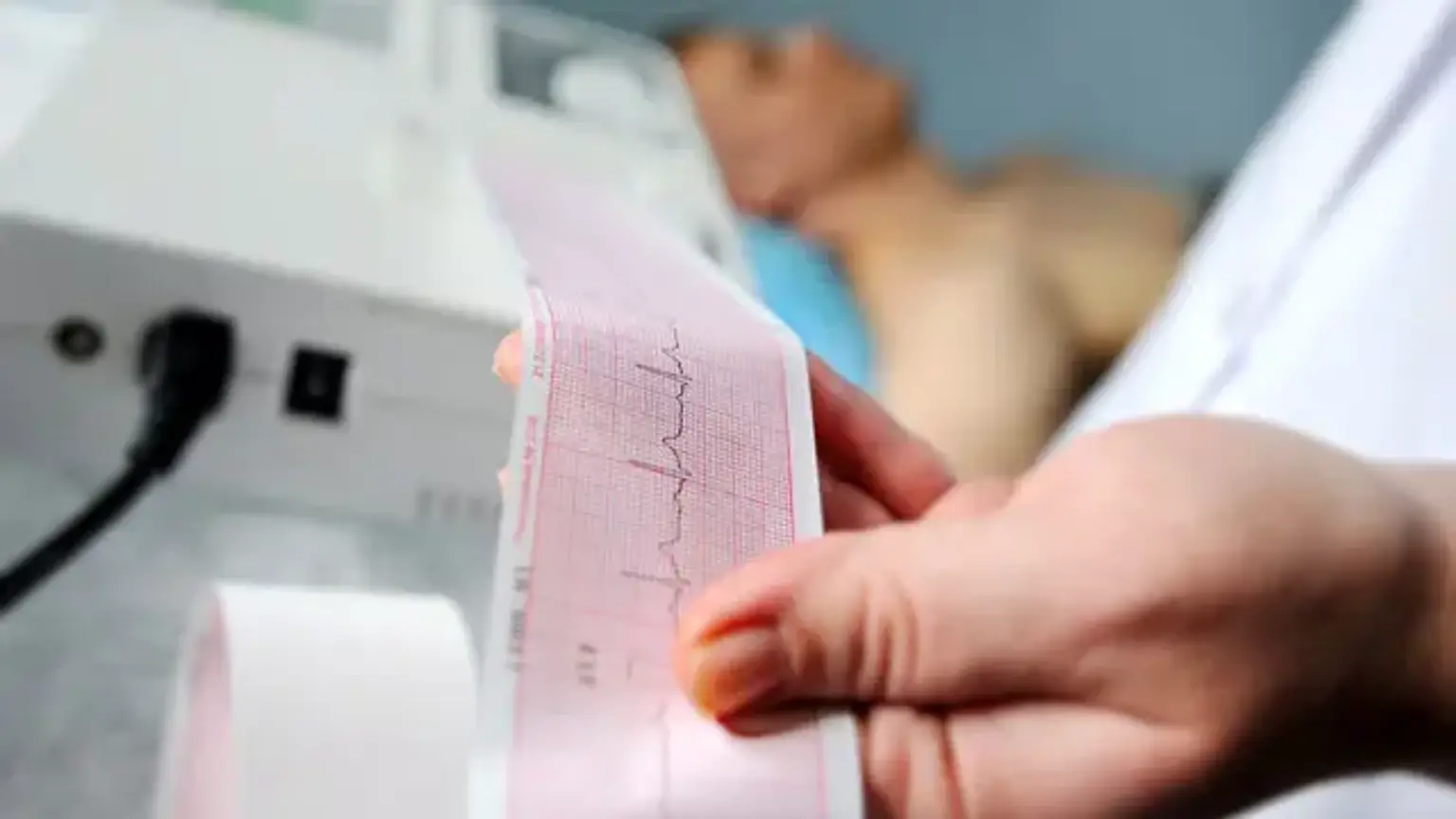
The evaluation of persistent sinus tachycardia at rest entails determining whether tachycardia is an appropriate response first, and then focusing on identifying the underlying cause. An electrocardiogram, 24-hour Holter recording, pulse oximetry, echocardiogram, arterial blood gas, lactic acid level, chest radiograph, D-dimer, chest computed tomography with angiography, ventilation-perfusion scan, cardiac enzymes levels, glucose level, electrolytes, complete blood count, and/or a toxicology screen may be included in the evaluation.
An electrocardiogram is frequently performed first to confirm the presence of sinus tachycardia and to rule out the presence of other tachydysrhythmias. The presence of inappropriate sinus tachycardia can be confirmed by a 24-hour Holter recording. Pulse oximetry can detect hypoxia quickly.
An echocardiogram can aid in the diagnosis of cardiac failure. Arterial blood gases are used to determine the presence of acidity and whether it is related to carbon dioxide levels or metabolic derangement. Lactic acid levels can be used to detect tissue hypoperfusion. A chest X-ray can help determine the source of an infection, cardiac failure, or a pneumothorax. A pulmonary embolus can be detected using D-dimer, chest computed tomography, and ventilation-perfusion scans.
Glucose and electrolyte levels can help identify metabolic derangements. A complete blood count can help identify the presence of anemia and infection. A toxicology screen can determine the presence of any prescription, illicit, and toxic substances that could elicit tachycardia, including cocaine or caffeine.
Treatment of Tachycardia
After reviewing your test results, your doctor will determine what is best for you.
If you have sinus tachycardia, they will help you identify the cause and make recommendations to lower your heart rate. These could include changes to one's lifestyle, such as reducing stress or taking medication to treat a fever.
If you have supraventricular tachycardia, your doctor may advise you to limit your caffeine and alcohol intake, get more sleep, or quit smoking.
Treatments for ventricular tachycardia may include medication to reset the heart's electrical signals or ablation, which is a procedure that destroys the abnormal heart tissue that is causing the condition. Your doctor may also use a defibrillator to stop rapid heartbeats.
A rapid heart rate does not always necessitate medical attention. However, it can sometimes be fatal. So, be safe and notify your doctor right away if you notice any type of irregular heartbeat.
Conclusion
Tachycardia, also known as tachyarrhythmia, is characterized by a heart rate that is faster than the normal resting rate. In adults, tachycardia is defined as a resting heart rate of more than 100 beats per minute. Heart rates that are higher than the resting rate can be normal (as during exercise) or abnormal (such as with electrical problems within the heart).
Sinus tachycardia can be caused by strenuous exercise, a fever, fear, stress, anxiety, certain medications, and street drugs. Anemia, an overactive thyroid, or damage from a heart attack or heart failure can also cause it.
Supraventricular tachycardia is more common in people who smoke, drink excessive amounts of alcohol, or consume a lot of caffeine. It has been linked to heart attacks in some cases. It is more prevalent in women and children.
The ventricular type is linked to abnormal electrical pathways present at birth (long QT), structural heart problems such as cardiomyopathy or coronary disease, medications, or electrolyte imbalance. The reason for this is not always clear.
You may experience dizziness, lightheadedness, shortness of breath, chest pain, and heart palpitations regardless of the type of tachycardia you have. In severe cases, you may become unconscious or suffer a cardiac arrest.
Electrocardiogram (ECG or EKG), Exercise stress test, or Magnetic source imaging may be used to diagnose tachycardia.
The identification and treatment of the underlying cause of tachycardia is the bedrock of management. Benign causes, such as physical activity or stress, do not always necessitate specific cardiac treatment. If the sinus tachycardia is caused by a medical condition that is likely to worsen clinically (e.g., sepsis, shock, hypoxia, metabolic acidosis, acute myocardial ischemia), the patient should be admitted for immediate evaluation. The underlying cause should be carefully addressed during treatment.