Testicular cancer
Overview
Testicular cancer is the most prevalent malignancy in males aged 15 to 45 years. It is also one of the most common curable cancers. It accounts for 1% of male tumors and 5% of urological malignancies. The prevalence of testicular cancer has been growing in recent years, gaining importance due to the long-term impact both the disease and its treatment can have on a patient's life. Over the last 40 years, the incidence of testicular cancer has more than doubled.
Testicular cancer is caused by a combination of environmental and genetic variables; typical risk factors include cryptorchidism, a family history of testicular cancer, a personal history of testicular cancer in the contralateral testis, age, and ethnicity. The first evaluation consists of a history and physical examination, tumor marker analysis, and scrotal ultrasound.
Testicular cancer definition
Testicular cancer occurs when abnormal cells in the testis begin to proliferate and expand uncontrollably. The testicles are an essential component of the male reproductive system.
Testicular cancer is distinct from other malignancies in that it typically affects younger men. Although it is very uncommon in general, testicular cancer is the most frequent kind of cancer among men aged 15 to 49. White men have a greater chance of having testicular cancer than men of other ethnic groups for unknown causes.
What are testicles?
The testicles (also known as testes; a solitary testicle is referred to as a testis) are a component of the male reproductive system. In mature males, the two organs are generally around the size of a golf ball. They are contained within a skin sac known as the scrotum. The scrotum is located underneath the base of the penis.
Testicles have 2 main functions:
- They make male hormones (androgens) such as testosterone.
- They make sperm, the male cells needed to fertilize a female egg cell to start a pregnancy.
Seminiferous tubules are long, thread-like tubes inside the testicles that produce sperm cells. They are subsequently kept in the epididymis, a tiny coiled tube located behind each testicle. This is the stage during which they mature.
During ejaculation, sperm cells are transported from the epididymis to the seminal vesicles through the vas deferens. They combine with fluids produced by vesicles, the prostate gland, and other glands to generate semen. This fluid then enters the urethra, the tube in the middle of the penis that allows urine and sperm to exit the body.
Epidemiology
Western and Northern Europe have the highest rate of testicular cancer (8.7 and 7.2 per 100,000 men, respectively). Western Asia has the greatest death rates, with most nations seeing a drop in mortality, most likely due to the combined impact of early identification by self-examination and integration of multimodal therapies.
Over the previous four decades, the total incidence in the United States has progressively climbed (6.3 per 100,000 persons in 2017 compared to 3.7 per 100,000 in 1975). Testicular cancer is more common in developed nations than in developing ones, and while Caucasian males have a greater prevalence, the incidence of testicular cancer among non-white and immigrant men in the United States is growing for unclear causes. Synchronous contralateral tumors are seen in 0.6 percent of patients, whereas metachronous contralateral tumors are found in 1.9 percent.
Etiology
Both genetic and environmental factors have been studied in the development of testicular cancers.
Epidemiological risk factors:
They include cryptorchidism, reduced spermatogenesis shown as subfertility or infertility, sexual development abnormalities, a family history of testicular cancers in first-degree relatives, the existence of a contralateral tumor or germ-cell neoplasia in-situ (GCNIS), and so on.
The most common environmental risk factors for testicular cancers can be summarized as below:
- Cryptorchidism —Risk increases by 2–4 times. Because cryptorchidism is more frequent on the right side, there is a slight rise in the risk of right-sided testicular cancer.
- Family history—relative risk increased 6–10 fold in brothers or sons of an affected man
- Infections - Human papillomavirus (HPV), Epstein-Barr virus (EBV), Cytomegalovirus (CMV), Parvovirus B-19, and Human immunodeficiency virus (HIV)
- Testicular trauma
- High maternal estrogen levels
- Carcinoma in situ (intratubular germ cell neoplasia)
- Prior history of testis cancer or extragonadal germ cell tumor
Genetic risk factors:
The genesis of testicular cancer has been linked to a number of genetic alterations. The isochromosome of chromosome 12's short arm – (i12p) – is pathognomonic for all forms of adult germ cell tumors (GCTs) and GCNIS. p53 mutations have been found in around 66% of GCNIS cases. Genetic variants in the PTEN tumor suppressor gene have also been linked to an increased risk of testicular cancer (TC).
Pathophysiology
Germ-cell cancers have been seen to arise as a result of a tumorigenic event in utero, which results in intratubular germ cell neoplasia. Intratubular germ-cell neoplasia is caused by gonocytes that have not differentiated into spermatogonia. These cells do not become invasive until hormonal changes occur throughout puberty.
Seminomas are made up of altered germ cells that are unable to differentiate. Embryonal carcinoma cells mimic undifferentiated stem cells in appearance and gene expression, and they have similarities with stem cells and intratubular germ-cell neoplasms. Extraembryonic differentiation occurs in choriocarcinomas and yolk sac tumors, while somatic differentiation occurs in teratomas.
Clinical presentation
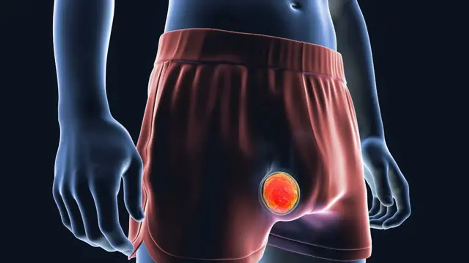
As an accidental discovery, testicular cancer often manifests as a unilateral lump or painless enlargement. Discomfort is a less frequent symptom of testicular cancer, with roughly one-third of patients experiencing dull pain. Acute pain is experienced by around 10% of the population. While trauma is not connected with testicular cancer, antecedent trauma may prompt examination or imaging of the testes, leading to a final diagnosis.
A thorough physical exam will indicate a solid intratesticular lesion. To recognize a solid intratesticular mass and separate intratesticular from extratesticular lesions, each testis should be softly gripped and rolled between the fingers. Because 0.6 percent of individuals have a synchronous contralateral testis tumor, it is critical to thoroughly evaluate the contralateral testis.
The existence of a testicular lesion should be verified using ultrasonography, which is an extension of the physical examination if the testis cannot be completely palpated due to the presence of a hydrocele. Patients who come with symptoms or indications of metastatic illness are infrequent. These are outlined below:
Clinical manifestations of testis cancer from metastatic disease
- Systemic symptoms: anorexia, malaise, weight loss
- Pulmonary metastasis: cough or shortness of breath
- Lymphatic metastasis: cervical or supraclavicular lymphadenopathy
- Retroperitoneal disease: Bulky retroperitoneal disease can present as back pain or may lead to compression on the gonadal veins leading to findings of varicocele
- Vascular obstruction or thrombosis leading to lower extremity edema
- Nausea, vomiting, or gastrointestinal hemorrhage from retroduodenal metastases
- Central or peripheral nervous system symptoms from the cerebral, spinal cord, or peripheral nerve root involvement
Because of its strong chemo-sensitivity with contemporary cisplatin-based chemotherapy, radio-sensitivity, and surgical excision with orchiectomy or retroperitoneal lymph node dissection, testicular cancer offers good cure rates.
Types of testicular cancer
The testicles are made up of many different types of cells, each of which has the potential to grow into one or more forms of cancer. It's critical to understand the sort of cell cancer began in and the type of cancer it is since they differ in how they're treated and their prognosis.
By examining the cells under a microscope, doctors can determine the sort of testicular cancer you have.
Germ cell tumors
More than 90% of testicular malignancies begin in cells known as germ cells. These are the cells responsible for the production of sperm. Seminomas and non-seminomas are the two most common forms of germ cell tumors (GCTs) in the testicles. These categories exist in about similar proportions. Seminoma and non-seminoma cells coexist in many testicular tumors. Because they develop and spread like non-seminomas, these mixed germ cell tumors are treated as non-seminomas.
Seminomas
Seminomas develop and spread at a slower rate than non-seminomas. These tumors are classified into two types: classical (or typical) seminomas and spermatocytic seminomas.
- Classical seminoma: More than 95% of seminomas are classical. These usually occur in men between 25 and 45.
- Spermatocytic seminoma: This rare type of seminoma tends to occur in older men. (The average age is about 65.) Spermatocytic tumors tend to grow more slowly and are less likely to spread to other parts of the body than classical seminomas.
Some seminomas can cause a rise in blood levels of a protein known as human chorionic gonadotropin (HCG). HCG is a tumor marker for some kinds of testicular cancer and may be detected with a simple blood test. It can be utilized for diagnosis as well as monitoring the patient's response to therapy.
Non-seminomas
Germ cell tumors of this sort are more common in men between the ages of 18 and 30. Non-seminoma cancers are classified into four types: embryonal carcinoma, yolk sac carcinoma, choriocarcinoma, and teratoma. Most tumors are a combination of various kinds (occasionally with seminoma cells as well), but this has little bearing on how most non-seminoma malignancies are treated.
Embryonal carcinoma:
These cells are seen in around 40% of testicular cancers, while pure embryonal carcinomas occur only 3% to 4% of the time. These tumors can resemble tissues from extremely early embryos when seen under a microscope. This form of non-seminoma grows quickly and spreads outside of the testicle.
Embryonal carcinoma can raise blood levels of alpha-fetoprotein (AFP), a tumor marker protein, as well as human chorionic gonadotropin (hCG) (HCG).
Yolk sac carcinoma:
These tumors are so-called because their cells resemble the yolk sac of a developing human fetus. This malignancy is also known as a yolk sac tumor, endodermal sinus tumor, infantile embryonal carcinoma, or orchidoblastoma.
Although this is the most prevalent kind of testicular cancer in children (particularly babies), pure yolk sac carcinomas (tumors that do not contain other types of non-seminoma cells) are uncommon in adults. When these tumors arise in youngsters, they are typically effectively treated. However, they are more concerned when they arise in adults, particularly if they are pure. Even if cancer has progressed, yolk sac carcinomas respond quite effectively to treatment.
This type of tumor almost always increases blood levels of AFP (alpha-fetoprotein).
Choriocarcinoma:
This is a relatively rare and rapidly developing kind of adult testicular cancer. Pure choriocarcinoma has a high propensity to spread to other regions of the body, including the lungs, bones, and brain. In a mixed germ cell tumor, choriocarcinoma cells are frequently seen with other forms of non-seminoma cells. Although the presence of choriocarcinoma is usually a cause for concern, these mixed tumors have a somewhat better prognosis than pure choriocarcinomas.
This form of tumor raises HCG levels in the blood (human chorionic gonadotropin).
Teratoma:
Teratomas are germ cell tumors that have sections that resemble each of the three layers of a developing embryo: the endoderm (innermost layer), mesoderm (middle layer), and ectoderm (outermost layer) (outer layer). Pure testicular teratomas are uncommon and do not raise AFP (alpha-fetoprotein) or HCG (human chorionic gonadotropin) values. Teratomas are most commonly found as components of mixed germ cell cancers.
Diagnosis
The evaluation of testicular cancer begins with a complete history and a thorough physical examination. As previously noted, history entails inquiring about clinical aspects. It is critical to inquire about any history of cryptorchidism, orchiopexy, or inguinal hernia repair as a child.
A father or brother with a family history of testicular cancer should be elicited. Until proven otherwise, every solid intratesticular lump seen during a physical examination should be considered testicular cancer. Examining data relating to metastatic disease, as previously mentioned, may also be detected.
When testicular cancer is suspected on physical examination, testicular imaging using trans-scrotal ultrasonography is the primary imaging modality used to diagnose it. When paired with a physical examination, ultrasound imaging gives approximately 100 percent sensitivity in the detection of testicular cancer.
When an ultrasound reveals a hypoechoic, solid, vascularized intratesticular lesion, testicular cancer is suspected, and different kinds of testicular cancer exhibit modest morphologic changes in imaging. Before any intervention, including orchiectomy, further testing should include serum tumor markers (AFP, HCG, and LDH). Adequate counseling on the likelihood of infertility, as well as the placement of a testicular prosthesis, should be provided. In individuals with bilateral testicular disease, sperm banking should be investigated.
Inguinal orchiectomy is both therapeutic and supplies tissue for histological analysis. Trans-scrotal biopsy should be avoided because to the possibility of local dissemination caused by the disruption of existing lymphatic drainage channels.
Testicular germ cell cancers spread through lymphatic pathways that are well-defined and predictable. The predominant landing zone for malignancies developing in the right testis is the infrarenal inter-aortocaval lymph nodes, followed by paracaval lymph nodes and para-aortic lymph nodes. Tumors from the left testis spread to the para-aortic lymph nodes first, then to the inter-aortocaval lymph nodes.
Tumors emerging from the right testis can have retroperitoneal spread from the right to the left side, although left-to-right dissemination inside the retroperitoneum is uncommon unless it is combined with bulky lymph node disease. Abdominopelvic cross-sectional imaging is critical for detecting retroperitoneal lymph node disease caused by primary testicular cancer and informing multimodal therapy.
All patients with testicular germ cell malignancies should get computed tomography abdominopelvic imaging (CT). Patients who have high blood tumor markers (AFP, -fetoprotein; -hCG, -subunit of human chorionic gonadotropin; LDH, lactate dehydrogenase) should be assessed further for staging by computed tomography (CT) of the chest, abdomen, and pelvis.
If testicular tumor markers are within the normal range, the rate of metastasis almost outside the retroperitoneum is very low; thus, adding a chest CT-scan to cross-sectional imaging of the abdomen and pelvis is highly unlikely to change the treatment plan, and a chest radiograph suffices when combined with abdominopelvic CT imaging.
Choriocarcinoma has been proven to spread by hematogenous channels, and patients with high levels of -hCG should have cross-sectional imaging of the brain to identify metastatic lesions caused by choriocarcinoma spreading to the brain.
Serum tumor indicators are helpful in the diagnosis and treatment of testicular cancer. Post-orchiectomy indicators are used in the staging and risk classification of testicular cancer, as well as in the evaluation of therapy response and disease return. LDH is present in a variety of organs across the body, with LDH-1 being expressed on chromosome 12p, which is overexpressed in germ cell cancers. Higher LDH levels may indicate a greater tumor burden.
In many germ cell cancers, AFP and -hCG levels are measured. -hCG is generated by syncytiotrophoblasts in germ cell tumors and may be seen in both non-seminomatous and seminomatous germ cell cancers. Choriocarcinomas, as well as around 15% of seminomas, frequently express -hCG. AFP is produced by the yolk sac, embryonal, and certain teratomas inside germ cell tumors, but it is never increased in pure seminoma or pure choriocarcinoma.
AFP and -hCG are seen in a variety of neoplasms, including germ cell tumors. -hCG levels are elevated in neuroendocrine tumors, kidney, lung, head & neck, bladder, and GI tract malignancies. In liver disorders, AFP levels have been observed to be high.
Management
Following imaging and measurement of tumor markers, all patients are classed as Clinical Stage 0, I, II, or III. Patients in stage 0 have germ cell neoplasia in situ, patients in stage I have tumor restricted to the testis, patients in stage II have lymph node involvement, and patients in stage III have distant metastases.
- Stage 0:
Testicular germ cell neoplasia in situ (GCNIS) has a 70% chance of evolving into TGCT after 7 years if left untreated. GCNIS should be managed collaboratively, with options including careful monitoring with ultrasonography, orchiectomy, and radiation. Despite the fact that low-dose radiation has similar cure rates to orchiectomy, scatter to the contralateral testis can result in up to 40% of men treated with radiotherapy requiring testosterone replacement therapy or experiencing infertility.
Contralateral GCNIS screening is advised for people with high-risk factors, such as contralateral cryptorchid testis, testicular volume of 12ml, and age of 40 years (30 percent risk of developing GCNIS). In such cases, screening for GCNIS in the contralateral testis should be combined with a biopsy, and radiotherapy/orchiectomy should be administered as needed. Regular ultrasonography, on the other hand, is preferred for males who want to keep their fertility.
- Stage I:
The initial therapeutic option is orchiectomy. The likelihood of relapse differs between seminoma and non-seminomatous tumors, as detailed below.
Seminoma
Without adjuvant therapy, around 15% to 18% of individuals with stage I illness will recur following orchiectomy. While surveillance is advised, two options are available for high-risk patients (rete-testis invasion and tumor size >4cm): single-agent carboplatin chemotherapy or surveillance. The probability of recurrence is 5% in the absence of substantial volume (>4cm) and rete-testis invasion, and no adjuvant treatment is recommended.
Invasion of both rete-testis and lymphovascular tissue results in a 25% chance of relapse, whereas neither is implicated in a 4% risk of relapse. Primary radiotherapy to the retroperitoneum and ipsilateral pelvis with 20 to 30 Gy has been the historical standard therapy for patients who are not agreeable to surveillance, and this treatment results in a 1% in-field recurrence rate, though longer-term cardiovascular side effects limit its widespread use.
Non-seminomatous tumors:
In tumors without lymphovascular invasion, the probability of recurrence without adjuvant therapy ranges from 14% to 22%. If lymphovascular invasion or embryonal predominance is present in the initial tumor, the chance of concealed metastatic illness increases. Without adjuvant therapy, there is a 50% chance of developing metastases if lymphatic or vascular invasion is present.
BEP (bleomycin, etoposide, and cisplatin) or EP (etoposide, bleomycin, and cisplatin) are the most often used chemotherapy regimens (etoposide and cisplatin). Because orchiectomy alone has a cure rate of 70%-80%, active surveillance without adjuvant treatment is suggested.
Although it is dependent on regular laboratory and cross-sectional imaging monitoring, this technique avoids the long-term consequences of chemotherapy and surgical recovery from initial retroperitoneal lymph node dissection. Despite the fact that most relapses occur during the first two years after diagnosis, longer-term monitoring is essential owing to the potential of late relapse. If a patient refuses to be monitored, one or two cycles of BEP treatment may be recommended.
A single cycle of BEP (bleomycin, etoposide, and cisplatin) has been shown to lower the probability of recurrence from 50% to 3%. While recurrence rates after first treatment are modest, those who do relapse after BEP are frequently chemoresistant. Nerve-sparing retroperitoneal lymph node detection (RPLND) delivers cure rates equivalent to chemotherapy in select patients at specialized referral facilities with competent surgeons, as well as a reduced requirement for monitoring cross-sectional imaging.
The argument for surgery is that contemporary nerve-sparing procedures can result in a high cure rate while preserving fertility. Retroperitoneal lymph node dissection is also used to treat occult metastatic teratoma, which is resistant to all chemotherapy regimens and affects up to 25% of males with occult metastatic illness.
According to studies, between 19% and 28% of men with clinical Stage I illness have pathologic Stage II disease in the retroperitoneum, and over 80% of these individuals can be treated with RPLND alone, avoiding chemotherapy and associated negative effects. Furthermore, long-term cancer-specific survival after RPLND (with or without adjuvant chemotherapy) is approximately 100%, and the chance of difficult-to-cure late recurrence is less than 2%.
Chemotherapy is required for all individuals who have brain metastases. According to a study conducted by the Global Germ Cell Cancer Group, main chemotherapy is an adequate treatment option in patients with brain metastases found after the first diagnosis of TGCT. Cranial radiation combined with systemic chemotherapy improved the overall prognosis in patients with brain metastases at recurrence.
Concurrent whole-brain radiation and chemotherapy have been linked to central nervous system damage (progressive multifocal leukoencephalopathy or cerebral atrophy) and should be avoided. When radiation is necessary, there is growing interest in the use of stereotactic radiosurgery to treat oligometastases or single brain metastases. In such circumstances, the relevance of neurosurgical procedures is uncertain. More research is required to accurately identify the value of neurosurgery in such circumstances.
Differential Diagnosis
Unless proven differently, a hard intratesticular tumor is indicative of testicular cancer. However, there are a few alternative diagnosis to consider when analyzing a testicular mass:
- Epididymo-orchitis
- Hematoma
- Inguinal hernia
- Hydrocele
- Spermatocele or epididymal head cyst
- Varicocele
- Lymphoma (the most common finding in bilateral testis lesions in older men)
- Metastasis from other cancers (eg, lung cancer, melanoma, prostate cancer)
- Syphilitic gumma
- Tuberculoma
- Ultrasonography aids in narrowing the diagnosis, and radical inguinal orchiectomy is the ultimate diagnostic procedure.
Prognosis
The histology, amount of distant tumor dissemination, and extent of tumor marker increase all have a significant impact on prognosis. The prevalence of metastases to visceral organs other than the lungs is the primary unfavorable prognostic characteristic for males with disseminated seminomas. When compared to a tumor that began in the testicle, a tumor that originated in the mediastinum has a poorer prognosis. Even individuals with broad metastases upon presentation, including those with brain metastases, may have a curable illness and should be treated as such.
Complications
Complications due to testicular malignancy can be broadly classified into two groups.
Complications secondary to the disease itself:
- Chronic fatigue
- Anxiety disorders
- Metastatic complications
- Venous thromboembolism
Complications secondary to the treatment:
- Hypogonadism, leading to depression, sexual problems, and decreased physical well-being
- Peripheral neuropathy (cisplatin use)
- Hearing loss (cisplatin use)
- Tinnitus (cisplatin use)
- Raynaud phenomenon (cisplatin use)
- Secondary malignancies
- Cardiovascular disease
- Infertility
- Infections
- Surgical complications(antegrade ejaculation failure, small bowel obstruction, etc.)
Conclusion
The testicles are the male reproductive glands that are found in the scrotum. Testicular carcinoma is a kind of cancer that arises from the cells in the testicles. Testicular cancer is the most frequent cancer in young men, yet it is also one of the most treatable. More than 95% of men diagnosed with testicular cancer survive the disease.
It most typically manifests as a painless hard mass within the scrotum or in the testicles, usually on one side and rarely on both sides. When cancer spreads to other parts of the body, such as the lungs, brain, abdomen, or neck, symptoms such as nausea, vomiting, stomach upset, cough, shortness of breath, weakness, sensory abnormalities, abdominal pain, lumps in the neck/groin regions, and back pain can arise.
Prompt evaluation is critical to guarantee early diagnosis and therapy and to reduce the burden of treatment in advanced disease. Medical evaluation, physical examination, and ultrasound of the testis are all required. Serum tumor markers and cross-sectional imaging are then examined. When there is a suspicion of metastatic illness, further imaging with computed tomography (CT) of the chest and abdomen may be performed for staging.
By removing the lump together with the testicle, both a definite diagnosis and beginning therapy can be obtained. The stage of the disease and the response to the initial treatment will determine if more surgery, radiation, or chemotherapy is required.