Thalassemia

Last updated date: 03-Mar-2023

Originally Written in English

Thalassemia

Overview

Thalassemia is a diverse collection of blood diseases that disrupt the hemoglobin genes, resulting in inefficient erythropoiesis. Anemia develops as a result of reduced hemoglobin synthesis, and regular blood transfusions are necessary to maintain hemoglobin levels. This exercise discusses the diagnosis and treatment of thalassemia, as well as the importance of an interprofessional team in the care of individuals with this illness.
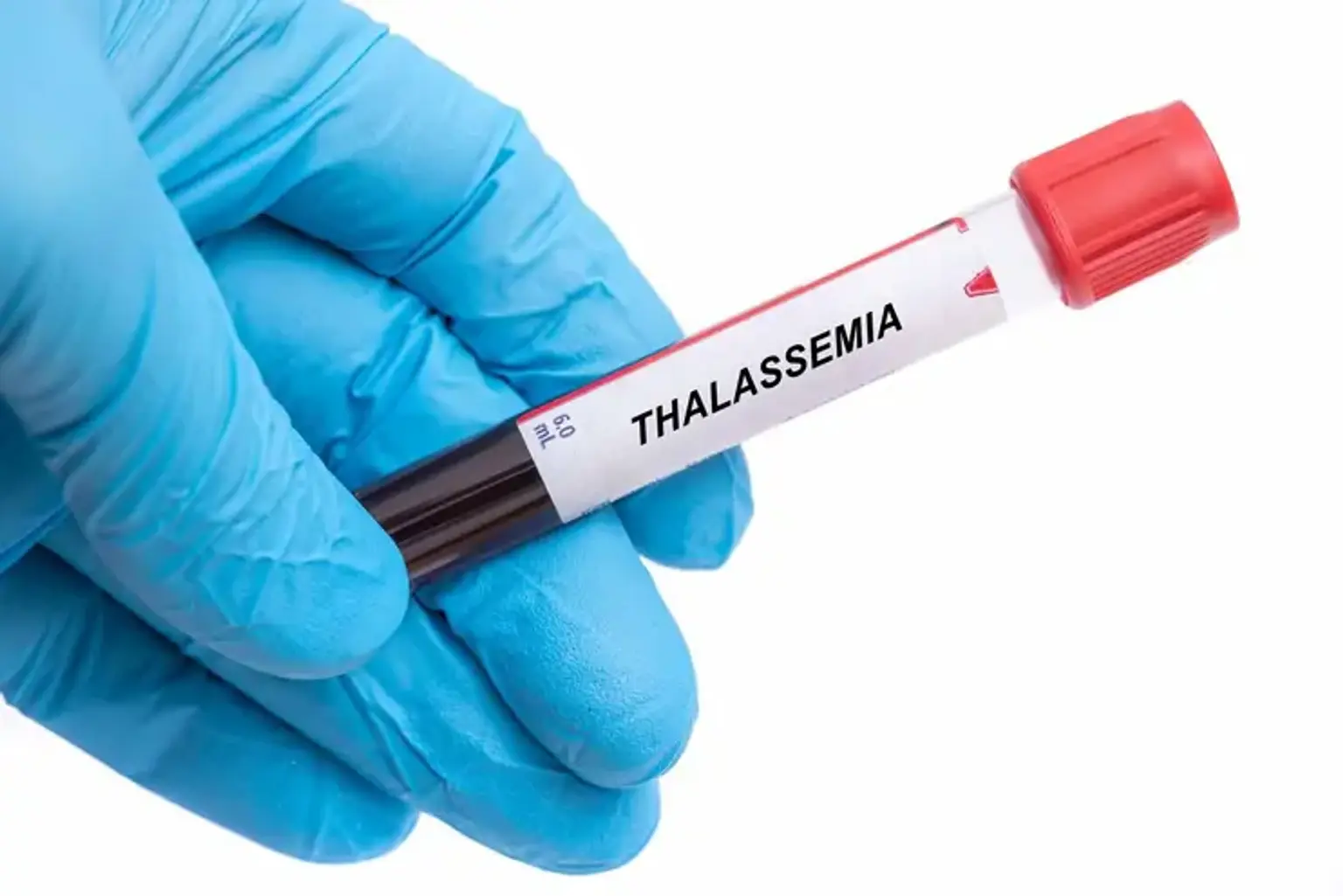
 

What is Thalassemia?

Thalassemia Identification

Thalassemia is a broad range of hereditary diseases caused by a reduction in the production of hemoglobin's alpha or beta chains (Hb). Hemoglobin is the component of red blood cells that transports oxygen. It is made up of two proteins, an alpha and a beta. If the body does not produce enough of one or both of these proteins, red blood cells may not form correctly and cannot carry enough oxygen, resulting in anemia that begins in childhood and lasts throughout life. Thalassemia is a hereditary condition, which means that at least one of the parents must be a carrier. It is caused by a genetic mutation or the loss of critical gene sequences.


What are the different types of thalassemia?

Types Thalassemia

There are many types of thalassemia:

Alpha thalassemia: is caused by the loss of the alpha-globin gene, which leads in diminished or nonexistent synthesis of alpha-globin chains. The alpha globin gene has four alleles, and illness severity varies based on the number of allele deletions. The most severe form is four allele deletion, in which no alpha globin's are produced and the excess gamma chains (present during the fetal period) form tetramers. It is harmful to life and causes hydrops fetalis. The mildest variant is one allele loss, which is typically clinically quiet.

Beta thalassemia: Point mutations in the beta-globin gene cause beta thalassemia. The zygosity of the beta-gene mutation divides it into three groups. Beta-thalassemia minor is caused by a heterozygous mutation (beta-plus thalassemia), in which beta chains are underproduced. It is typically asymptomatic and mild. A homozygous mutation (beta-zero thalassemia) of the beta-globin gene results in the complete lack of beta chains in beta thalassemia major. Clinical symptoms include jaundice, growth retardation, hepatosplenomegaly, endocrine problems, and severe anemia that necessitates life-long blood transfusions. The condition in between these two forms is known as beta-thalassemia intermedia, and it is characterized by mild to moderate clinical symptoms.

One mutated gene: Mild indications and symptoms due to one faulty gene Thalassemia mild is the medical term for this ailment.

Two mutated genes: The signs and symptoms will range from mild to severe. This is known as thalassemia major, often known as Cooley anemia. Babies with two defective beta hemoglobin genes are normally healthy at birth, but sickness emerges after 6 months of life when fetal hemoglobin (Hb-gamma) disappears and is replaced by adult Hb.

Excess unpaired alpha-globin chains combine and form precipitates in beta-thalassemia, causing red cell membrane damage and intravascular hemolysis. This early loss of erythroid precursor cells results in inefficient erythropoiesis and, eventually, extramedullary hematopoiesis.

Alpha thalassemia coinheritance: Beta-thalassemia patients with alpha thalassemia have a milder clinical history due to a less severe alpha-beta chain imbalance.

Coexistence of sickle cell trait: The presence of sickle cell trait in the context of beta-thalassemia is a significant hemoglobinopathy that leads to sickle cell disease symptoms. Unlike the sickle cell trait, where the predominant Hb is HbA, the major Hb in the coexistence condition is HbS, which accounts for more than 60% of Hb depending on the type of the disease (beta-zero or beta-plus0.)

Hemoglobin (HbE) is another prevalent Hb variation found in Southeast Asia. It has a link to a beta-thalassemia phenotype, since patients with thalassemia in this area are frequently found to have HbE.

Transfusion-requiring and non-transfusion-requiring thalassemia are two new terms being used more often in clinical settings, and all fundamental classifications fall into these two kinds depending on whether or not frequent blood transfusions are required.


Epidemiology

Epidemiology of Thalassemia

Alpha thalassemia is more frequent in Asian and African people, but beta thalassemia is more common in the Mediterranean population, however it is also seen in Southeast Asia and Africa. In certain areas, the prevalence might be as high as 10%. Because there is no efficient screening mechanism in place, the real number of thalassemia patients in the United States is unclear.


What is the etiology of thalassemia?

etiology of thalassemia

Thalassemia is autosomal recessive, which means that both parents must have the disease or be carriers for it to be passed on to the next generation. It is caused by Hb gene mutations or deletions, which result in the underproduction or lack of alpha or beta chains. Over 200 mutations have been discovered as the cause of thalassemia. Alpha thalassemia is caused by deletions of alpha-globin genes, whereas beta-thalassemia is caused by a single mutation in the beta-globin gene's splicing site and promoter regions on chromosome 11.

What are the symptoms of thalassemia?

symptoms of thalassemia

The appearance of thalassemia varies greatly depending on the kind and severity. A thorough history and physical examination might reveal various indicators that are not always evident to the patient. The following discoveries are noteworthy:

  • Skin

Skin pallor owing to anemia and jaundice due to hyperbilirubinemia caused by intravascular hemolysis might be seen. As the initial presenting symptom, patients frequently describe weariness owing to anemia. Ulcerations might be discovered during an examination of the extremities. Bronze skin can occur from chronic iron deposition caused by many transfusions.

  • Musculoskeletal

Extramedullary hematopoiesis causes malformed facial and other skeletal bones, as well as the chipmunk face look.

  • Cardiac

Chronic transfusion-induced iron deposition in cardiac myocytes can disturb heart rhythm, resulting in a variety of arrhythmias. Overt cardiac failure can occur as a result of persistent anemia.

  • Abdominal

Chronic hyperbilirubinemia can cause bilirubin gallstones to form, resulting in the classic colicky discomfort of cholelithiasis. Chronic iron deposition and extramedullary hematopoiesis in these organs can also cause hepatosplenomegaly. Chronic hemolysis caused by poorly controlled hematopoiesis results in splenic infarctions or autophagy.

  • Hepatic

Hepatic involvement is widespread in thalassemia, owing to the recurrent requirement for transfusions. Chronic iron deposition or transfusion-related viral hepatitis can cause chronic liver failure or cirrhosis.

  • Slow Rates of Growth

Anemia can slow a child's development rate, and thalassemia can cause puberty to be delayed. Particular attention should be paid to the child's age-appropriate growth and development.

  • Endocrinopathies

Iron excess can cause iron deposition in numerous organ systems of the body, resulting in impaired system function. Iron deposition in the pancreas can cause diabetes mellitus, and iron deposition in the thyroid or parathyroid glands can cause hypothyroidism and hypoparathyroidism, respectively. Chronic arthropathies result from deposits in joints. Iron preferentially accumulates in the substantia nigra of the brain, manifesting as early-onset Parkinson's disease and other physiatry issues. These symptoms are classified as hemochromatosis.

How is thalassemia diagnosed?

Thalassemia blood test

Several laboratory assays for screening and diagnosing thalassemia have been developed:

Complete blood count (CBC): A CBC is frequently the first test performed in a suspected case of thalassemia. After ruling out iron deficiency as the cause of anemia, a CBC revealing low hemoglobin and low MCV is the first evidence of thalassemia. The Mentzer index (mean corpuscular volume divided by red cell count) may be calculated. A Mentzer index less than 13 indicates thalassemia, while a Mentzer index more than 13 indicates anemia due to iron shortage. 

A blood smear (also known as a peripheral smear or a manual differential): Is performed next to check further red cell qualities. Thalassemia can be identified on a peripheral blood smear by the following findings:

  • Microscopic cells (low MCV)
  • Cells that are hypochromic
  • Size and form variations (anisocytosis and poikilocytosis)
  • Increased reticulocyte percentage
  • Cells of interest
  • The Heinz bodies 

Iron tests (serum iron, ferritin, unsaturated iron-binding capacity (UIBC), total iron-binding capacity (TIBC), and transferrin percent saturation) are also performed to rule out iron deficiency anemia as a reason.

Erythrocyte porphyrin levels: To differentiate an uncertain beta-thalassemia minor diagnosis from iron shortage or lead poisoning, erythrocyte porphyrin levels can be measured. Beta-thalassemia patients have normal porphyrin levels, but individuals with the latter disorders have high porphyrin levels.

Hemoglobin electrophoresis: Hemoglobinopathy (Hb) testing determines the type and quantity of hemoglobin in red blood cells. Hemoglobin A (HbA), which is made up of both alpha and beta-globin chains, accounts for 95% to 98% of hemoglobin in humans. Hemoglobin A2 (HbA2) accounts for 2% to 3% of total hemoglobin, while hemoglobin F accounts for less than 2% of hemoglobin in adulthood. The equilibrium of beta and alpha hemoglobin chain production is disrupted by beta-thalassemia. Patients with beta-thalassemia have higher percentages of HbF and HbA2 and no or extremely low HbA. Beta-thalassemia minor is characterized by a moderate increase in HbA2 and a mild reduction in HbA. HbH is a less frequent type of hemoglobin seen in some instances of alpha thalassemia. HbS is the hemoglobin found in sickle cell disease patients.

Hemoglobinopathy (Hb) testing: Is used for prenatal screening when parents are at high risk for hemoglobin abnormalities, as well as for state-mandated newborn hemoglobin testing.

DNA analysis: These tests are used to validate mutations in the genes that produce alpha and beta globin. Although DNA testing is not standard, it can be used to assist diagnose thalassemia and identify carrier status if necessary. Because having relatives with thalassemia mutations enhances a person's chances of carrying the same mutant gene, family investigations may be required to determine carrier status and the sorts of mutations present in other family members.

Amniotic fluid genetic testing: is important in the rare case where a fetus is at elevated risk for thalassemia. This is especially critical if both parents have a mutation since it raises the likelihood that their kid may inherit a mix of defective genes. This results in a more severe type of thalassemia. In high-risk families, prenatal diagnosis with a chorionic villi sample at 8 to 10 weeks or amniocentesis at 14 to 20 weeks' gestation can be performed. 

Multisystem evaluation: Because of their frequent participation in illness development, all associated systems should be evaluated on a regular basis. Depending on the clinical suspicion and case description, biliary tract and gallbladder imaging, abdominal ultrasonography, cardiac MRI, and serum hormone measures are a few examples of tests that can be performed or repeated.


How is thalassemia managed or treated?

Thalassemia treatment

Treatment for thalassemia is determined on the kind and severity of the disease:

Mild thalassemia (Hb 6–10 g/dl):

The signs and symptoms of thalassemia are often moderate, and little, if any, therapy is required. Patients may require a blood transfusion on occasion, notably after surgery, childbirth, or to assist manage thalassemia problems.

Thalassemia of moderate to severe severity (Hb less than 5 to 6 g/dl):

  • Frequent blood transfusions: More severe cases of thalassemia frequently need regular blood transfusions, potentially every several weeks. The objective is to keep Hb at 9 to 10 mg/dl to give patients a sense of well-being while also controlling erythropoiesis and suppressing extramedullary hematopoiesis. Washed, packed red blood cells (RBCs) at roughly 8 to 15 mL cells per kilogram (kg) of body weight over 1 to 2 hours are advised to prevent transfusion-related problems.
  • Chelation therapy: As a result of prolonged transfusions, iron begins to accumulate in numerous organs of the body. To eliminate excess iron from the body, iron chelators (deferasirox, deferoxamine, deferiprone) are administered concurrently.
  • Transplantation of stem cells: In certain circumstances, such as infants born with severe thalassemia, stem cell transplant (bone marrow transplant) may be a possibility. It has the potential to remove the requirement for lifelong blood transfusions. [8] This technique, however, has its own set of problems, which the doctor must consider against the advantages. Graft vs. host disease, long term immunosuppressive medication, graft failure, and transplant-related death are all risks.
  • Gene therapy: Is the most recent innovation in the treatment of severe thalassemia. It entails extracting the patient's autologous hematopoietic stem cells (HSCs) and genetically altering them using vectors expressing the normal genes. After the patients have undergone the necessary conditioning to kill the existing HSCs, these are reinfused into them. Normal erythropoiesis results from the genetically engineered HSCs producing normal hemoglobin Normal erythropoiesis results from the genetically engineered HSCs producing normal hemoglobin chains.
  • Genome editing techniques: Editing genomic libraries, such as zinc-finger nucleases, transcription activator-like effectors, and cluster-regulated interspaced short palindromic repeats (CRISPR), with the Cas9 nuclease system, is another emerging strategy. These strategies aim to replace particular mutant sites with the normal sequence. The disadvantage of this technology is that it cannot create a big enough number of repaired genes to cure the condition.
  • Splenectomy: Patients with thalassemia major frequently have splenectomy to reduce the number of transfusions necessary. Splenectomy is usually recommended when the yearly transfusion demand exceeds 200 to 220 mL RBCs/kg/year with a hematocrit value of 70%. Splenectomy not only reduces the number of transfusions necessary, but it also inhibits the spread of extramedullary hematopoiesis. Immunizations after splenectomy are required to avoid bacterial infections such as Pneumococcus, Meningococcus, and Haemophilus influenzae. Because post-splenectomy sepsis can occur in youngsters, this treatment is postponed until they are 6 to 7 years old, and then penicillin is administered for prophylaxis until they reach a particular age.
  • Cholecystectomy: Cholelithiasis can occur as a result of increased Hb breakdown and bilirubin accumulation in the gallbladder. If it becomes symptomatic, patients should have a cholecystectomy at the same time as a splenectomy.


Exercise and diet:

There have been reports that drinking tea helps to reduce iron absorption from the digestive system. As a result, for thalassemia sufferers, tea may be a healthy drink to consume on a regular basis. When combined with deferoxamine, vitamin C aids in iron excretion from the intestines. However, consuming significant amounts of vitamin C without concurrent deferoxamine administration increases the risk of deadly arrhythmias. As a result, the advice is to utilize modest doses of vitamin C in conjunction with iron chelators (deferoxamine).


Conclusion

Thalassemia is a hereditary blood illness in which the body produces an aberrant form or an insufficient amount of hemoglobin. Hemoglobin is a protein found in red blood cells that transports oxygen. Anemia is caused by the disorder's destruction of a significant number of red blood cells.