Thymus cancer
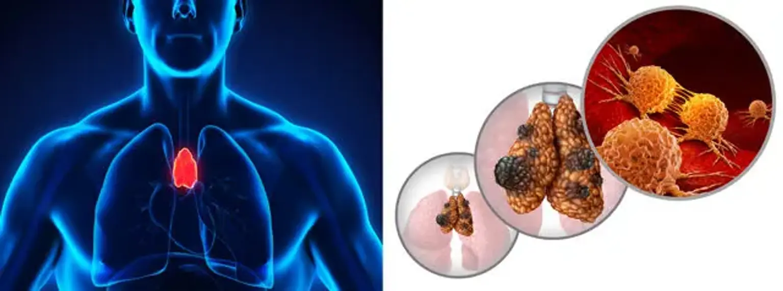
The thymus gland is part of the chest organ located under the breastbone. It forms part of the lymphatic system in the immune system and releases white blood cells referred to as lymphocytes. These cells are responsible for protecting the body by fighting off foreign organisms.
However, the thymus gland is sometimes susceptible to two different types of cancers, including thymoma and thymic carcinoma. Although rare, thymus cancer can develop outside the thymus surface. Unlike thymoma, thymic carcinoma tends to be highly aggressive and difficult to address.
The thymus is a bilobed encapsulated gland. Each thymus lobe contains a superior and inferior horn and extends laterally to each phrenic nerve. The thymus receives blood from the internal mammary, inferior thyroid, and pericardiophrenic arteries. It is emptied into the superior vena cava by tributaries from the innominate vein (SVC).
The thymus is essential for the development of the adaptive immune system and typically involves into a fibrofatty tissue after puberty. There is a strong link between myasthenia gravis and thymomas.
Thymic tumors are a diverse category of tumors that range from very benign thymomas to extremely aggressive carcinomas. Over 90% of all thymic tumors occur in the anterior mediastinum, with the remainder occurring in the neck or other mediastinal areas, particularly the aortopulmonary window and retrocardiac area, which are common sites for ectopic thymic tissues and may explain why simple thymectomy fails to improve Myasthenia Gravis in some cases (MG).
Incidence and Mortality
Thymus cancer is a relatively uncommon tumor, accounting for 0.2 to 1.5 percent of all malignancies. Thymoma has an overall incidence of 0.13 cases per 100,000 person-years. The five-year survival rate for inoperable, locally advanced cancer is 36%; for metastatic thymoma and thymic carcinoma, the five-year survival rate is 24%.
Thymomas and thymic carcinomas are the most prevalent anterior mediastinal masses. They account for around 20% of all mediastinal neoplasms. They are most frequent between the fourth and sixth decades of life. There is no sexual or racial preference.
Causes of Thymus Cancer
There are no specific thymus cancer causes. However, medical experts believe that it’s linked to a rare genetic health condition known as multiple endocrine neoplasia type 1 (MEN1). This means that people with this condition are at a higher risk of developing thymus cancer. Furthermore, thymoma is often associated with various autoimmune diseases.
Thymomas are strongly linked to myasthenia gravis and other paraneoplastic disorders include complete red cell aplasia, polymyositis, systemic lupus erythematosus, Cushing syndrome, and syndrome of inappropriate antidiuretic hormone production.
Pathophysiology
A thymoma is a kind of malignant epithelial tumor that most usually occurs in the prevascular mediastinum. It can also be present in the neck, the pulmonary hilum, the thyroid, the lung, the pleura, and the pericardium. On the surface, it appears to be a well-circumscribed, tan, hard mass ranging in size from microscopic to more than 30 cm in diameter. It is lobulated with fibrous stromal bands and, occasionally, cystic alterations.
In contrast to thymomas, thymic carcinomas lack a lobulated morphology. They have carcinoma-like cytoarchitecture and lack immature T cell lymphocytes.
Thymic carcinomas and thymomas can occur at the same time. Thymomas can sometimes grow into carcinomas, although it takes 10 to 14 years for this to happen. They are big, hard, penetrating masses with multiple regions of cystic alteration and necrosis on the surface. It is encapsulated for only 15% of the population.
Thymoma
Thymoma is a form of tumor that develops from the thymus' epithelial or lining cells. Thymic neoplasms are tumors of the thymus that include thymomas and thymic carcinomas. Thymoma is a term used to describe tumors of the thymus that develop slowly and typically do not extend beyond the thymus.
Many people with thymoma will develop a condition known as a paraneoplastic syndrome. A paraneoplastic condition develops before or is concomitant with the detection of a thymoma. These disorders occur in conjunction with the development of cancer but are not a direct outcome of the disease, as a lump or pain could be.
They appear to be an indirect effect of cancer and may or may not improve when the underlying disease is treated. Myasthenia gravis, a muscular disorder, is the most usually related ailment with thymoma. A thymoma affects 20% of people with myasthenia gravis.
Thymoma is a term used to describe tumors of the thymus that develop slowly and typically do not extend beyond the thymus. Thymic carcinomas are cancers of the thymus that develop rapidly and can spread to other organs.
A thymoma affects less than one person in every 1.5 million. This indicates that around 400 persons in the United States get thymoma each year. Thymic carcinomas are extremely uncommon, accounting for just 0.06 percent of all thymic malignancies.
Up to half of the thymomas are asymptomatic, which means they generate no symptoms or signs and are diagnosed when a physician does a chest imaging study for another reason. When symptoms do emerge, they may include chest discomfort, shortness of breath, and a cough.
Thymoma classifications
There are several classifications to describe thymomas:
- Type A thymoma. This condition is also known as spindle cell thymoma or medullary thymoma. People with type A thymoma have an excellent probability of recovery. Almost all persons with this kind live for at least 15 years after being diagnosed.
- Type AB thymoma. Type AB thymoma, often known as mixed thymoma, is similar to type A thymoma. Type AB thymoma, on the other hand, has lymphocytes in the tumor. People with type AB thymoma have an excellent probability of recovery as well. Approximately 90% of persons with this kind live for at least 15 years following diagnosis.
- Type B1 thymoma. Lymphocyte-rich thymoma, lymphocytic thymoma, mainly cortical thymoma, and organoid thymoma are all names for this kind of thymoma. This form of thymoma has a large number of lymphocytes, yet the thymus cells appear to be healthy. People with type B1 thymoma have a fair probability of recovery as well. Approximately 90% of persons with this kind live for at least 20 years following diagnosis.
- Type B2 thymoma. Cortical thymoma and polygonal cell thymoma are two more names for type B2 thymoma. This form of thymoma, like type B1, contains a high number of lymphocytes. The thymus cells, on the other hand, do not appear to be in good health. Approximately 60% of persons with this kind live for at least 20 years following diagnosis.
- Type B3 thymoma. Epithelial thymoma, atypical thymoma, squamoid thymoma, and well-differentiated thymic carcinoma are all names for type B3 thymoma. This form of thymoma has few lymphocytes and aberrant thymus cells. Approximately 40% of persons with this kind live for at least 20 years after being diagnosed.
Difference Between Thymoma & Thymic Carcinoma
Thymoma differs greatly from thymic carcinoma. While noncancerous thymus cells resemble thymoma cells, thymic carcinoma cells do not. Thymoma varies from thymic carcinoma in terms of how it spreads (metastasis). Thymoma cells develop slowly and seldom spread to other regions of the body.
Thymic carcinoma cells, on the other hand, proliferate quickly and spread throughout the body. Indeed, there is a good possibility that thymic carcinoma will have spread by the time it is identified. As a result, thymic cancer usually necessitates a more involved treatment plan. It should also be noted that thymoma is more prevalent than thymic carcinoma.
Thymic carcinoma manifests variably. It is more aggressive than thymoma and is frequently coupled with an invasion of other mediastinal structures. Cough, chest discomfort, phrenic nerve palsy, or SVC syndrome are common symptoms. Extrathoracic metastases, which include the kidney, extrathoracic lymph nodes, liver, brain, adrenal glands, thyroid, and bone, are observed in less than 7% of individuals with thymic cancer.
Thymus cancer symptoms
Patients with thymomas or thymic carcinomas present in one of three ways:
- An accidental imaging discovery in an asymptomatic patient
- A patient who is experiencing symptoms as a result of the mass's local compressive effects within the thoracic cavity (e.g., dyspnea, cough)
- A patient experiencing symptoms as a result of a paraneoplastic syndrome
In most cases, the condition is often detected during other tests and examinations. However, if the condition is associated with symptoms, then it can include the following;
- Breathing difficulties
- Prolonged cough
- Chest pain
- Loss of appetite
- Weight loss
- Swallowing difficulties
- Hoarse voice
Some thymoma patients may develop superior vena cava (SVC) syndrome. Pleural and pericardial effusions in thymoma patients are usually caused by widespread illness. Patients with thymomas may also have paraneoplastic symptoms.
Myasthenia gravis
Myasthenia gravis is the most frequent paraneoplastic condition. These paraneoplastic symptoms might appear at any point during the diagnosis and treatment of thymomas. Myasthenia gravis is an autoimmune illness characterized by an immunological assault on acetylcholine receptors located in the neuromuscular junction of skeletal muscle.
Diplopia, ptosis, dysphagia, weakness, or weariness are common symptoms in these people. Approximately half of all thymoma patients have myasthenia gravis, and their condition is frequently less progressed since their symptoms lead to an earlier diagnosis. The severe symptoms of myasthenia gravis are reduced after a thymectomy.
Diagnosing Thymus Cancer
To diagnose thymus cancer, doctors often conduct several tests and procedures depending on the underlying symptoms. They include;
- Physical examination
This is an overall body examination to evaluate thymus cancer. It can include checking for various signs and symptoms associated with the condition, including lumps and anything unusual. If necessary, the doctor can inquire about the overall health history and previous diseases or treatments.
- Chest x-ray
Doctors use an x-ray technique to visualize and take pictures of the organs and bones within the chest. It’s a form of energy beam that can easily pass through the body, creating images of the inner body parts.
- Computerized tomography (CT) scan
This is an imaging technique that creates a collection of detailed images of various sections within the body. It can be the chest images taken at varying angles. A computer connected to the x-ray machine is then used to display the images captured. Sometimes, the doctor can inject dye through the vein or ask the patient to swallow. This is to make the internal organs and tissues more visible and clear.
- Magnetic resonance imaging (MRI)
This procedure uses radio waves, strong magnetic fields, and computer technology. It creates a series of detailed images of the inner parts of the body, including the chest.
- Positron emission tomography (PET) scan
Doctors use this imaging method to check for the cancerous cells within the body. It involves the use of a small amount of radioactive glucose injectable into a vein. After that, the PET scanner rotates throughout the body, creating images of areas where glucose has been used in the body. The spots where there is a presence of the cancerous cells will be bright in the pictures. This is because they are highly active and absorb more glucose, unlike normal cells.
- Biopsy
Doctors can perform a biopsy to diagnose thymus cancer. Sometimes, they can recommend a biopsy before or after a surgical procedure using a small needle to take out some cell samples. This procedure is referred to as fine-needle aspiration (FNA) biopsy. If the doctor uses a wide object to extract a sample of cells, then the process is referred to as a core biopsy.
After extracting the sample, the doctor will use a microscope to view and analyze the cancerous cells. In case they diagnose any cancerous cells, they will run more tests to identify the type of cells in the tumor. For thymoma, there can be more than one type of cancerous cell. Therefore, the doctor will decide to remove a section or the entire part of the tumor through a surgical operation. Depending on the extent of the condition, the lymph nodes and surrounding tissues can also be removed.
In addition to imaging, the patient should have a variety of laboratory tests performed, including germ cell markers (beta-hCG and AFP) and lymphoma indicators (LDH and CBC), as both germ cell tumors and lymphomas are in the differential diagnosis of anterior mediastinal masses.
Pulmonary function tests and a cardiac stress test should also be conducted on any patient who is considering surgical resection to evaluate if they have enough cardiopulmonary reserve to survive a major thymus excision.
Tissue is required for a conclusive diagnosis of a thymic tumor. This can be acquired by surgical resection or with a percutaneous or open biopsy if the tumor is unresectable, neoadjuvant treatment is required, or surgery is contraindicated owing to age or comorbidities. The percutaneous approach is performed under CT guidance with fine-needle aspiration or a core needle biopsy.
Stages of Thymus Cancer
Doctors can sometimes use diagnostic results from various tests and procedures to determine the stage of cancer. Staging refers to the process of determining whether cancer has advanced and spread to other body parts. Generally, it’s highly essential to understand the current stage as this helps plan for the treatment.
In addition, thymus cancer can spread in various ways, including;
Tissue: The cells can spread from the thymus where it starts and invades the surrounding areas.
Lymph system: The cancerous cells can spread from the thymus and get into the lymph system. It can also travel via the lymph vessels to various body parts.
Blood: The tumor cells can spread from the thymus into the blood and travel via the blood vessels to various parts of the body.
Thymus cancer treatment
Chemoradiotherapy, corticosteroids, immunotherapy, tyrosine kinase inhibitors, and surgical excision are all used to treat thymic cancers. The use of multidisciplinary tumor boards is critical in selecting the best treatment option for malignant neoplasms. There has been no randomized controlled study that gives clear management strategy recommendations.
Once the doctor diagnoses cancer, they will use the results to develop a suitable treatment plan. Thymus cancer treatment will also depend on the specific stage of the condition. It can include one or a combination of treatment forms such as;
- Surgery
A surgical procedure to take out the tumor is one of the most common forms of treating thymoma. The aim of surgery is to remove all the resectable cancerous cells visible. However, some patients may still require radiation therapy after the operation. This helps destroy the remaining tumor cells and prevent the condition from returning (adjuvant treatment).
Some individuals have the non-resectable illness, which is characterized by a tumor entering the nominate vein, phrenic nerve(s), heart, and major arteries. These individuals require multimodal treatment, which includes preoperative chemotherapy and postoperative radiation.
- Radiation therapy
Radiation therapy is a type of cancer treatment that utilizes high-energy x-rays and other forms of radiation. It destroys the cancerous cells and prevents them from advancing and spreading to distant organs. Radiation therapy treatment comprises two types, which include;
External radiation therapy: This involves the use of a machine located outside the patient’s body to transmit radiation to the affected area.
Internal radiation therapy: This involves the use of radioactive contents that are sealed in seeds, needles, catheters, or wires. They are directly placed inside the affected area or near the tumor.
Doctors administer radiation therapy depending on the type of cancer and the current stage. However, external radiation therapy is suitable for addressing both thymoma and thymic carcinoma.
- Chemotherapy
Chemotherapy is another type of cancer treatment. It involves the use of drugs to prevent or slow down the growth of cancerous cells. This could be by destroying the cells or preventing them from multiplying.
Doctors usually administer chemotherapy orally into the mouth or by injecting it into the vein or muscle. Once in the body system, the drugs get into the bloodstream and move to various parts of the body, reaching all the cancer cells. This procedure is known as systemic chemotherapy.
Alternatively, the doctor can insert the drug directly into the body organ, cerebrospinal fluid, or through the body cavity like the abdomen. This process is known as regional chemotherapy since the drug will only destroy the cells within the area.
While there are several ways of administering chemotherapy, the best way usually depends on the type of cancer and the stage to address. Sometimes, doctors can use chemotherapy to shrink the tumor before the main treatment, which can be radiation therapy or surgery. This process is known as neoadjuvant chemotherapy.
- Hormone therapy
Doctors recommend hormone therapy to treat thymus cancer. It removes hormones triggering cell growth from the body or blocks their normal activities that might facilitate growth. Hormones are usually secreted by various glands in the body, after which it enters the bloodstream and circulates throughout the body.
Some of these hormones can trigger certain cancerous cells to grow. Therefore, if diagnostic results indicate that the cells are associated with hormones, the doctor can recommend drugs, radiation therapy, or surgery. This helps minimize hormone production or prevent them from working normally.
Hormone therapy alongside drugs known as corticosteroids can be useful in treating thymoma or thymic carcinoma.
Follow up
Long-term care for thymus cancers usually depends on various factors. It includes the patient’s age, general health state, and the type of treatment used to address the issue. If the treatment is a surgical procedure, it will depend on whether the operation eliminated the cancerous cells. Other factors include the type of tumor cells present and the current stage of the condition.
After a successful treatment, regular check-ups and follow-up visits are essential to monitor and prevent possible side effects. This also helps ensure that the tumors do not come back after treatment.
Thymus cancer patients are still at a high risk of the condition returning. This can sometimes result in anxiety among the patients. To address this, doctors often recommend opting for counseling services to help patients suffering emotionally.
Differential Diagnosis
The differential diagnosis for thymomas and thymic carcinomas include:
- Thymic carcinoid tumors (seen in patients with multiple endocrine neoplasia syndrome type 1)
- Thymic cysts (differentiated from solid tumors by MRI),
- Non-Hodgkins lymphomas,
- Ectopic parathyroid glands (identified by a sestamibi scan),
- Thyroid goiters, and
- Paragangliomas.
To narrow the differential, suitable laboratory procedures such as germ cell markers (AFP and hCG) and lymphoma markers must be ordered (LDH and CBC with differential). Imaging investigations can assist in distinguishing between these masses; for example, an MRI can distinguish between a cystic and solid mass, a sestamibi scan can show ectopic parathyroid tissue, and a thyroid scan can detect a mediastinal thyroid goiter.
Prognosis
Thymomas are neoplasms that develop slowly. The invasiveness of the tumor is the most reliable unfavorable prognostic sign. Thymic carcinomas, on the other hand, are more aggressive and have a worse prognosis than thymoma. The prognosis is determined by the stage of the disease and the tumor's total resectability.
Annual imaging after therapy is suggested, despite the fact that no clinical trials have demonstrated that such imaging is beneficial. A CT chest is advised every six months for two years, yearly for five years in situations of thymic cancer, and 10 years in cases of thymomas. Thymic tumors typically return as pleural nodules, with a mean duration of 8 to 68 months after therapy.
Thymic tumors are associated with an increased chance of developing secondary cancer (approximately 17 percent to 28 percent risk after thymectomy). There is an elevated risk of B cell non-Hodgkin lymphoma, gastrointestinal malignancies, and soft tissue sarcomas.
Complications
Complications include radiation pericarditis, radiation pneumonitis, and pulmonary fibrosis. These typically develop after postoperative radiation and can result in patient harm. As a result, doctors must carefully examine a risk-benefit analysis before considering adjuvant radiation.
Conclusion
Thymus cancers are a rare health condition that begins in the thymus. However, people with diseases such as Rheumatoid Arthritis, Systemic lupus erythematosus (SLE), or myasthenia gravis can get cancer.
The two main types of thymus cancer include thymoma and thymic carcinoma. Thymoma is common and mainly affects individuals aged 40 to 60, while thymic carcinoma can develop at any age.
A screening CT scan of the chest should be performed on all individuals with myasthenia gravis to search for thymoma or thymic hyperplasia. Thymectomy has been reported to improve symptoms in these people.
Patients with thymoma or thymic carcinomas will need to visit medical and oncology specialists. In individuals with myasthenia gravis, a neurology consult should be scheduled both before and after surgery. In individuals with myasthenia gravis, anesthesiology should be contacted prior to any scheduled thymectomy.
To select effective treatment techniques, thymomas should always be treated by a multidisciplinary team. A thoracic surgeon should be engaged in all cases of anterior mediastinal masses and thymomas; however, other experts, such as medical and radiation oncologists, as well as a neurologist, may be required in patients with established myasthenia gravis.
These neoplasms frequently occur asymptomatically and are discovered inadvertently. The symptoms are caused by local effects of the tumor or paraneoplastic diseases. Further imaging and laboratory techniques are used to narrow the differential diagnosis.