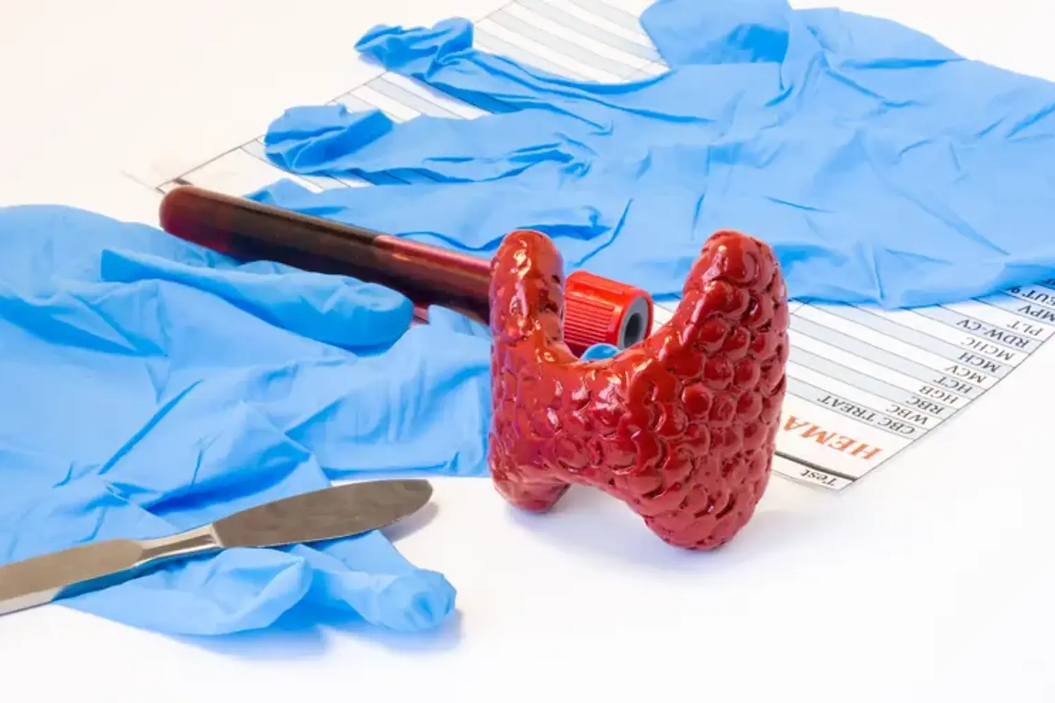
Thyroidectomy
Overview
Thyroid cancer arises from the thyroid gland, which is part of the endocrine system. Hormones produced by the thyroid gland govern body temperature, heart rate, and metabolism. The most prevalent kinds of thyroid cancer, papillary and follicular, respond quite well to therapy. The majority of thyroid malignancies are treatable. The most frequent therapy for thyroid cancer is surgery. Depending on the size and location of the tumor, your surgeon may remove a portion of the thyroid gland (lobectomy) or the entire gland (thyroidectomy).
What is Thyroidectomy?
Thyroidectomy is a well-documented surgery for removing the thyroid gland. It is a popular technique in modern medicine and can be used to treat cancer, benign illness, or hormonal disease that is resistant to conventional treatment.
Thyroidectomy is a difficult treatment to execute safely and successfully due to the delicate architecture of the anterior neck, the vital importance of neighboring tissues, and restricted working spaces. Thyroidectomy as a surgery has progressed along with anatomic understanding and surgical procedures. Billroth and Kocher pioneered the conventional thyroidectomy in the 1870s and reported an 8 percent mortality rate, which was considered a considerable success at the time.
Anatomy of the thyroid gland
Thyroid hormone, a regulatory hormone with numerous vital physiologic activities, is secreted by the thyroid gland, an endocrine gland. Both hypothyroid and hyperthyroid states cause a non-specific constellation of symptoms, and thyroid dysfunction is a factor in the differential diagnosis for a similarly large number of symptoms. Despite this, hypothyroidism is traditionally associated with cold sensitivity, dry skin, lethargy, constipation, pretibial edema, and weight gain.
Weight loss, heat sensitivity, osteoporosis, muscular weakness, muscle tremor, brittle hair or hair loss, and atrial fibrillation are all symptoms of hyperthyroidism. The thyroid gland is surrounded by four parathyroid glands, the major function of which is to regulate blood calcium levels through the release of parathyroid hormone (parathermone, PTH).
The thyroid gland has two lateral lobes and a central isthmus with or without a pyramidal lobe when completely formed (40-50 %). The thyroid is located in the deep cervical fascia's middle layer and is connected to the thyroid and cricoid cartilages via the anterior suspensory ligament from its superior-medial aspect. Berry's ligament connects the posterior-medial aspect to the first and second tracheal rings, as well as the cricoid cartilage. The thyroid gland is covered anteriorly by the sternohyoid and sternothyroid muscles. The Zuckerkandl tubercle protrudes from the posterior and lateral aspects of the thyroid lobes.
The thyroid gland is a highly vascular organ. The superior thyroid artery, a branch of the external carotid artery, and the inferior thyroid artery, a branch of the thyrocervical trunk, are the principal arterial contributions.
Types of Thyroidectomy
- Hemithyroidectomy — Entire isthmus is removed along with 1 lobe. Done in benign diseases of only 1 lobe.
- Subtotal thyroidectomy — The most common surgery for multinodular goitre was to remove the majority of both lobes, leaving behind 4-5 grams (equivalent to the size of a normal thyroid gland) of thyroid tissue on one or both sides.
- Partial thyroidectomy —Removal of gland in front of trachea after mobilization.
- Near total thyroidectomy — Except for a little quantity of thyroid tissue (on one or both sides) around the recurrent laryngeal nerve entrance point and the superior parathyroid gland, both lobes are excised.
- Total thyroidectomy — The whole gland is extracted. Done in situations of papillary or follicular thyroid cancer, as well as medullary thyroid carcinoma. This is now the most common procedure for multinodular goiter.
Indications for Thyroidectomy
Thyroid nodules, hyperthyroidism, obstructive or substernal goiter, differentiated (papillary or follicular) thyroid cancer, medullary thyroid cancer (MTC), anaplastic thyroid cancer, primary thyroid lymphoma (surgery is limited to obtaining tissue biopsy), and metastases to the thyroid from extrathyroidal primary cancer can all be treated with thyroidectomy (most commonly renal cell and lung cancer).
Thyroid nodules are a global phenomena that affects 1% of men and 5% of women clinically. The vast majority of nodules are benign, with about 5% signifying malignancy. Thyroid nodules can be found by high-resolution ultrasound in up to 68 % of randomly selected people who have a screening ultrasound, with a preference for women and the elderly.
A goiter is an abnormal development of the thyroid gland that can be diffuse or nodular. Goiter is associated with iodine deficiency and is so endemic in iodine-deficient parts of the world. A goiter can be identified in around a quarter of the population in asymptomatic, iodine-deficient individuals, with an increasing prevalence in elderly populations. The vast majority, however, will not become surgical candidates or acquire thyroid nodules that need surgery.
Goiter can also arise in areas where there is no substantial iodine deficit. Goiter is often multinodular in certain areas and may be due to thyroid autoimmune illnesses such as Hashimoto's thyroiditis or Graves' disease.
Thyroidectomy may be justified in both malignant and benign diseases with high selectivity. Thyroid cancer, toxic multinodular goiter, toxic adenomas, goiter with compressive symptoms, Graves disease that is either not responsive to medical treatment or for whom medical management is not recommended, such as those attempting to become pregnant, are examples of indications.
The majority of thyroid nodules do not require removal. Fine needle aspiration (FNA) is frequently used to differentiate between benign and malignant nodules in nodules that are at high risk of cancer. When nodules are larger than 1 cm in size, non-functional (referred to as a "cold" nodule), and/or exhibit worrying ultrasonography results, they often fulfill the requirements for biopsy. Several associations have published treatment strategies for thyroid nodules.
Contraindications
Because of the risk of intraoperative or postoperative thyroid storm, uncontrolled severe hyperthyroidism (ie, Graves disease) is a relative contraindication to surgery. Although thyroidectomy can be performed during pregnancy for malignancy, many experts recommend deferring surgery until after birth if feasible due to anaesthetic risks to the fetus. Surgery during pregnancy is indicated for severe malignancies or airway impairment. If elective thyroid surgery is performed during pregnancy, it should ideally be done during the second trimester.
How should I prepare for thyroid surgery?
- Thyroid imaging:
For first thyroid imaging, ultrasonography is the gold standard. In certain circumstances, other imaging modalities such as computed tomography (CT) or magnetic resonance imaging are used to determine the degree of advanced illness. In the presence of aggressive thyroid cancer, such as anaplastic carcinoma or advanced or recurring papillary thyroid carcinoma, positron emission tomography can be used.
In the case that the thyroid extends below the thoracic inlet, a chest CT scan can help determine whether a sternotomy approach is necessary. Extension of the nodule beyond the aortic arch or into the posterior mediastinum, presence of mediastinal nodal disease, capsular calcification (implying difficult soft tissue dissection), and conical shape of the goiter are all associated with the need for sternotomy. Even with these risk factors, sternotomies are extremely rare.
- Laboratory testing:
A blood thyroid-stimulating hormone (TSH) level is required for all patients to identify whether they are euthyroid, hyperthyroid, or hypothyroid prior to surgery. Thyrotropin receptor antibodies (TRAb) should be collected in individuals with serologic evidence of hyperthyroidism to rule out Graves disease.
If the TRAb is negative but there is a nodule or nodules on ultrasound, thyroid scintigraphy should be performed to rule out a toxic adenoma or toxic multinodular goiter. Thyroid peroxidase antibodies (TPO) can be collected in the context of hypothyroidism to rule out Hashimoto thyroiditis. Testing for suspected medullary thyroid cancer may include calcitonin, carcinogenic embryonic antigen (CEA), and genetic testing for medullary thyroid cancer, similar to that used for multiple endocrine neoplasia type 2.
- Laryngeal examination:
Thyroid surgery can cause vocal cord paresis or paralysis if the recurrent laryngeal nerve (RLN) is injured. Pre-operative voice evaluation is advised, with dedicated, flexible fiberoptic laryngoscopy considered if vocal anomalies are discovered. Outpatient thyroidectomy is usually regarded as safe for selected patients and an experienced surgeon, although the ultimate decision is up to the operating surgeon.
The patient is positioned on the operating table supine, and general anesthesia is administered. If such monitoring equipment is intended, the patient is intubated with a nerve-monitoring endotracheal tube (ETT). The ETT should be appropriately positioned with the recurrent laryngeal nerve monitoring sensors situated between the vocal cords, and the monitor's operation should be validated following intubation according to the device manufacturer's recommendations.
The patient is then put with the neck hyperextended; a shoulder roll may be used to aid in this posture. Depending on the surgeon's inclination, the head may be taped or padded to the bed. Extending the neck and resting the head on a donut cushion will also aid with surgical exposure. The thyroid cartilage, cricoid cartilage, thyroid lobes, and sternal notch are all palpated to identify anatomic markers.
The sternal notch and the mandible serve as markers to define the anatomic midline. The cricoid cartilage and thyroid notch have been identified. If feasible, a surgical incision is made in a natural skin fold about 2 cm above the sternal notch. A normal incision is 4-6 cm long; larger incisions may be necessary for massive thyroidectomy or if concomitant lateral neck dissection is required. The neck is next scrubbed with surgical scrub, and the patient is draped with care to keep the anatomic markers visible.
If such monitoring equipment is employed, a recurrent laryngeal nerve monitor is linked to the leads from the ETT and proven to be operational. The ETT uses EMG input from the vocalis muscles to monitor the RLN's function. When changes in nerve output to the vocalis muscles occur or when the surgeon uses a nerve probe to activate the RLN, the monitor may sound an alert. Postoperative vocal cord paresis or paralysis affects 83 to 85 percent of individuals who had intraoperative signal loss.
Surgical Technique
The skin, subcutaneous tissue, fat, and platysma are all incised. Following that, skin flaps are raised deep to the platysma and superficial to the sternohyoid muscle. Superior and inferior flaps are lifted to the level of the thyroid cartilage and sternal notches, respectively. To minimize needless bleeding, the anterior jugular veins and the superficial network of veins under the platysma should be identified and preserved.
By incising the median raphe, the strap muscles (sternohyoid and sternothyroid) are lateralized until the thyroid capsule is discovered. The strap muscles may infrequently necessitate the horizontal separation to increase exposure during the excision of a big multi-nodular goiter. If these muscles must be divided, it should be done as high as feasible to retain the innervation of the strap muscles by the ansa cervicalis.
When doing a complete thyroidectomy, it is best to start on the smaller side if the diagnosis is proven malignant or on the bigger side if the condition is benign. In the case of an intra-operative complication or nerve injury necessitating an early termination of the treatment, this enables for the removal of the most critical tissue prior to the surgery's conclusion. It should be emphasized that there are several techniques of thyroidectomy, just one of which is discussed in depth here.
Sharp dissection of the thyroid gland's surface loose areolar tissue advances laterally until the carotid sheath is discovered. This can be accomplished by precise finger dissection or through tissue retraction and bipolar cautery. This describes the dissection's lateral extent. The lobe is moved gently toward the midline, and the central thyroid vein is located, ligated, and split (this is sometimes performed after both poles are addressed). After that, the lobe is retracted anteromedially to reveal the superior thyroid artery and vein.
Once skeletonized, they are ligated and separated as near to the gland as feasible in order to minimize harm to the superior laryngeal nerve (SLN). In all thyroidectomies, some surgeons want to firmly identify the SLN. The superior parathyroid glands should be identified and preserved throughout this dissection. Following the identification of the inferior parathyroid glands, the inferior thyroid vein is ligated and split as it approaches the thyroid. The lobe is then retracted medially to reveal the recurrent laryngeal nerve.
The recurrent laryngeal nerve is almost often found within a few millimeters of the inferior thyroid artery, however it can be superficial or deep. It should be observed that as the gland is dragged anteriorly and medially to allow nerve dissection, the nerve is pulled onto the body of the thyroid, forming a genu. Once the nerve has been found and freed, it returns to its anatomic position. Once found, the nerve can be carefully dissected to its entrance point into the larynx at the cricothyroid joint, while the thyroid lobe is dissected free of the recurrent laryngeal nerve.
Parathyroid glands can also act as markers during the search for the recurrent laryngeal nerve. The superior gland is often situated along the posterior portion of the thyroid capsule in a plane deep (dorsal) to the RLN plane. The inferior glands vary in position but are assumed to be anterior (ventral) to the RLN plane. Once these components have been identified and conserved, the gland can be raised and Berry's ligament separated or cauterized using bipolar cautery to liberate the gland.
Berry's ligament should be separated as close to the trachea as feasible without entering the trachea. Elevating the gland to the midline is recommended. The isthmus can be tied off with a surgical knot or separated using a device such as a harmonic scalpel during a hemithyroidectomy. The first dissected lobe can be removed to maximize working space in the neck during a complete thyroidectomy, or it can be left in place so that the entire thyroid can be removed en bloc.
In some cases, the surgeon may consider transmitting intraoperative frozen sections. Frozen sections are less useful in Bethesda criteria 3 and 4 nodules because to advancements in molecular testing. They may be considered in Bethesda 5 nodules (those suspected of malignancy) to determine whether a complete thyroidectomy should be performed, particularly in elderly patients or those with comorbidities, and in noncompliant patients with surgeon concern for follow-up.
The surgical field is checked for hemostasis prior to closure. The separated strap muscles and platysma are re-adjusted with absorbable suture such as 3-0 Vicryl, followed by skin re-adjustment. Many surgeons choose to insert a drain to track and trend outflow. Drains implanted after a thyroidectomy with neck dissection can also be used to check for chyle leak, which can cause the drain to appear milky.
If a parathyroid gland is found to be devascularized intraoperatively, it should be re-implanted to reduce the risk of postoperative hypoparathyroidism. A tiny part of the material can be analyzed by pathology using a frozen slice prior to re-implantation to confirm the tissue is actually parathyroid tissue. Alternatively, put the material in a small cup of saline. Fat floats, whereas parathyroid tissue sinks because to its larger density. After that, the gland should be morselized and placed in a nearby sternocleidomastoid or strap muscle. To indicate the re-implantation pocket, a surgical clip should be put over it.
This enables for simpler identification if the patient develops hyperparathyroidism later on and the material must be removed. If a parathyroid gland seems significantly enlarged intraoperatively in comparison to the other parathyroid glands, and if a parathyroid adenoma is suspected, an intraoperative PTH level may be taken. The surgeon may then elect to remove the suspect parathyroid gland. Prior to resection, a frozen section of this gland may be sent for pathology confirmation.
What is recovery like after thyroid surgery?
You may experience a temporary sore throat, neck pain, difficulty swallowing, or a weak voice following your thyroidectomy or thyroid lobectomy. Your food will be restricted for the evening of your operation, but it should be back to normal the next day in most circumstances. We'll plan a follow-up visit, give you instructions for at-home recuperation, and go through any recommended medicines before you leave the hospital.
Most individuals are able to return home the day after surgery, but it takes around two weeks to recuperate. Heavy lifting and other duties that might strain your neck should be avoided for up to three weeks after surgery. Soaking or cleaning the incision site is also avoided for at least one week to allow it to heal properly. Showering is usually permitted after around one day.
The pain at the location of your incision will go away after a few days, although it may last a week or more. Contact our office if you detect rapid swelling in your neck, which might indicate an infection.
Your calcium level may decline after surgery due to disruption of the parathyroid glands, which maintain calcium balance. You may get numbness and tingling in your fingers or around your lips if it drops. We'll check your calcium levels with blood tests and provide you recommendations on how to take calcium supplements if necessary.
Complications
- Hemorrhage: severe cases may cause airway compression and be life-threatening
- Hypoparathyroidism: results in hypocalcemia, which can be symptomatic and life-threatening if left untreated. Up to one-third of individuals following complete thyroidectomy will experience temporary hypocalcemia after surgery. To avoid problems, it is critical to follow a consistent calcium management plan following a whole or complete thyroidectomy.
- Injury to the recurrent laryngeal nerve causes hoarseness and, in certain cases, aspiration. This is usually transient, but in 1% of cases, it might be permanent.
- Injury to the superior laryngeal nerve causes a shift in voice pitch. The reported rates of injury range from 0% to 58%.
- Post-surgical infection: approximately 6% of cases.
- Esophageal injury
- Tracheal injury
- Horner syndrome
- Dysphagia
Conclusion
Thyroidectomy is the surgical removal of the entire or a portion of the thyroid. It is the primary therapy for the vast majority of thyroid tumor patients. Thyroid surgery options may include partial thyroid removal (also known as a thyroid lobectomy or hemithyroidectomy), complete thyroidectomy, or total thyroidectomy plus lymph node removal.