Tibial Osteotomy

Last updated date: 17-Jun-2023

Originally Written in English

Tibial Osteotomy

High tibial osteotomy (HTO), also known as tibial osteotomy, was first suggested by Jackson and Waugh and has been popularized by Coventry since the 1960s as a treatment for medial compartment osteoarthritis of the knee with varus distortion. HTO has two goals: to alleviate knee pain by moving weight-bearing stresses to the barely affected lateral compartment in varus knees and to prevent the need for a knee replacement by halting medial joint compartment degeneration. Although the use of HTO has decreased in recent years due to advancements in knee arthroplasty, it is clear that proper patient selection, careful surgical planning, and a variety of operative procedures can all contribute to positive HTO treatment outcomes. The selection of opening vs. closing wedge HTO, graft choice in opening wedge HTO, a form of fixation, comparative benefits over unicompartmental knee arthroplasty, and the effect of HTO on future knee arthroplasty are the main areas of HTO controversy.


Tibial Osteotomy Indications and Contraindications

A successful HTO relies on proper patient selection. The most common cause of HTO is primary or secondary medial compartment degenerative arthritis. A person between the ages of 62 and 65 with isolated medial osteoarthritis, a varus deformity, good range of motion, and no ligamentous instability is a suitable candidate for HTO.
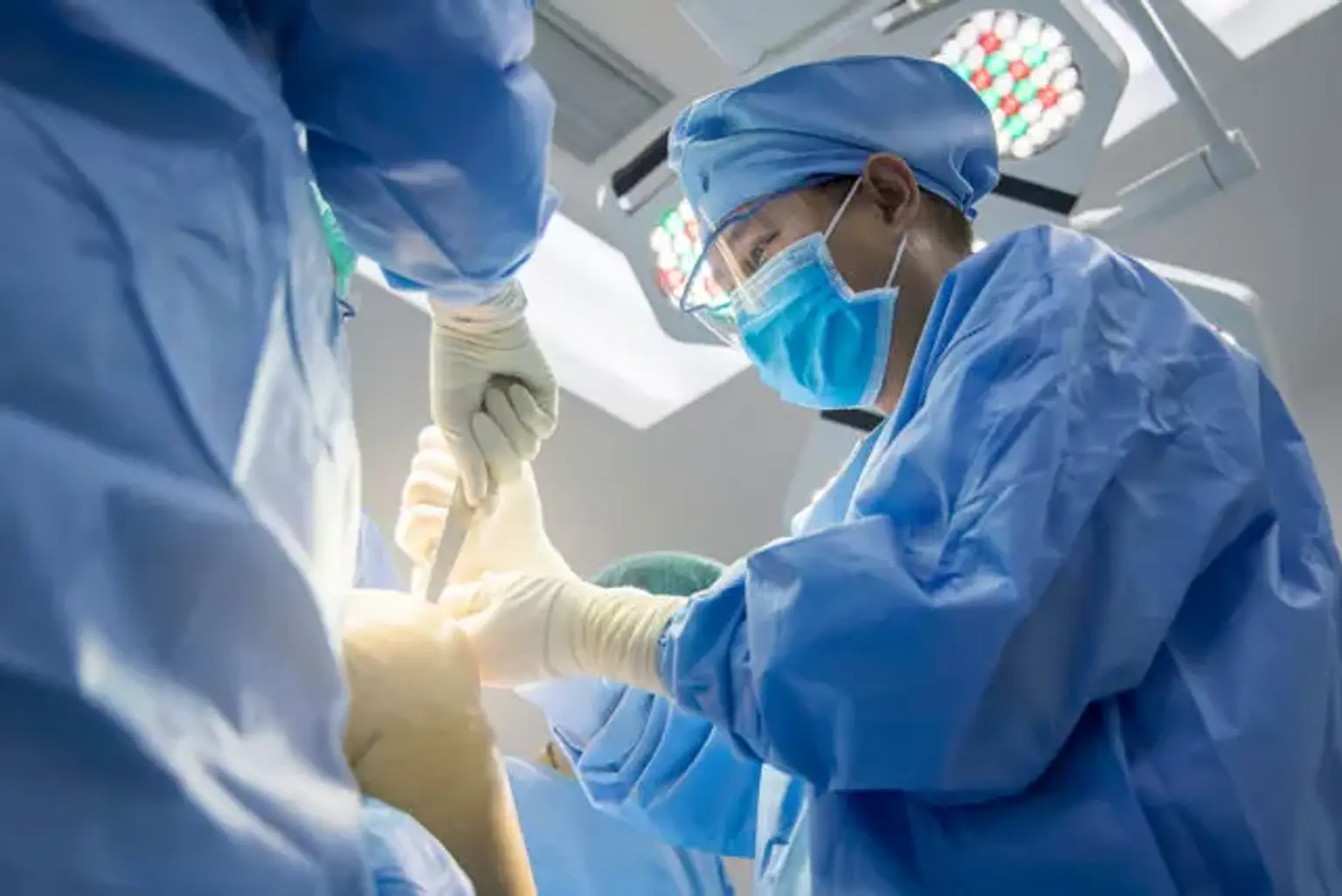
Severe joint destruction, 65 years of age, extensive patellofemoral arthritis, 90 degrees of ROM, 15 degrees of flexion contracture, joint problems and 1 cm lateral tibial push, 20 degrees of correction, and rheumatoid arthritis are all poor prognostic markers.

Although most writers agree that HTO is better than unicompartmental knee arthroplasty for overweight patients, the impact of BMI on HTO outcomes is yet unknown.


Preoperative Planning

Patient Assessment

Tibial Osteotomy Patient Assessment

Before choosing surgery, the patient's age, profession, degree of exercise, past history of knee surgical procedure, and expectations should all be considered. For heavy smokers, closing wedge HTO may be more effective than opening wedge HTO in lowering the risk of nonunion.

Physical examination should be used to determine the range of motion, degree of deformity, ligamentous laxity, and leg length disparity. Valgus HTO can be utilized to treat modest or moderate medial instability caused by osteoarthritis of the medial compartment. The condition of the hip joint can have an impact on the ipsilateral knee's medial osteoarthritis. During the stance phase, hip abduction raises stress on the lateral compartment of the knee, causing the stabilizers (gluteus maximus, tensor fascia lata, and biceps femoris) to become involved, resulting in increased forces on the lateral knee. As a result, weakening or limitation of the hip abductor muscle, as well as ankylosis of the hip joint, should be addressed before HTO.


Radiographic Assessment

For preoperative radiographic examination, a variety of views should be obtained, including bilateral weight-bearing anterior-posterior views in full stretch, tunnel views with the knee in 30° of flexion, Rosenberg views with the knee in 45° of flexion, lateral views, and skyline views. From the anterior-posterior views, the degree of medial osteoarthritis and bone loss can be assessed, and patellar height can be estimated from the lateral views using the Insall-Salvati, Blackburne-Peel, or Caton-Deschamps index. In cases of severe patella alta, a combination of tibial tubercle osteotomy and closing/opening HTO may be required. Full-length radiographs of the lower extremity, which show the alignment of the hip, knee, and ankle joints, can be used to examine lower limb alignment. Intraosseous diseases, meniscal tears, ligamentous lesions, osteochondral abnormalities, osteonecrosis, and subchondral edema can all be detected with magnetic resonance imaging.


Correction Angle Calculation

Correction Angle Calculation

The mechanical axis, a line connecting the dots, is 0° in normal lower extremities, with the center of the hip aligned with the center of the knee and the center of the ankle. In most studies, the optimal postoperative lower limb alignment is 4°-5° valgus from the mechanical axis or 9°-10° anatomical valgus. The postoperative mechanical axis should travel through the lateral one-third of the tibial plateau, according to Fujisawa et al. According to jakob and Jacobi, the mechanical axis should be corrected depending on the thickness of the cartilage in the medial compartment: if one-third of the medial cartilage is destroyed, the mechanical axis should be moved 12-15% lateral from the tibial plateaus' center. The axis should pass 22-25 percent lateral if two-thirds of the cartilage is removed, and 32-35 percent lateral if all of the cartilage is lost. The weight-bearing line (a line connecting the center of the femoral head and the center of the tibiotalar joint) is located at 62 percent between the medial and lateral compartments of the proximal tibia, which is slightly lateral to the lateral tibial spine and 4°-5° valgus from the mechanical axis, as explained by Dugdale et al. Under-correction can lead to a recurrence of the varus deformity, while overcorrection can lead to poor cosmetic and functional consequences.

The correction angle (alpha) is described in closing wedge HTO as an angle produced by a line drawn from the 62 percent point of the tibial plateau width to the center of the femoral head and another line to the middle of the ankle. The proximal osteotomy line is 2-3 cm inferior to the joint line and parallel to the articular surface. The angle is used to calculate the distal osteotomy line, and the wedge bone between the osteotomy lines is excised.

The opening wedge HTO is planned in the same way that the closing wedge HTO is. The proximal osteotomy line runs from a point 3-4 cm inferior to the medial knee joint line to the tip of the fibular head, from which the angle draws another line of the same length obliquely. During operation, the line that passes between the endpoints of each line is used for exposure.

Attempts should be made to maintain the prior anatomical posterior tibial slope in HTO for medial compartment osteoarthritis without knee instability.

The use of navigation systems has been shown in certain recent clinical investigations to improve the precision, accuracy, and reproducibility of HTO.


Medial Opening Wedge Osteotomy

Advantages and Disadvantages

Medial Opening Wedge Osteotomy

A single osteotomy and a few dissections are all that are required for a medial opening wedge osteotomy. The procedure does not require a fibular osteotomy, which has been linked to neurovascular problems, or lateral tibia bone excision. As a result, the normal anatomical tibial bone structure is preserved following the treatment, allowing for knee replacement conversion. Shortening of the lower limbs can be avoided by identifying and adjusting the level of correction on the coronal and sagittal planes intraoperatively.

The procedure, however, has been linked to a high rate of nonunion, a protracted period of weight-bearing limitation, and leg-lengthening. Other drawbacks include morbidity at the harvest site, loss of correction due to insecure fixation, and an increase in posterior tibial slope due to the metal plates' anterior position.


Candidates to Medial Opening Wedge Osteotomy

In knees with a 2 mm leg length disparity, a simultaneous posterior cruciate ligament injury, patella alta, or medial collateral ligament instability, medial opening wedge osteotomy may be more successful than closing wedge HTO.


Surgical Technique

Tibial Osteotomy Surgical Technique

A tourniquet is administered to the patient, who is positioned supine on a radiolucent operating table. An arthroscopy can be utilized to identify and treat intra-articular lesions at the same time. Below the joint line, a 7-cm vertical cut is made between the medial side of the tibial tuberosity and the posteromedial aspect of the tibia. To reveal the superficial medial collateral ligament, the pes anserinus is separated from the tibia. To protect the neurovascular structures posterior to the cut line, the exposed ligament is detached from the bone and a blunt retractor is introduced posterior to the medial collateral ligament and the tibia. Subperiosteal dissection is conducted from the tibial tuberosity to the posteromedial portion of the tibia after finding the medial boundary of the patellar tendon. Two guide wires are inserted 3-4 cm below the medial joint line and obliquely traversed 1 cm below the lateral articular border of the tibia to the tip of the fibular head. An oscillating saw or an osteotome is used to execute a tibial osteotomy directly below the guide wires after confirming the right placement with a fluoroscope. Make that the osteotomy line runs parallel to the posterior tibial slope on the sagittal plane and continues from the tibial tuberosity along the posteromedial aspect of the tibia to 1 cm medial to the lateral tibial cortex. The mobility of the osteotomy site is checked and therefore the osteotomy is opened with a valgus force. If the opening of the osteotomy seems insufficient, use 2 or 3 stacked osteotomes to minimize the danger of intraarticular fractures. Subsequently, a calibrated wedge is inserted until the osteotomy is opened to the wanted extent. 

Ensure with fluoroscopy when a long alignment rod or wire cable is centered over the hip joint and the ankle joint, it lies at 62.5% of the width of the tibial plateau.

Internal fixation of a steel plate is done once the appropriate degree of correction has been attained. Metal plates come in a variety of shapes and sizes, such as the Puddu plate, Tomofix, Aesculap plate, and plates with or without a spacer. The most frequent of them are spacer plates, and the metal block should be the same as the calibrated wedge. Using fluoroscopic guidance, the proximal fixation screws should be used, and the defect should be repaired with an iliac crest autograft, allograft, or a bone replacement. Corticocancellous autografts or allografts are used for defects less than 10 mm, while bone grafting is optional for minor defects.


Lateral Closing Wedge Osteotomy

Advantages and Disadvantages

Lateral Closing Wedge Osteotomy

For treatment around the maximum point of deformity, lateral closing wedge osteotomy is successful. Because of the wide contact surface of cancellous bone at the osteotomy site, early weight-bearing and rehabilitation, and the utilization of quadriceps femoris muscle action, the procedure allows for a quick bone union. Furthermore, the articular surface can be seen through the surgical incision, and the danger of corrective loss is minimal.

However, the operation necessitates a fibular osteotomy or proximal tibiofibular joint release, which might lead to neurovascular problems. Shortening of the lower limb can result from lateral bone excision. Furthermore, due to proximal tibial deformity and lateral condyle bone loss, stem impingement or metal augmentation is inevitable in a subsequent complete knee replacement.


Candidates to Lateral Closing Wedge Osteotomy

Patients with heavy smoking, diabetes, or long-term use of steroids, all of which have been linked to a higher risk of nonunion, may consider lateral closing wedge osteotomy. Furthermore, when patella infera is prominent, the patient does not want bone grafting, or anterior cruciate ligament restoration is required, the treatment can be conducted.


Surgical Technique

Tibial Osteotomy Surgical Technique

Although there are several variations of lateral closing wedge osteotomy, they all follow the same principles. A transverse, longitudinal, or L-shaped cut can be made on the skin. Because tibial osteotomy and partial excision of the inferomedial fibular head can be performed along the same incision line, a longitudinal cut allows for easier conversion to knee replacement, but a transverse cut minimizes the possibility of peroneal nerve palsy. Although the osteotomy can be done below the tibial tubercle, the proximal tibial tubercle osteotomy has more advantages: the metaphyseal osteotomy promotes early bone union, correction can be done adjacent to the deformity, and the quadriceps femoris muscle force on the osteotomy site improves knee stability. To expose the proximal tibiofibular joint, a 10-cm vertical cut is made and the tibialis anterior muscle of the proximal tibia is lifted. Joint disruption, fibular osteotomy at a point 16 cm distal to the fibular head, and removal of the fibular head are all options for treating the proximal tibiofibular joint.

To preserve the neurovascular systems, a retractor is put under the lateral edge of the patellar tendon. Another retractor is implanted along the posterolateral aspect of the tibia. The osteotomy is started 2 cm below the tibias' lateral articular surface. To avoid the patellar tendon, the proximal osteotomy should be done parallel to the articular surface and the distal osteotomy should be cut obliquely. An angular cutting guide can be used to cut the lateral bone wedge. To limit the likelihood of intraarticular fractures, the lateral cortex and wedge should be correctly removed with a saw, rongeur, curette, or osteotome, while leaving 3-6 mm of the opposite cortical hinge intact. When the osteotomy is closed, the medial tibial cortex should be perforated with several holes to act as a hinge. To induce plastic deformation, the hinge should be progressively closed. When the proximal cortex of the distal component is overlapped by the distal cortex of the proximal fragment, overcorrection might occur.

Using fluoroscopy, confirm that the wedge has been completely removed. The alignment should be verified after the osteotomy has been closed with valgus force. If the rod is 62 percent of the width of the tibial plateau, an optimal correction, overcorrection of the mechanical plane by 4°-5°, can be regarded attained. 1 or 2 staples from lateral to a medial position anterior to the fibula can be used for fixation. A contoured plate or locking plate can be used for firmer fixation.


Tibial Osteotomy Other Indications

1. Varus malalignment in meniscectomized knee

Long-term loading can cause osteoarthritis in meniscectomized knees. The load on the joint and the deterioration to the cartilage and graft would be reduced if the lower limb alignment was restored. As a result, HTO and meniscal transplants can be done at the same time in young meniscectomy patients.

2. Varus malalignment and chondral resurfacing

After a cartilage repair, such as microfracture, mosaicplasty, osteochondral allograft transplantation, or autologous chondrocyte implantation, lower limb malalignment might lead to poor consequences. The use of chondral resurfacing with HTO at the same time has not been proved to be a viable strategy for improving clinical outcomes. However, a few doctors feel that correcting the varus alignment during chondral resurfacing will help to improve treatment outcomes.


Rehabilitation after Tibial Osteotomy

Rehabilitation after Tibial Osteotomy

Early joint exercises should be begun once adequate fixation has been obtained following medial opening wedge osteotomy. For 6 weeks, partial weight-bearing exercises with the knee supported in a hinged brace that allows 0°-90° of motion should be performed. After the knee brace is removed, weight-bearing should be gradually raised over 6 to 12 weeks. The lower limb alignment on full-length radiography of the lower extremity at 6 months postoperatively should be monitored for maintenance of correction and bone union by radiography on a monthly basis. Partially weight-bearing workouts are permitted shortly following lateral closing wedge osteotomy. Weight-bearing can be gradually increased depending on the condition of the bone union while wearing a hinged knee brace for 6 weeks following surgery.


Tibial Osteotomy Complications

Tibial Osteotomy Complications

After HTO, fractures of the medial or lateral tibial cortical hinge, as well as intra-articular fractures, are prevalent, disrupting stability and healing at the osteotomy site, as well as articular surface congruence. In these circumstances, locking metal plates or extra screws and metal plates can offer stale fixation to reduce the risk of nonunion and loss of correction. Nonunion following HTO has been documented to occur in 0.8-4.5 percent of cases. Nonunion is caused by a high degree of correction in HTO, cigarettes, and inadequate fixation. Early joint activities are permitted following HTO due to recent advancements in internal fixation procedures. In the past, after closing HTO, long-term cast immobilization was unavoidable; at this time, patellar infera due to patellar tendon contracture could occur with a prevalence of 7.5-9%. Furthermore, significant patella infera has a detrimental impact on HTO prognosis and makes TKA conversion harder. Nerve injury during HTO causes 3-17 percent of common peroneal nerve palsy, and fibular shaft osteotomy can help reduce this damage. External fixation is associated with a 2-54% infection incidence, whereas internal fixation is associated with a 5% infection rate. Fixation failure, loss of correction, pseudoarthrosis, deep venous thrombosis, pulmonary embolism, and compartment syndrome are all possible consequences.


Conclusion

HTO is a treatment used to treat medial knee arthrosis in active patients who are young or middle-aged. The success of HTO is dependent on adequate patient selection, osteotomy types, and surgical procedures. The surgery has drawbacks, including limited motion during the bone union period and the chance of delayed or nonunion. After bone union, however, active activities are permitted, and short-term follow-up clinical results have been reported to be excellent in 80-90 percent of patients. Furthermore, the effective outcome of HTO can be maintained for up to 10 years, delaying the need for TKA conversion. As a result, I believe HTO should be suggested for the symptomatic relief of degenerative arthritis of the knee in young, active individuals.