Torn meniscus

Last updated date: 10-Jul-2023

Originally Written in English

Torn meniscus

Overview

Each side of your knee has one meniscus - the medial meniscus on the inside and the lateral meniscus on the outside. Your menisci operate as shock absorbers for your lower leg, cushioning the impact of your upper leg. They also aid to stabilize your knee joint and maintain your knee motions smooth.

Meniscus tears are common among athletes, but they can also occur as a result of aging and wear and strain. When people say they have 'torn cartilage' in their knee, they typically imply they have a meniscus injury. They are graded differently based on the severity of the damage. If your injury is serious, you may injure other components of your knee in addition to your meniscus. You might, for example, sprain or tear a ligament in your knee, such as the anterior cruciate ligament.
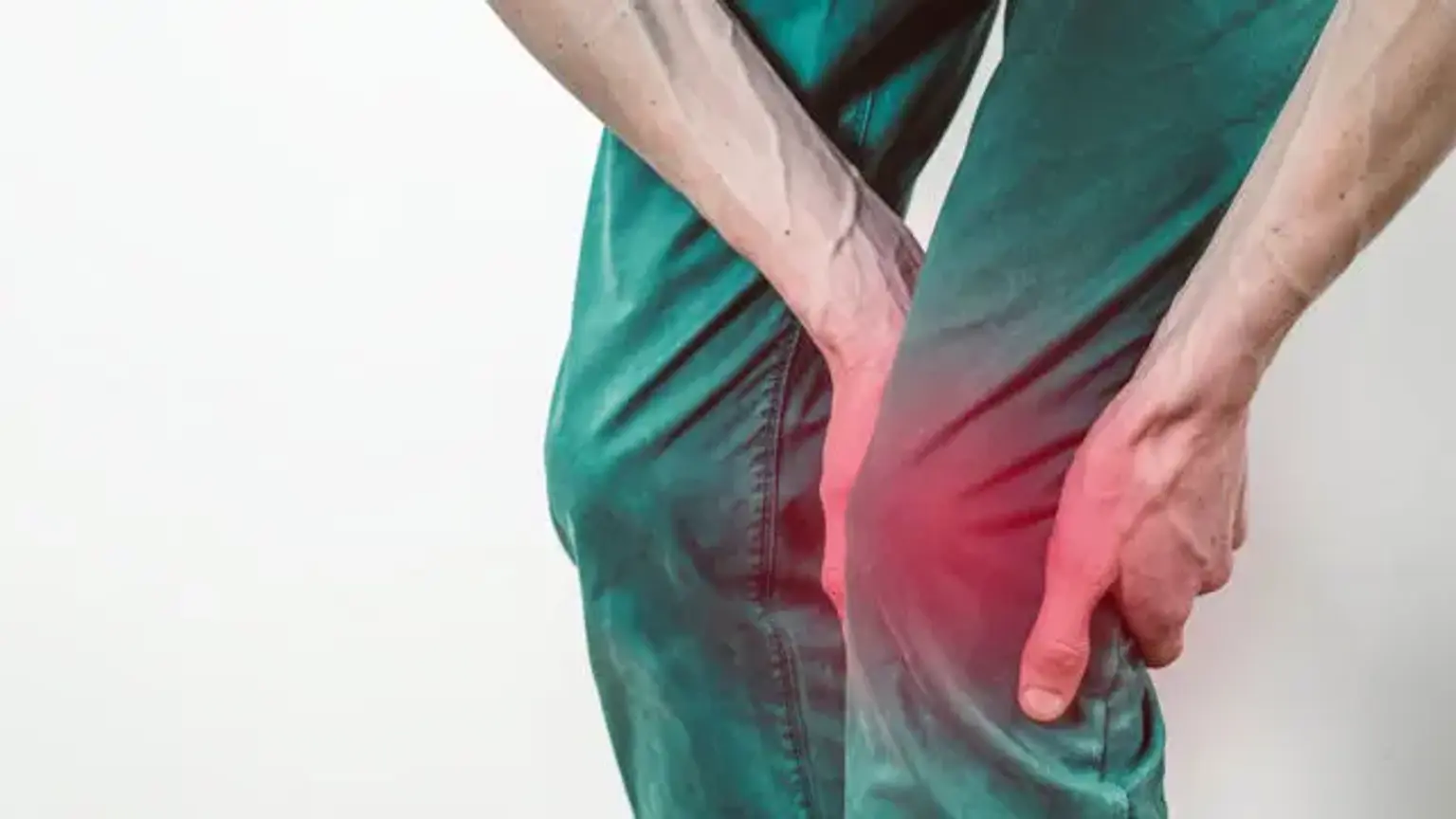
 

Torn meniscus definition

Torn meniscus definition

Anatomy

The menisci are C-shaped fibrocartilage wedges that sit between the tibial plateau and the femoral condyles. The bigger, semilunar medial meniscus is more securely connected than the smaller, more circular lateral meniscus. Both menisci's anterior and posterior horns are attached to the tibial plateaus.

The transverse ligament joins the two menisci anteriorly; the meniscofemoral ligament stabilizes the posterior horn of the lateral meniscus to the femoral condyle posteriorly. The coronary ligaments link the tibia to the peripheral meniscal rim. The lateral meniscus has no connection to the lateral collateral ligament (LCL), despite its near closeness.

The joint capsule clings to the whole perimeter of each meniscus, although the medial meniscus is more securely attached. The popliteal hiatus is formed by a disruption in the connection of the joint capsule to the lateral meniscus, allowing the popliteus tendon to pass through to its femoral attachment point. Popliteus contraction during knee flexion pushes the lateral meniscus posteriorly, preventing entrapment inside the joint space. There is no direct muscle attachment to the medial meniscus. The medial meniscus may migrate a few millimeters, whereas the lateral meniscus, which is less stable, may move at least one centimeter.

The meniscus has a thick fibrocartilage microanatomy formed of cells and an extracellular matrix of collagen fibers in a network. The cells are called fibrochondrocytes because they resemble a cross between fibroblasts and chondrocytes. These cells are in charge of the creation and upkeep of the extracellular fibrocartilaginous matrix.

Collagen is the most prevalent component of the menisci, with type I collagen accounting for the majority, while types II, III, V, and VI are also present. Collagen fibers are mostly organized longitudinally or circumferentially, with some interlaced radial and oblique strands. The circumferential fibers are closely connected to the functional capacity of the menisci to dissipate compressive stresses. The remaining fibers serve mostly as links, increasing structural stiffness and aiding in the prevention of longitudinal splitting. Proteoglycans, glycoproteins, and elastin are other components of the extracellular matrix.

A tensile force is conveyed to the menisci when a compressive force is applied to the knee joint. In extension and flexion, the femur seeks to expand the menisci anteroposteriorly and mediolaterally. Shrive et al investigated the effects of a radial incision in the menisci's peripheral rim under loading. The force was applied through the menisci and articular cartilage in joints with intact menisci; however, a lesion in the peripheral rim interrupted the usual mechanics of the menisci and enabled spreading when a load was applied.

The load was now transferred directly to the articular cartilage. Given these findings, it is critical to retain the peripheral rim after partial meniscectomy in order to avoid irreparable damage of the structure's hoop tension capabilities.


Blood supply

The menisci's blood supply is restricted to their periphery. The medial and lateral geniculate arteries connect to form a parameniscal capillary plexus that supplies the knee joint's synovial and capsular tissues. Vascular penetration through this capsular connection is restricted to 10-25 percent of the medial and lateral meniscal rims' peripheral diameters. 

Vascular ingrowth is required for effective meniscal healing and surgical repair. The meniscus is divided into zones depending on blood supply: the red zone is the well-vascularized perimeter, the red-white zone is the intermediate section with vascularity peripherally but not centrally, and the white zone is the core avascular portion. Arnoczky suggested a categorization system for lesions in respect to the meniscal vascular supply in 1982, as follows:

  • A red-red tear is an injury that results in a lesion inside the blood-rich periphery; both sides of the tear are in tissue with a functioning blood supply, which promotes recovery.
  • A red-white tear encompasses the peripheral rim and center component of the tear; in this case, one end of the lesion is in tissue with a high blood supply, while the other end is in avascular tissue.
  • A white-white tear is a lesion that is only seen in the avascular center area of the skin; the prognosis for healing in such a rip is poor.

The reports outline ways for creating a vascular access channel from the peripheral vasculature in order to increase the likelihood that tissue in the core area would heal itself.


Role of the meniscus

The meniscus is an avascular structure with a very minimal blood supply. However, it is an essential component of the biomechanical function of the knee. The meniscus is in charge of enhancing the congruence of the knee joint's articulating surfaces. Aside from that, the meniscus is important for shock absorption and load transfer when walking and other activities. Furthermore, it aids in knee joint stability, restricting flexion and extension of the knee joint at extreme angles, and giving proprioception. 


Epidemiology

Meniscal rips occur at a rate of 61 per 100,000 in the general population and up to 8.7 per 1,000 in the active-duty military population, according to research conducted in the United States. Given the etiology of tears, it's not surprising that infantry-related jobs, vocations requiring frequent squatting/kneeling, and sports like soccer, rugby, football, basketball, baseball, skiing, and wrestling all raise the risk of meniscal tears.

Male gender and age beyond 40 years old are also linked to an increased incidence of meniscal tears. Medial meniscal tears are more prevalent than lateral meniscal tears, probably due to the medial meniscus's reduced mobility as a result of its relationship to the MCL. Furthermore, patients with ACL-deficient knees are more likely to develop medial meniscal tears, especially if ACL restoration is delayed for more than a year after the initial injury.

Meniscal tears are infrequent in youngsters under the age of ten. However, the existence of a discoid meniscus, which is an anatomic variation most commonly found in the lateral meniscus and involves an extra central extension of the meniscus across the tibial articular surface, may predispose someone to tears at a younger age.


Meniscal tears causes

Isolated meniscal tears develop as a result of rotational or shearing pressures applied to the tibiofemoral joint, particularly when an increased axial strain is applied to the menisci. Positions with increasing degrees of closed kinetic chain flexion (kneeling, squatting), lifting/carrying heavyweights, and activities involving fast acceleration/deceleration, change of direction, and jumping are examples of such circumstances.

A traumatic impact to the knee can also cause solitary meniscal tears, tears that occur concurrently with bone lesions, or injury to the key stabilizing ligaments of the knee, such as the ACL and MCL. Tears are created with less power in people with degenerative alterations of the menisci, which are commonly found in adults over the age of 40, sometimes with associated osteoarthritis (OA).


Pathophysiology

Pathophysiology of torn meniscus

When meniscal rips are detected on magnetic resonance imaging (MRI), they are classified based on their form and location, as high-intensity intrameniscal signals interact with at least one articular surface on otherwise black-appearing meniscal tissue. Horizontal (cleavage) rips run parallel to the tibial plateau through the meniscus's mid-substance. In the absence of a specific inciting incident, they are more likely to occur in adults over the age of 40 who have underlying degenerative alterations.

Longitudinal (vertical) rips run parallel to the long axis of the meniscus and perpendicular to the tibial plateau. Radial tears, on the other hand, originate from the inner free edge of the meniscus and run perpendicular to both the tibial plateau and the long axis of the meniscus. Complex tears are made up of a mix of horizontal, longitudinal, and vertical tears. Displaced tears, on the other hand, include either total detachment of a section of the meniscus or flipping of a segment of the meniscus that is still linked to the rest of the meniscus.

Bucket-handle tears are radial tears with partially detached pieces that migrate centrally over the remaining menisci, whereas parrot-beak tears are full longitudinal tears that move centrally over the remaining menisci. Flap tears are pieces of horizontal tears that have partially separated. Tears within the outer 1/3 vascular zone of the menisci are referred to as "red-red," whereas those with central edges extending into the inner 2/3 avascular zone are referred to as "red-white."

Tears that are confined to the inner two-thirds of the avascular zone are referred to as "white-white" tears. Tears in the red zone have the best chance of healing spontaneously with conservative care or successfully following meniscal restoration.

  • Traumatic tears

Traumatic rips in the meniscus most commonly occur during strong twisting of the knee and are prevalent among football, basketball, and soccer players, although they can occur during any activity that involves knee twisting. Repetitive kneeling or rising from a crouching position while lifting might cause a tear less often.

  • Degenerative (atraumatic) tears

Degenerative or atraumatic tears are more common in elderly people and are caused by biology as well as degeneration and disintegration of the meniscal structure. People who have degenerative tears may have twisted their knee, causing the injury to worsen. A degenerative tear, on the other hand, has a totally different tear pattern than a traumatic rip. This is significant because the therapy for a degenerative tear may differ significantly from the treatment for a traumatic tear.


Symptoms of torn meniscus

Symptoms of torn meniscus

The clinical appearance of a patient with a meniscal tear varies depending on the type of injury and the severity of associated tibiofemoral injuries. An ACL rupture with a probable medial meniscal tear is linked with the sensation of a "pop" with acute effusion of the knee following high-impact exercise or trauma. In contrast, effusion that occurs gradually over the course of 24 hours is more likely to be caused by a single meniscal tear.

Symptoms can sometimes be subtle, with low-grade effusion and stiffness developing over many days in the lack of an inciting event. Typically, pain is described at the anteromedial or anterolateral joint line. Depending on the nature and amount of the meniscal lesion, symptoms such as locking, clicking, catching, occasional inability to completely extend the knee, or a sense of the knee giving way may occur.

Inspection of the knee for edema, palpation of the joint line, standing and supine range of motion (ROM), muscular strength testing, and specific tests should all be part of the physical examination. Anteromedial and anterolateral joint line discomfort is 83 percent sensitive and 83 percent specific for a meniscal tear when done with the patient's knee at the edge of the table at 90 degrees of flexion.

Depending on the kind of tear and the quantity of effusion, there may be discomfort and impairments in either flexion or extension ROM. It is doubtful that there are any deficiencies in open kinetic chain knee flexion/extension strength tests. Still, due to higher compressive stresses over the menisci, antalgic gait or greater discomfort with single or double-leg squatting is conceivable. There are various intriguing specific tests for detecting meniscal tears.

The Thessaly test, in which the patient stands on one leg, squats to 20 degrees of flexion, and rotates the knee internally/externally by active adduction/abduction of the hip, is 75 percent sensitive and 87 percent specific. McMurray's test has a sensitivity of 61% and a specificity of 84%.


It entails gently stretching the supine patient's knee from a completely flexed to a 90-degree flexion position while retaining full external or internal rotation. Apley's compression test, which involves passively flexing the prone patient's knee to 90 degrees and then externally/internally rotating with axial compressive force through the knee, is 20% sensitive and up to 80% to 90% specific.

After a meniscus tear, simple walking and other activities that do not demand twisting, pivoting, fast changes of direction, and so on are often well tolerated. Tears can develop over time, although the pace of advancement is usually slow and unpredictable. The guide is pain. When a tear is aggravated, there are usually accompanied signs of greater discomfort.


Diagnosis

Diagnosis of torn meniscus

When a meniscal tear is suspected, imaging should begin with AP, lateral, oblique, dawn, and weight bearing views to rule out concurrent bone diseases, loose bodies, and osteoarthritis. MRI is the best form of imaging for diagnosing and characterizing meniscal injuries when arthroscopy is used as the gold standard. MR imaging has been shown to be 93% sensitive and 88% specific for medial meniscal tears, and 79% sensitive and 96% specific for lateral meniscal tears.


Torn meniscus treatment

Torn meniscus treatment

The RICE guidelines for acute soft tissue injury should be followed in the treatment of the intensely painful, edematous knee in which a meniscal rupture is suspected (rest, ice, compression, and elevation). To relieve pain and swelling, oral analgesics and nonsteroidal anti-inflammatory drugs (NSAIDs) may be administered. Bracing or knee sleeves can be utilized for protection and compression, and early pain-free knee and ankle ROM training can assist to reduce mobility loss and manage edema.

It is permissible to do a 4-6 week course of relative rest and physical therapy for uncomplicated tears restricted to the outer third of the meniscus and degenerative tears to assess if spontaneous healing and recovery to the desired level of function will occur. Regardless of conservative treatment, individuals with chronic pain, edema, or mechanical symptoms should be evaluated for surgical surgery. 

If feasible, meniscal repair is favored over meniscectomy for injuries requiring arthroscopic surgery because removal of meniscal tissue increases the risk of hastened osteoarthritis owing to decreased cushioning and greater force transmission across articular cartilage surfaces.

Rips in the red zone of the meniscus, tears less than 2cm, vertical longitudinal tears, and acute tears are all factors that improve the chance of effective meniscal healing. Depending on the location of the tear and the kind of surgery, rehabilitation after meniscal repair often includes limits in knee flexion ROM and weight-bearing status for the first 6 weeks postoperatively. Meniscal allograft transplantation is regarded as a salvage operation for symptomatic patients under the age of 50 who have a meniscus-deficient compartment but an otherwise stable knee.


Torn meniscus surgery

Arthroscopic meniscal surgery

Two tiny incisions will be created during a knee arthroscopy. An arthroscope (or "scope") will be passed through one of them to give a saline solution to the knee joint, which will enlarge it. The camera on the scope allows the surgeon to view into the joint.

A surgical instrument is then inserted in through the other incision to perform one of the two basic operative treatments for meniscus tears:

  • Trimming away the torn piece of meniscus
  • Repair of the meniscus using sutures.


Exercises for torn meniscus

If you have a torn meniscus in your knee, you may benefit from a physical therapy exercise regimen to help you recover. Working with a physical therapist (PT) can assist you in regaining maximum knee range of motion and strength, as well as returning to your typical ideal level of activity. Physical treatment for a meniscus tear may potentially help you avoid knee surgery, according to research.

Your physical therapist may use a range of methods and treatments to relieve your knee pain or swelling, as well as to improve the way the muscles surrounding your knee contract and support the joint. Exercises should be a big part of your knee therapy after a meniscus injury. Physical therapy activities, both in the clinic and at home, can help you properly heal from a meniscus injury.


Recovery from torn meniscus

The length of time it may take to recover completely from surgery will depend on the severity of the damage and the degree of meniscal surgery required. If a meniscus tear is repaired, restricted weightbearing with crutches may be necessary for four to six weeks. If the torn section of the meniscus is removed, however, protective weightbearing may only be needed for a few days.

To obtain a good outcome, a well-directed rehabilitation strategy is required. Early therapy will focus on restoring complete knee mobility and minimizing edema caused by surgery. Following this, the major focus will be on regaining muscular strength. The rehabilitation following surgery will be carefully guided by the treating physician, physical therapist, or sports trainer.


Prognosis 

It is predicted that following a meniscus injury, he will be able to return to play. The timing varies and is determined by the nature of the injury, treatment, and rehabilitation program. Athletes can often return to their sport as soon as 2-3 weeks after arthroscopic partial meniscectomy or 6-8 weeks after meniscal repair. In the long run, meniscal repairs fail to heal in 5-10% of patients. When rips are treated in individuals who are also undergoing ACL reconstruction, the failure rate is reduced. Meniscal injuries can predispose to osteoarthritis in the long run due to higher stresses on the articular surfaces of the knee.

Meniscal rips and lateral meniscal extrusion increase joint space reduction in middle-aged people. Patients with medial meniscal tears experienced an extra mean medial space loss of 0.05 mm/year, regardless of whether extrusion occurred, whereas those with lateral tears experienced an additional mean lateral space loss of 0.09 mm/year. 


Conclusion 

Meniscal tears typically arrive to the emergency room with discomfort or trouble moving the knee joint. An interprofessional team comprised of an emergency department physician, nurse practitioner, orthopedic surgeon, physical therapist, and radiologist is most suited to handle these tears.

The RICE principles and pain management should be used as the first line of therapy for an intensely painful, edematous knee with a suspected meniscal rupture. Bracing or knee sleeves may also be utilized for protection and compression, and early pain-free knee and ankle ROM training may help to reduce mobility loss and aid in edema control.

While conservative therapy has a function and may be warranted in individuals with severe osteoarthritis or minor tears, partial meniscectomy remains the most often done operation for meniscal injuries. While its efficacy is debatable, its short learning curve and acceptable short-term results have discouraged many orthopedic surgeons from switching to more effective procedures.

Meniscal repairs have risen in favor over the last three decades, proving to be a far more effective option than partial meniscectomy. Meniscal repair is becoming increasingly suggested in all repairable rips, particularly in young and physically active patients. Long-term trials of partial meniscal implants have also demonstrated outstanding results, although their usefulness in acute circumstances requires more investigation. Meniscal regeneration research appears to be promising, and regenerative medicine appears to be the way forward.