Total Mastectomy
Overview
For patients who are candidates for a preventive mastectomy, a total mastectomy is a successful procedure. This procedure eliminates a greater proportion of breast tissue than subcutaneous mastectomy. Total mastectomy with quick reconstruction has better aesthetic outcomes than subcutaneous mastectomy.
Total Mastectomy definition
A mastectomy is a surgical surgery in which all or portion of the breast is removed. The phrase is derived from the Greek word mastos, which means "woman's breast," and the Latin term ectomia, which means "excision of." Mastectomy is divided into four types: partial, simple, modified-radical, and radical. Skin-sparing mastectomy and nipple-areolar sparing mastectomy are two more variants in terminology or method that are frequently used in conjunction with breast reconstruction.
Anatomy and Physiology
The breast is located on the anterior thoracic wall and is located above the pectoralis major muscle. The superior border of the adult female breast extends inferiorly to the inframammary crease or fold, approaching the level of the second or third rib. The sternal border is the breast's medial limit. The breast extends laterally to the mid-axillary line. Posteriorly, roughly two-thirds of the breast overlies the pectoralis major muscle, with the remainder overlying the serratus anterior and upper section of the oblique abdominal muscles.
The axillary tail of Spence refers to the region of the upper breast that extends superior-laterally toward the axilla. The breast is divided into four quadrants, allowing for uniformity in the reporting of physical examination or breast imaging results.
Upper inner, upper outer, lower inner, and lower outer are the four quadrants. The upper outer quadrant of the breast contains the bulk of the breast tissue, including the axillary tail of Spence. As a result, it is the most prevalent site of breast cancer.
The breast is made up of mammary tissue and is surrounded by subcutaneous fat and skin, as well as superficial and deep fascial layers. The superficial layer of fascia penetrates deep into the dermis and covers the anterior breast before extending over the medial and lateral breast. The deep layer of superficial fascia covers the breast's posterior surface and is located prior to the pectoralis major fascia.
Suspensory ligaments of Cooper are fibrous bands of connective tissue that run across the breast parenchyma and enter perpendicular to the dermis from the deep layer of superficial fascia. Breast ptosis is caused by a lack of strength in these ligaments. Breast tissue is made up of epithelial parenchymal parts as well as stromal tissue.
The epithelial component accounts for around 10 to 15% of total breast volume, with the remaining made up of stromal parts. The breast stroma is made up of 15 to 20 lobes, which are further subdivided into 20 to 40 lobules. Lobules are made up of tubuloalveolar glands that are branching. Adipose tissue can be seen in the gaps between the separate lobes. Each lobe drains into a large lactiferous duct that runs all the way to the nipple.
Several veins traveling medially and laterally, as well as several deeper penetrating vessels that must pass through chest wall muscles before reaching the breast, give blood to the breast. The internal mammary artery gives birth to a number of perforating branches in the middle. These two to four anterior intercostal perforating arteries enter the breast tissue as medial mammary arteries after passing through the pectoralis major.
Lateral branches of the posterior intercostal arteries and branches of the axillary artery, including the lateral thoracic artery and pectoral branches of the thoracoacromial artery, feed the breast with blood. To reach the breast, the lateral vessels loop across the superior and lateral borders of the pectoral muscle.
Venous drainage normally follows arterial supply, with the majority of the drainage going toward the axilla. The perforating branches of the internal thoracic vein, tributaries of the axillary vein, and perforating branches of the posterior intercostal veins provide the majority of the venous drainage. Because lymphatic channels often follow the path of blood vessels, knowledge of venous drainage is vital. This lymphatic outflow is important because it represents a possible conduit for cancer spread via lymphatic and venous channels.
95 percent of the time, breast lymphatic drainage occurs through the axilla. Anatomists and surgeons differ slightly in their descriptions of lymph node groupings. Typically, axillary nodes are defined by their connection to the pectoralis minor muscle. Level I lymph nodes are those placed lateral to or below the lower border of the pectoralis minor muscle and often include the external mammary, axillary vein, and scapular lymph node groups.
The central lymph node group and probably some of the subclavicular nodes are found deep to the pectoralis minor muscle in level II lymph nodes. The subclavicular lymph nodes are located medial or superior to the top border of the pectoralis minor muscle in level III axillary nodes. Rotter's or interpectoral nodes, which are positioned between the pectoralis major and minor muscles, are also routinely identified by surgeons.
These Rotter's nodes may drain farther into the central or subclavicular node groups, indicating a putative "skip channel" for tumor cells to spread from the breast to level III nodes while avoiding levels I and II. The internal mammary chain, the lateral and medial intramammary areas, the interpectoral region, and the subclavicular lymph node basin are other lymphatic drainage locations.
Indications
A breast cancer diagnosis is the most common reason for a mastectomy. In most cases, the cornerstone of treatment for breast cancer is targeted surgical treatment (mastectomy or breast-conserving surgery), which can be combined with neoadjuvant or adjuvant therapy, such as radiation, chemotherapy, or hormone antagonist drugs, or a combination of these.
Tumor parameters such as size and location, as well as patient desire, play an important role in the decision-making process, considering that in many cases, survival rates among patients following mastectomy or lumpectomy with adjuvant radiation therapy are comparable.
In brief, breast cancers can have both invasive and non-invasive histologies. Invasive ductal carcinoma is the most frequent kind of breast cancer, accounting for roughly 85 percent of all invasive breast cancers. In contrast, invasive lobular carcinoma and other uncommon histologies, such as breast sarcoma or lymphoma, are significantly less prevalent.
Breast non-invasive carcinomas include ductal carcinoma in situ and lobular carcinoma in situ. The latter is frequently viewed as a risk factor for future breast cancer and may be better classified as a benign precursor lesion.
Patients with Paget's disease of the breast may also be candidates for mastectomy. Paget's disease is an uncommon kind of breast cancer in which neoplastic cells are seen in the nipple-areolar complex epidermis. While the illness may be restricted to one location, 80 to 90 percent of individuals will have an accompanying malignancy elsewhere in the affected breast.
The usual method to surgical therapy of Paget's disease has been total mastectomy with axillary sentinel node biopsy. When combined with whole breast radiation therapy, central lumpectomy with total excision of the nipple-areolar complex has been shown to be successful for local control in patients with no other cancers in the breast.
Due to the amount and spread of illness, mastectomy may be indicated in individuals whose disease is multifocal or multicentric inside the breast. Furthermore, individuals with advanced locoregional illness, such as big primary tumors (T2 lesions larger than 5 cm) and skin or chest wall involvement, may benefit from mastectomy in many cases.
Due to tumor load inside the dermal lymphatic pathways and more widespread involvement of the underlying breast parenchyma, patients with inflammatory breast cancer are treated with mastectomy in addition to systemic chemotherapy and radiation treatment.
Patients who have had breast-conserving surgery (lumpectomy or partial mastectomy) and have margin involvement with tumor cells may be candidates for mastectomy if margin re-excision fails or is not technically or aesthetically possible. Clear or negative margins after removal of an initial tumor are an important component in lowering the likelihood of recurrence. Mastectomy is also recommended for individuals with recurrent breast cancer who have previously had lumpectomy and radiation therapy.
In rare instances, mastectomy may be an option for risk reduction or prophylaxis in individuals who do not have a cancer diagnosis. Patients who are found to have a harmful BRCA genetic mutation are at an elevated risk of developing breast cancer throughout their lives. Carriers of the BRCA1 or BRCA2 mutations have an 80 to 85 percent lifetime chance of developing breast cancer.
Contraindications
Mastectomy may be performed safely and easily in most cases if medically required. There are a few critical variables that should be considered as contraindications to surgery. These are frequently divided into two categories: systemic and locoregional. Mastectomy may be contraindicated in patients who have been diagnosed with distant metastatic illness.
Furthermore, due to the burden of their general health and low performance status, weak or elderly patients with major medical co-morbidities or systemic organ failure may not be candidates for surgery. Patients who are at a high risk of death as a result of surgery or anesthesia are not suitable for surgery. Mastectomy may be comparatively contraindicated in patients with advanced locoregional illness at the time of diagnosis if there is skin or chest wall involvement and concerns about the capacity to seal the surgical incision or obtain a negative surgical margin.
In certain cases, neoadjuvant chemotherapy, radiation, or endocrine therapy may be beneficial in reducing the volume or extent of local illness and allowing for surgery.
Equipment
Sharp dissection using a scalpel or scissor method can be used to conduct a mastectomy. Another option is to employ an energy device like electrocautery or one of the several ultrasonic devices to separate the breast from the superficial tissues and remove the posterior attachments from the chest wall.
Standard general surgical equipment, such as retractors and suction, is frequently used. A temporary, closed suction drain (i.e., Jackson Pratt drain) may be put in the wound bed at the surgeon's discretion to reduce the pace of seroma development. The most often used suture for closing the mastectomy defect and incision is absorbable suture.
Axillary sentinel lymph node biopsy is frequently used for staging purposes after final breast surgery. Most surgeons utilize a dual tracer approach, which commonly involves the use of technetium sulfur colloid, which is injected into the breast preoperatively by the radiology department's nuclear medicine department.
Many surgeons will inject a crucial blue dye, such as methylene blue or isosulfan blue, into the breast intraoperatively to help identify axillary sentinel lymph nodes. To identify any nuclear tracer absorption in the axilla and to guide dissection and exploration in this region for biopsy, a gamma probe is necessary.
Personnel
Breast cancer care is difficult, requiring a multidisciplinary and interprofessional care team. The mastectomy is often performed by a general surgeon or a breast/oncologic surgeon. Perioperative nursing staff, anesthetic providers, and surgical technologists or surgical assistants are all engaged in mastectomy surgeries.
A radiologist or nuclear medicine technician is typically engaged in perioperative technetium sulfur colloid injection as part of sentinel lymph node mapping for patients who require axillary node biopsy at the time of mastectomy. A plastic surgeon often works alongside a general or breast surgeon to execute the right reconstructive surgery, which is based on many patient characteristics, in patients having reconstructive operations.
Furthermore, the majority of patients who get a mastectomy have cancer, and their post-operative care team includes medical oncology and probably radiation oncology. Following a mastectomy, patients may require home healthcare nursing assistance as well as physical or occupational therapy to maximize the recovery of capacity for daily activities.
Preparation
Mastectomy is often an elective operation, and patients are expected to report to the hospital or surgical facility on the day of their procedure. Pre-operative antibiotics are recommended for patients undergoing mastectomy, with or without axillary surgery or reconstruction, to lower the risk of surgical site infection; this consensus statement was issued by the American Society of Breast Surgeons in 2017 and was amended. Unless the patient is allergic to penicillin or has a history of methicillin-resistant Staphylococcus aureus infection, a first-generation cephalosporin is the antibiotic of choice for prophylaxis.
After anesthesia is administered, the patient is placed in a supine posture in the operating room, and the breast, chest wall, axilla, and upper arm are exposed. Many surgeons will include the contralateral breast in the surgical field. The surgical site is sterilely treated using an agent to diminish the presence of skin flora and the risk of surgical site infection. For surgical antisepsis, alcohol-based skin preps such as chlorhexidine gluconate are commonly used.
Surgeons should prepare patients for their surgery and discuss the expected postoperative course and care with them perioperatively. Many surgeons choose to insert a drain during mastectomy to remove any fluids that may build in the wound bed and to enhance flap adhesion to the chest wall. Patients benefit from drain care instruction as well as keeping an accurate journal of outflow. Patients should also be counseled about their postoperative restrictions, such as lifting, driving, and any other limits in the initial recovery period.
Technique
While surgical excision of the breast dates back to the 18th century, William Halsted detailed his radical mastectomy approach in 1894, stating that "suspected tissue should be removed in one piece." This aggressive procedure is a total en bloc resection of the breast, including the pectoralis major muscle and regional lymphatics.
With this procedure, a large quantity of skin was lost, and a skin transplant was frequently required for covering of the chest wall defect. Women were left with major malformations and impairments as a result of this treatment. As a result, numerous improvements to the procedure have been made in order to lessen the morbidity of the surgery.
David Patey improved the Halsted radical mastectomy in the 1940s by maintaining the pectoralis muscle, and his outcomes were good for less postoperative problems, such as discomfort, lymphedema, and upper extremity movement impairments.
In 1972, John Madden defined the current criteria for a mastectomy. This method entails creating an elliptical incision around the breast, including the nipple areolar complex, and preserving the tumor location as a focal reference. The mammary tissue is separated from the skin flaps and resected across the pectoralis major fascia, while both pectoral muscles are preserved.
As a result, minimal damage of neighboring neurovascular and lymphatic tissues is required. Madden's modified radical mastectomy procedure originally included level I-III axillary lymphadenectomy for staging purposes, and this was assumed to have a therapeutic advantage as well. Total mastectomy, in contrast to this modified radical mastectomy approach, refers to the surgical removal of entire breast tissue. However, there is no need for axillary node dissection.
Some patients who opt to have breast reconstruction may be candidates for skin-sparing and nipple-sparing mastectomy as reconstructive procedures have improved and grown more popular. Whatever technique is used, oncologic principles take precedence above aesthetic considerations. Skin-sparing mastectomy, as the name implies, is intended to retain a healthy skin envelope for rapid breast reconstruction, if acceptable margins can be established.
The nipple-sparing mastectomy removes the mammary tissue and pectoral fascia while leaving the nipple-areolar complex and the whole skin envelope of the breast intact. Only patients having urgent breast reconstruction are subjected to this surgical method. It is crucial to maintain blood flow to the nipple areolar complex using this method to avoid flap ischemia or collapse.
All of the abovementioned mastectomy procedures generate uniform flaps by dissecting just above the superficial layer of the breast's superficial fascia. The appropriate flap thickness has been hotly debated, with the ultimate objective of eliminating all feasible breast tissue while retaining skin viability.
As previously stated, regardless of the style or location of skin incision employed, mastectomy dissection should continue until the anatomic borders of the breast. While the surgeon retracts the breast tissue away from the surrounding tissue, the flaps are lifted and retracted at a right angle to the chest wall. The skin flap is elevated superiorly to the clavicle, laterally to the anterior boundary of the latissimus dorsi, medially to the sternal border, and inferiorly to just below the inframammary crease.
Once the overlaying flaps have been dissected, the breast is excised with the pectoralis major muscle, working from the superomedial to the inferolateral border. A closed suction drain is frequently implanted in the wound bed behind the skin flaps, and the incision is closed in two layers with absorbable suture.
Complications
In most cases, patients handle mastectomy successfully, with little morbidity and death. However, a number of difficulties are conceivable. Seroma or hematoma development, wound infection, skin flap disintegration or necrosis, and lymphedema are examples of these. A seroma is a collection of fluid in a surgically produced cavity caused by vascular and lymphatic transection. To reduce the incidence of seroma development, most surgeons employed closed suction drains beneath the skin flaps. Approximately 8% of people following breast surgery develop surgical site infection.
Staphylococcus aureus and Streptococcus epidermis are the most prevalent organisms implicated, and infections should be treated with an appropriate antibiotic, with or without wound opening. Similarly, flap necrosis affects roughly 8% of patients and is associated with insufficient blood flow to the flap, wound closure under strain, obesity, and style of incision (vertical versus transverse). Necrosis is treated with debridement and, if necessary, skin transplant covering. Lymphedema has been less prevalent since the introduction of modified mastectomy methods.
With a reported prevalence of more than 20%, axillary lymph node dissection is the most major risk factor for the development of lymphedema. Lymphedema develops in 3.5 to 11% of individuals who receive sentinel lymph node biopsy. Early intervention with physical therapy and decompressive massage techniques in individuals who acquire lymphedema can assist prevent development and, in some situations, minimize lymphedema.
Conclusion
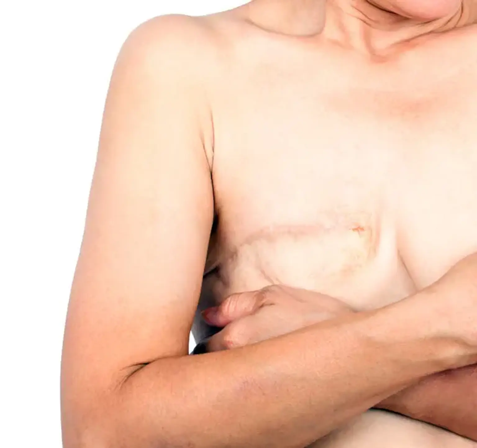
A complete mastectomy is a surgical surgery that removes the entire breast, including the breast tissue, nipple, areola, and skin, to cure breast cancer. This method, also known as simple mastectomy, may be useful for a patient with severe cancer who is unsuitable for a lumpectomy or partial mastectomy. For increased peace of mind, some patients choose complete mastectomy over breast-conserving surgeries such as lumpectomy.
Others choose to have the surgery done as a preventive step even though they have not been diagnosed with breast cancer - because they have a genetic predisposition or are otherwise at high risk of getting the disease.