Transurethral Incision of The Prostate (TUIP)
The surgical method of transurethral incision of the prostate (TUIP) was established in the mid-nineteenth century. The first descriptions originate from 1834 when Guthrie proposed that the bladder neck be mechanically dilacerated. There are no clinical statistics on the number of interventions or Guthrie's outcomes, however. During the same time period, Bottini presented a diathermy-based approach. However, TUIP was not widely seen as a feasible alternative for the management of prostate adenoma at the time.
Keitzer first introduced the TUIP procedure as an endoscopic option for addressing sub-vesical blockage in younger patients with tiny prostate adenomas. The technique differs from transurethral resection of the prostate (TURP) in that it removes the obstruction by reducing the constrictive tonus following prostatic incision (and not by tissue removal, as in the case of TURP).
Since its introduction, TUIP has steadily gained traction in the therapeutic arsenal, owing to its ease of use, low risk of complications, and long-term effectiveness. Furthermore, most authors believe that this strategy is simple to master. Orandi presented the first long-term outcomes, recommending TUIP as a possible therapeutic option in young patients with moderate prostate sizes or concomitant diseases that preclude resection. In the absence of severe obstructive symptoms, however, the intervention is not indicated.
Prostate Anatomy and Physiology
The prostate is one of the male's major pelvic organs. There are seven lobes in total: two posterior, two lateral, one subcervical, one middle, and one anterior.
An understanding of embryology is required to comprehend the development of these seven lobes and their links to the surrounding structures. The cloacal ectoderm is influenced by testosterone production from the fetal testes to release narrow epithelial tubules into the surrounding mesenchyme to produce the prostate gland in the 12-week-old embryo. The different lobes of the adult prostate develop from five primordial ducts that are distinguished by their proximity to the urethra.
The top of the urethra gives rise to the anterior lobes, which generally atrophy before birth. The ducts on either border of the verumontanum give rise to the lateral lobes. They unite to produce the anterior commissure when they expand laterally and anteriorly in adults. Between the bladder neck and the ejaculatory ducts, the anlage of the middle lobe originates from the bottom of the posterior urethra. These tubules grow, merge with, and become indistinguishable from lateral lobe tissue.
A set of tubules in the urethral segment distal to the verumontanum give rise to the posterior lobe. These acini form behind the ejaculatory ducts and grow posteriorly. A fibromuscular septum separates this lobe from the lateral lobes and lasts into adulthood.
The sub-trigonal glands of Home, located directly proximal to the vesical exit, are the source of the contentious median bar blockage in a hypertrophied adult prostate.
The inferior vesical artery, a terminal branch of the internal iliac artery, provides blood flow. Additional branches from the middle rectal, superior hemorrhoidal, and deferential supply the inferior section of the prostate. A urethral artery and a capsular artery branch off the prostatic artery. The primary supply of the middle lobe is provided by the urethral group, which comes from lateral to medial at the 4 o'clock and 8 o'clock points. The prostatic vessels, particularly the urethral group, grow in size as the prostate hypertrophies, supplying the enlarged tissue.
Secretory and motor fibers coming from the sacral and hypogastric plexus innervate the prostate. With the urethral branches of the prostatic artery, they reach the prostatic urethra. Within the prostate, Timefeew identified a wide-mouthed plexus of medullated fibers. Inflammation produced by inspissation of secretions and poor drainage of hyperplastic ducts may excite these nerve fibers to produce pain. The hypogastric plexus arises from the posterior surface of the bladder and the rectum, contributing to the chronic prostatitis clinical syndrome.
The deep branch of the perineal nerve, a division of the pudendal nerve, also innervates the external urethral sphincter. Referred pain to the sacral, perineal, low back, and rectal areas is common in patients.
TUIP Indications
TUIP is appropriate in patients with a prostate volume of less than 30 cm3 (estimated by transrectal ultrasonography) and no median lobe, per the European Association of Urology guidelines. The European Association of Urology guidelines is based on a review of numerous randomized clinical trials that compared TUIP to TURP, the current therapeutic gold standard, in terms of efficacy, intra- and postoperative complication rates, and long-term benefits.
Some publications claim that TUIP can also be conducted with satisfactory outcomes in the treatment of adenomas with a volume more than 30 cm3 in young people or in patients who have other medical issues that make other treatments contraindicated. TUIP was first introduced as a surgical treatment option for patients with tiny, obstructive adenomas who were not suitable for TURP or open operation. Young males with strong obstructive symptoms but no markedly enlarged prostates made up this target group.
Patients with tiny prostates have stromal hyperplasia and respond less positively to TURP than men with large prostate adenomas with glandular hyperplasia, although having identical symptoms. TUIP has been suggested by several writers for patients with massive adenomas. Deep incisions were made in these cases, which always resulted in the opening of the venous sinuses and perforation of the capsule, resulting in extravasation and uncontrolled venous hemorrhage. Fulguration was commonly required, which elevated the risk of postoperative complications substantially. The blockage of the bladder neck after trans-vesical adenectomy is another indication of TUIP. A median cyst of the prostatic urethra is a rather uncommon indication for TUIP.
Transurethral Incision of the Prostate TUIP Procedure
Anesthesia and Equipment
This technique has unquestionable benefits: it is simple to learn, quick, and inexpensive, with excellent practical results if the indication is accurate. The procedure is usually performed under spinal anesthesia, but other writers favor general anesthetic. Local anesthetic is being used by an increasing number of authors. This can be a significant benefit for patients who are at risk of anesthetic complications and for departments looking to cut costs. The resectoscope or the rigid urethrocystoscope with a 30° telescope are the items used. Cold knife, Collings’s loop, resection loops, vaporization electrodes, laser fibers, and other auxiliary instruments that allow this procedure can all be employed for the incision.
The cutting instrument of choice must favor the lowest-priced procedure, with the literature indicating that neither tool nor technique gives superior results than the other. As a result, the use of costly equipment for TUIP is unnecessary, and these resources should be dedicated to procedures where they can make a significant therapeutic benefit. Orandi is the first to describe the surgical procedure, which he modified from Aboulker and Steg's prostate divulsion concept. As a result, he hypothesized that endoscopic prostate incision could be as effective as divulsion but with fewer risks. Initially, a perineal urethrotomy was used to gain access to the bladder.
Making Prostatic Incisions
A resectoscope is used to produce cuts at 5 and 7 o'clock, beginning distally from the ureteral orifices, continuing to the prostatic urethra, and finishing proximal to both sides of the verumontanum. Orandi adapted the method for younger patients who desired to keep their ejaculation, producing superficial cuts confined to the prostatic urethra without extending to the bladder neck. Other authors adopted this modification, performing short and shallow incisions at 5 and 7 o'clock, primarily at the level of the prostatic urethra and contacting the fibrous capsule. In most patients, this resulted in a significant rise in maximum urine flow rate and a substantial decrease in symptoms while preserving normal ejaculation.
Several authors refined the procedure, describing the use of a single incision for TUIP. Single incisions were typically made at 5 o'clock, 6 o'clock, or 7 o'clock, extending to both sides of the verumontanum. Most authors observed a considerable decrease in the incidence of retrograde ejaculation as well as patient outcomes comparable to the classical procedure when employing a single incision on the midline. Other researchers believe that the outcomes are the same regardless of whether the incision is single or multiple. At this time, there is no clinical evidence to support a recommendation in favor of single or several incisions.
Based on the available data, single incisions are most usually performed at 6 o'clock, whereas multiple incisions are most frequently performed between 5 and 7 o'clock. Other writers have documented using Nd:Yag or Ho:Yag lasers to make incisions, but there have been no comparative studies to verify their superiority. Although some doctors claim to use a greater number of incisions in various locations around the periphery of the prostate, there is no clinical evidence to support this claim. The link between TUIP and prostate posterior resection at 4 and 8 o'clock represents an attractive technical alternative.
According to the authors, this approach prevents adhesions from forming between the incised portions of the prostate, making deep incisions up to the prostatic capsule easier to maintain. Other doctors used endoscopic resection to create a conduit in the prostate tissue, allowing them to retrieve prostate tissue specimens for histological analysis. They found a greater reintervention rate due to postoperative urinary retention, although having an efficacy equivalent to TUIP. Because it was easier to place the urethral catheter at the end of the procedure, there was less bladder neck contraction, and there was less retrograde ejaculation, minimal transurethral prostate resection combined with bladder neck incision was found to be superior to standard TURP. However, this was a one-off experience that was not supported by other evidence.
A technical alternative proposes limiting incisions to the bladder neck, arguing that extending them toward the verumontanum or distal to it is pointless. The advantages include a shorter surgery time and catheterization period, as well as identical long-term outcomes. Whatever technical alternative is chosen; the incision should be made all the way to the prostatic capsule in terms of depth. This structure is easily identifiable due to its endoscopic appearance, which differs significantly from that of prostatic tissue.
Finishing the Procedure
An incision that extends beyond the prostatic capsule increases the risk of rectum damage or irrigation fluid extravasation. The side of the bladder neck will be examined at the completion of the procedure, with the endoscope positioned distal to the prostatic urethra. If there is no obvious obstruction of the bladder neck, the procedure can be completed by inserting a urethro-vesical catheter, which will be kept in place for one to three days, depending on local anatomical features and other factors. Some writers claim that by avoiding the use of a urethro-vesical catheter, postoperative hemorrhage is greatly decreased while functional results are maintained. There is, however, no clinical data to support the recommendation of a minimum catheterization period.
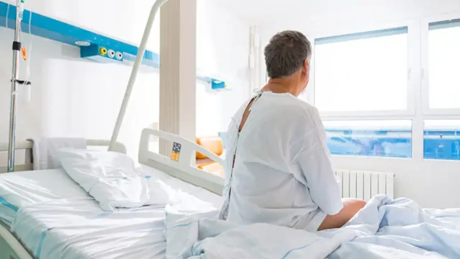
What Happens Immediately After the Procedure?
Following the operation, you will likely experience some bleeding from the prostate site, and you will have a catheter emptying the bladder with fluid passing through it to clean the bladder and avoid the formation of clots.
Although some people lose blood for longer, the urine normally clears of blood within half a day. The need for a blood transfusion following a bladder neck incision is uncommon.
When you are ready, you'll be able to drink and eat on the same day as the surgery. Following surgery, the catheter is usually withdrawn two to three days later. For the first one to two days after catheter removal, it's not uncommon for your pee to become bloody again. Several weeks following surgery, some blood may be observed in the urine.
Passing pee might be uncomfortable and inconvenient at first. Tablets or injections can reduce any pain, and frequent urination normally resolves within a few weeks. Passing pee frequently, urgency, and getting up at night to pass urine, on the other hand, can take many months to entirely resolve in some patients. This is because these symptoms are caused by excessive bladder activity, whereas obstruction symptoms (hesitancy and inadequate flow) improve almost quickly.
If you are unable to pass pee and are uncomfortable, please notify the designated nurse. Some patients are unable to pass urine at all following the operation, and in these cases, a new bladder catheter is usually inserted. This helps the bladder to restore and the internal bloating to subside. If this occurs, you will be sent home with the catheter in place and will be seen again in a week for proper second catheter removal. The typical length of stay in the hospital is 1-2 days.
TUIP Complications
In general, any procedure that is regarded as a suitable alternative to a standard treatment must have comparable efficacy while posing fewer dangers. The most common TURP consequences are hydro-electrolytic disorders and intraoperative hemorrhage. Erectile dysfunction and retrograde ejaculation are two sexual function changes that might arise following TURP. A key advantage of TUIP over TURP is a substantially reduced incidence of retrograde ejaculation, which is especially essential in young patients but should be evaluated in the elderly as well. If a 55–95 percent incidence of retrograde ejaculation is recognized for TURP, this value drops drastically during TUIP, even falling below 12%.
Hydro-electrolytic diseases are not observed in TUIP since the operating period is relatively brief, and they are not described in any major study. TUIP is a faster procedure than TURP, and the amount of liquid required for lavage is much less. Patients with TUIP rarely experience severe bleeding that necessitates blood transfusions. According to Dorflinger et al., blood transfusions were needed in 12% of patients with TURP but not in any patient with TUIP. Patients with TUIP had a transfusion rate of 1.5 percent, whereas those with TURP had a rate of 19.5 percent, according to Edwards. 6 patients (nearly 1%) received blood transfusions in Orandi's study.
The rate of retention due to clots was 1.2 percent in the TUIP group and 2.4 percent in the TURP group, according to Edwards. Dorflinger evaluated 43 sexually active patients who were randomly assigned to one of two groups: TUIP or TURP. Only 2 of 19 patients with TUIP had retrograde ejaculation one year after the operation, opposed to 13 of 24 patients with TURP. Larsen found that 3 of 7 TUIP patients had retrograde ejaculation, whereas 8 of 8 TURP patients had this problem. According to Christensen, the TUIP group had a 14 percent incidence of retrograde ejaculation, while the TURP group had a 38 percent incidence.
Other Things to Know
As long as you had regular erections before the surgery, a prostate incision should not have a negative impact on your sexual life. Sexual activity can be resumed as soon as you feel comfortable, which is normally three to four weeks after the procedure.
Starting pelvic floor strengthening exercises as quickly as possible after the operation is beneficial since it enhances the control when you get home. Contact the ward staff or the specialist nurses if you have any questions about these exercises. Although the urine flow improves quickly, the symptoms of an overactive bladder (frequency and urgency) can take up to three months to resolve.
Most patients need a 3-to-4-week rehabilitation period at home before they become able to return to work. Before returning to any activity, we recommend taking three to four weeks off, particularly if it is physically demanding. Heavy lifting should be avoided at this point.
Conclusion
Chronic prostatitis, middle lobe hypertrophy of the prostate, or a combination of the two can all be treated surgically with the transurethral incision. Since the procedure's inception, no deaths have been documented as a result of it.
Chronic infection and benign prostatic hypertrophy have been suggested as probable contributors to prostate cancer. Others have proposed a link between chronic obstruction at the vesical exit and transitional cell carcinoma of the urinary bladder. Further research should include a 25- to 30-year follow-up period to establish the incidence of these two cancers in TUIP patients.
Some surgeons have been hesitant to perform a TUIP for fear of missing prostate cancer. The age range of the patients ranged from 40 to 71. The majority are under the age of 50, and the real incidence of carcinoma in this age range is extremely low. Murphy documented one patient under the age of 50 with prostate cancer, who was 49 years old, in a series of cases. To rule out this risk, all patients get a rectal examination prior to surgery, with any worrisome nodules aspirated or biopsied. Patients who have undergone surgery will be monitored as closely as possible in the future to see if this is an issue. To date, no TUIP patient has developed prostate cancer after surgery. A yearly rectal examination is performed on all patients.