Trophic ulcer
Last updated date: 15-May-2023
Originally Written in English
Trophic ulcer
Overview
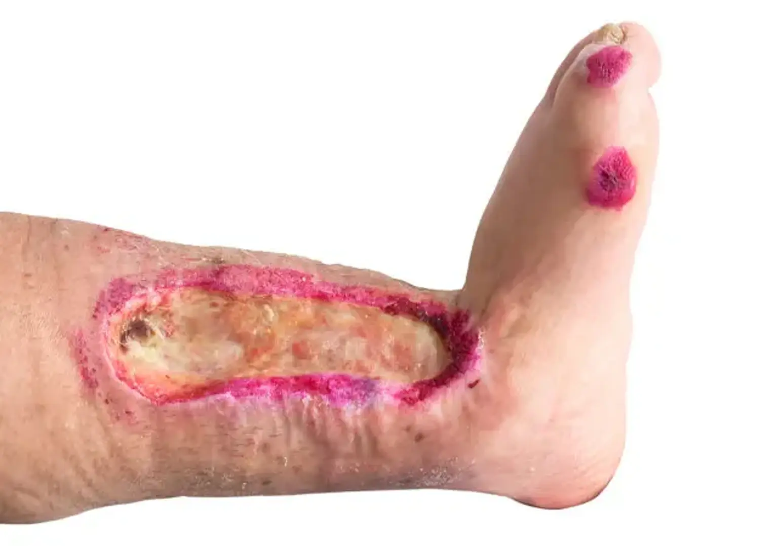
Lower limb trophic ulcers afflict around 1–1.3% of the population and are the most serious and unpleasant consequence of chronic venous insufficiency. Although the pathophysiology of trophic ulcers is unknown, sufficient oxygen transport to tissues is undeniably important in wound healing. Hyperbaric oxygenation can now be used in patients with difficult-to-heal wounds thanks to recent technology improvements.
Though the general care of all trophic ulcers would be similar, the management would have to be tweaked on a case-by-case basis based on the aetiology and pathophysiology of the lesion. Because each ulcer's pathophysiology is unique, knowing the pathology might aid us in better addressing the disease.
Trophic ulcer definition
The term "Trophic" is derived from the Greek word Trophe, which means "nutrition." Tropic ulcers are defined as "an ulcer caused by inadequate nourishment of the part" by the American Heritage Medical Dictionary 2007. The 2009 edition of Mosby's Medical Dictionary describes trophic ulcer as "a pressure ulcer induced by external damage to a portion of the body that is in poor health due to illness, vascular insufficiency, or loss of afferent nerve fibres."
Trophic ulcers develop as a result of untreated leg vein issues. It is an unhealing wound that requires immediate medical attention. If superficial vein issues, which are the most common cause, are not addressed, the lower shin expands and venose blood flow is disrupted, which is when all of the aesthetic faults appear - pigmentation, hard and uncomfortable skin. Deep veins may be affected as the condition worsens.
People who experience frequent edema as a result of enlarged veins should be extra cautious. These folks frequently experience itchy skin on their shins, as well as discoloration in the ankle region.
People with persistent skin alterations, as well as those who have had deep vein thrombosis, should be extremely vigilant, because even the smallest lesion, such as a mosquito bite, can grow into an infection or a poorly healing skin defect known as thropic ulcer.
Trophic ulcers can spread quickly and are difficult to cure. Also, if it is not treated properly, the condition can spread — the trophic ulcer becomes larger, begins to smell, and hurts, affecting the patient's quality of life and self-esteem.
Etiology
Ulceration from vascular reasons is frequently complex, and it can be caused by both arterial and venous illness. Hypertension and peripheral artery atherosclerosis cause vascular disease with ischemic ulcers. Venous ulcers are caused by chronic venous insufficiency and the associated venous hypertension.
Ulceration can also be caused by Vasculitis, such as Buerger disease (thromboangiitis obliterans) or Takayasu disease. The former is characterized by arterial or ischemic ulcers, whereas the latter is characterized by cutaneous illness such as pyoderma gangrenosum or erythema nodosum.
When an ulcer fails to respond to appropriate medical and wound care, the possibility of an underlying cancer should be examined. Nodulo-ulcerative basal cell carcinoma, squamous cell carcinoma, keratoacanthoma, nodular melanoma, tumor stage mycosis fungoides, lymphomatoid granulomatosis, lymphomatoid papulosis, angiosarcoma, and cutaneous metastases from internal malignancy are examples of cutaneous malignancies that can masquerade as ulcers. These manifestations must be recognized by healthcare practitioners, who must then give appropriate therapeutic action.
Pathophysiology
Either progressive atherosclerosis or arterial embolization can result in arterial (or ischemic) ulceration. Both cause skin ischemia and ulceration.
Venous (or stasis) ulceration is caused by venous hypertension caused by insufficient calf muscle pump activity and the emergence of either primary (with no clear underlying cause) or secondary (as observed after deep venous thrombosis) valvular incompetence. Two concepts have been offered to explain venous ulcers after the development of venous hypertension.
According to the first, capillary bed distention develops as a result of enhanced stasis. This causes fibrinogen to flow into the surrounding dermis. A fibrinous pericapillary cuff forms over time, obstructing the flow of oxygen and other nutrients or growth factors to the afflicted tissue. The hypoxic damage that results causes fibrosis and finally ulceration.
The alternative theory contends that increased venous pressure and leukocyte activity harm the endothelium. Proteolytic enzymes and free radicals are produced, escaping through the leaky vessel walls and causing harm and ulceration to the surrounding tissue.
Furthermore, investigations have revealed a link between obesity, chronic venous illness, and popliteal venous compression (PVC). This illness may provide light on previously inexplicable venous manifestations. Other causes for the higher frequency of vascular ulcers in obese people include intra-abdominal venous compression.
Rasmussen et al discovered impaired lymphatic function early in the development of venous leg ulcers using near-infrared fluorescence lymphatic imaging, as well as bilateral dermal backflow in the presence of chronic venous insufficiency, including in patients without ulcers in the contralateral limb.
Clinical Presentation
Chronic leg or vascular ulcers commonly appear as arterial, neurotrophic, or venous ulcers. They differ in terms of location, appearance, bleeding, and related pain and findings.
Arterial ulcers
Arterial ulcers are frequently found distally and on the dorsum of the foot or toes. They have uneven edges at first, but they may grow to a more defined look. Grayish, unhealthy-looking granulation tissue might be seen near the ulcer's base. These ulcers bleed extremely little or barely at all when manipulated, such as debriding. The patient may complain of distinctive discomfort, particularly while recumbent at night, which is eased by extremity dependency. On inspection, hairlessness, pale skin, and missing pulses are found to be typical with chronic ischemia.
Neurotrophic ulcers
Neurotrophic ulcers have a punched-out look with a deep sinus. These are frequently observed as underlying calluses or pressure points (eg, plantar aspect of the first or fifth metatarsophalangeal joint). They are frequently encased in chronic inflammatory tissue. Probing or debriding may result in profuse bleeding. These ulcers are generally painless because these individuals usually have a neuropathy that causes hypesthesia and a loss of positional perception or 2-point discrimination.
Venous ulcers
Venous ulceration (seen below) is a common occurrence in the "gaiter" area of the legs. This area extends circumferentially around the lower leg, from the mid-calf to just below the medial and lateral malleoli. Stasis ulcers are larger but shallower than typical ulcers, with a wet granulating base and an uneven border. When manipulated, this base exudes venous blood. The skin around these lesions may show indications of stasis dermatitis. Patients frequently describe slight discomfort that is alleviated by elevation.
Diabetic ulcers
Diabetic ulcers are caused by a variety of reasons. Mechanical alterations in the shape of the bone architecture of the foot, or the combination of any of the ulcers previously listed, are examples of such variables. Ligaments are predisposed to stiffness due to nonenzymatic glycosylation. Neuropathy results in the loss of protective feeling as well as the lack of coordination of muscle groups in the foot and leg, all of which increase mechanical stressors during ambulation.
Diagnosis
Imaging Studies
- When noninvasive testing show that the pedal perfusion is inadequate, imaging examinations of the lower extremities should be performed to determine the amount of blockage and to evaluate the distal runoff.
- When it is necessary to see the vessels of the lower extremities, angiography should be performed. The study of choice is a femoral runoff research. It shows the filling of leg veins all the way down to the ankle. If the lesion is distant enough, the plantar arch may also be visible. This study is important to both the plastic surgeon who provides coverage and the vascular surgeon who does revascularization.
- MRA (magnetic resonance angiography) can also be used to evaluate lower extremity disease. When compared to traditional angiography or surgery, Yucel et colleagues discovered that MRA was 94% accurate in evaluating lower extremity arteries. When compared to traditional angiography, Owen and colleagues discovered that MRA recognized all runoff vessels and was, in fact, more sensitive than conventional arteriography for identifying both runoff vessels and arterial stenosis.
Imaging tests for venous disease can also reveal important preoperative issues.
- Doppler duplex scanning has a sensitivity of more than 75% for detecting venous reflux, whereas descending venography has a sensitivity of about 40%. According to Neglen and Raju, combining duplex scanning with air plethysmography aids in distinguishing severe venous illness from moderate venous disease.
- To gain precise anatomic information, ascending venography may also be utilized. This test can detect axial channel patency, perforator incompetence, blockage, and deep venous thrombosis.
Other Tests
See the list below:
- If an ulcer is reoccurring, the etiology is unknown, and all invasive and noninvasive procedures have been completed, a biopsy is required to establish a diagnosis and further understand the disease's genesis. As is always the case, chronic wound treatment can be improved by knowing the real cause and thereby treating the underlying problem.
- Assess the vascular supply to the ulceration location in order to predict the chances of good wound healing. There are several approaches for assessing the sufficiency of the pedal circulation.
- Ankle-brachial indices (ABIs) and toe digital pressures with pulse volume recordings can give useful information on foot perfusion. The findings are also predictive of wound healing, albeit they may be deceptive in diabetics and people with calcified noncompressible arteries. A higher ankle pressure than 55 mm Hg indicates appropriate leg perfusion. According to research, venous ulcers require a greater ABI to heal than arterial ulcers. An ankle systolic pressure of 50 mm Hg or less or digital pressures less than 30 mm Hg support the diagnosis of critical limb ischemia.
- Transcutaneous oxygen tension may be measured; however, the least amount below which wound healing does not occur has a wide range. Most experts believe that a pressure of 30-35 mm Hg is enough for healing more than 90% of lesions.
Vascular ulcers
Chronic leg or vascular ulcers commonly appear as arterial, neurotrophic, or venous ulcers. They differ in terms of location, appearance, bleeding, and related pain and findings.
Lower extremity ulcers are a common reason for visits to the podiatrist, wound care expert, primary care physician, vascular surgeon, or dermatologist, especially in those over the age of 65.
The vast majority of vascular ulcers are chronic or recurring in nature. They cause significant morbidity, including job impairment, in people with peripheral vascular disease. The patient and the health-care system bear a tremendous burden in the treatment of chronic vascular ulcers. Furthermore, nonhealing ulcers put the patient at a considerably increased risk of lower extremity amputation.
Workup of vascular ulcers
- Imaging studies
When noninvasive testing show that the pedal perfusion is inadequate, imaging examinations of the lower extremities should be performed to determine the amount of blockage and to evaluate the distal runoff.
When it is necessary to see the vessels of the lower extremities, angiography should be performed. The study of choice is a femoral runoff analysis. MRA (magnetic resonance angiography) can also be used to evaluate lower extremity disease.
Doppler duplex scanning has a sensitivity of more than 75% for detecting venous reflux, whereas descending venography has a sensitivity of about 40%. To gain precise anatomic information, ascending venography may also be utilized.
Management of vascular ulcers
- Medical therapy
Wound care research has led to an increase in the use of interactive and active dressings rather than passive dressings that cover and absorb. Interactive hydrocolloid dressings offer wound healing with a controlled microenvironment. Active dressings offer compounds that aid in the healing process, such as growth factors.
- Surgical therapy
When determining whether to perform surgical therapy for chronic vascular ulcers, consider which is more appropriate for the patient:
- revascularization and/or coverage of the wound,
- ligation of incompetent venous perforators, or
- primary amputation and rehabilitation.
When the ulcer is caused by venous reflux in the superficial venous system, it is treatable using minimally invasive techniques widely utilized by vascular surgeons. Saphenofemoral junction disconnection, stripping of the long saphenous vein to below the knee, calf varicosity avulsions, and saphenopopliteal junction disconnection are among the procedures performed. Although the rate of wound healing for individuals treated with surgery is not substantially greater than for those managed conservatively, the reduced rate of wound recurrence is a benefit.
Ligation of superficial venous perforators has been demonstrated to lower the 4-year recurrence rate of vascular ulcers from 56% in ulcers treated alone with compression to 31% in ulcers treated with compression and surgery.
Management of trophic ulcers
Medical Therapy
The most recent wound care research has led to a rise in the usage of interactive and active dressings rather than passive dressings that cover and absorb. Interactive hydrocolloid dressings offer wound healing with a controlled microenvironment. Active dressings offer compounds that aid in the healing process, such as growth factors.
Occlusive dressings are the most common type of interactive dressing. They are significant sources of moisture and create a suitable milieu for the formation of new tissue. There are various advantages to using wet occlusive dressings. Exudate is regulated, whereas epithelial cell motility is promoted. To facilitate debridement, eschar is liquefied and fibrin is lysed. Infection is treated with leukocyte-rich wound fluid. These dressings are also thought to give symptomatic alleviation, such as pain and pruritus reduction.
Dressings that supply chemicals active in the healing process, such as growth factors, have recently received a lot of attention. Normal wounds heal by epidermal division and migration inside a neovascularized mesh of granulation tissue, resulting in a cover of new skin; chronic wounds, on the other hand, generally demonstrate incomplete healing due to insufficient perfusion or wound infection.
Wounds caused by venous hypertension are often treated with multilayer compression dressings that help pooled blood return to the central circulation. These dressings have had positive outcomes, with 73 percent of lesions healing without the need for further treatment.
Topically injected growth factors are intended to help the chronic wound generate healthy granulation tissue or epidermal cell activity for better healing. Several growth factors have been investigated in this regard. Platelet-derived growth factor has been demonstrated to reduce the size of chronic ulcers by up to 70% compared to placebo, most likely by accelerating provisional wound matrix formation.
Epidermal growth factor supplementation was linked to the recovery of 8 of 9 previously untreated lesions. Fibroblast growth factor has also been investigated, but no beneficial findings have been obtained.
In contrast to the preceding observations, de Carvalho et al discovered that, while positive healing outcomes were seen after growth factors (from platelet-rich plasma or in the form of epidermal growth factor) were used against venous ulcers, the results tended to be of little statistical significance.
Chronic wounds, such as cellulitis, may be accompanied with active infection. Furthermore, a persistent wound may serve as a breeding ground for bacteremia and sepsis. In these circumstances, systemic antibiotics should be administered. Alternatively, the wound may get infected without causing systemic symptoms. Reduce the bacterial count in these wounds by using topical techniques to promote wound healing.
Surgical Therapy
Surgical therapy is an important aspect of treating nonhealing wounds. Debridement or incision of the damaged tissue is frequently required for wounds with necrosis or infection. The objective is to create a clean, granulating substrate on which to insert a split-thickness skin graft (STSG) for closure. In some cases, the wound bed may be incapable of supporting a skin transplant or debridement, or illness may have exposed a structure such as a joint or bone.
Under these circumstances, tissue flaps, either local or free, may be employed to cover the wound. These flaps can be used in conjunction with or independently of arterial revascularization or venous repair treatments. Even moderately sized ulcers frequently heal largely as a result of revascularization.
Basic surgical principles should be followed as always. To allow the normal wound healing cycle to restart, all chronic wounds must be debrided and converted into acute wounds. A formal debridement also eliminates the biofilm that has accumulated throughout the chronic period. To begin the inflammatory phase of wound healing, colonized bacteria are eliminated and cytoprotective cytokines are released.
When the ulcer is caused by venous reflux in the superficial venous system, it is treatable using minimally invasive techniques widely utilized by vascular surgeons. Saphenofemoral junction disconnection, stripping of the long saphenous vein to below the knee, calf varicosity avulsions, and saphenopopliteal junction disconnection are among the procedures performed.
Although the rate of wound healing for individuals treated with surgery is not substantially greater than for those managed conservatively, the reduced rate of wound recurrence is a benefit.
- Contraindications
Surgical treatment of vascular ulcers can be achieved in a variety of ways; adapt the option to meet the expectations of both the patient and the surgeon. In one patient, primary coverage and/or revascularization may be the best option, whereas amputation with rehabilitation may be the best option in another. Evaluate contraindications to treating an ulcer with an STSG, pedicled or free flap depending on the chances of survival of the covering tissue vs the dangers of doing the treatment, each of which has varied degrees of morbidity.
Existing infection or the likelihood of developing infection at the surgical site; perfusion of the surgical site; the condition of the surrounding tissue, such as edema or ischemia; the patient's rehabilitation potential; any existing comorbid conditions; or habits of the patient that preclude graft or flap survival are all factors to consider when evaluating an ulcer to determine the likelihood of successful coverage.
Conclusion
The therapy of patients with trophic ulcers and associated complications is challenging not only because it is a recurring and resistant disease, but also because the pathophysiology of the ulcer may change from case to case. Methodically and methodically analyzing and ruling out concurrent disorders aids in addressing each patient's particular needs and, as a result, reducing potentially fatal consequences such as amputation.
With a high prevalence of diabetes and leprosy in our nation, most wound care experts are confronted with the repercussions of neuropathy and angiopathy.