Ultrasound Guided Injections

Last updated date: 03-Mar-2023

Originally Written in English

Ultrasound Guided Injections

Overview

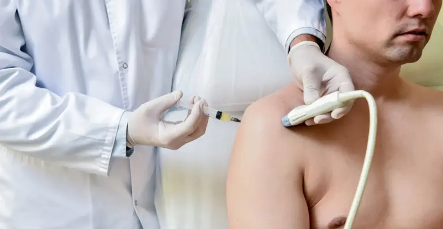
Modern medical technology has made it easier and more accurate to diagnose and treat a wide range of orthopedic illnesses and injuries. Consider ultrasound-guided injections as an example. To treat a patient's symptoms, ultrasound-guided injections are typically utilized to administer drugs such as painkillers, anti-inflammatories, or joint lubricants (hyaluronic acid) into highly specific areas. Numerous of these injections occur at typical injury locations, such as joints like the knee, hip, or shoulder, and maybe a component of a more all-encompassing pain management strategy.


What is Ultrasound

Waves are how sound moves. Ultrasound is sound that travels at a frequency greater than what our ears can detect.

By assembling images from the return of sound waves as they bounce back from whatever obstacle they encounter, ultrasound scans are performed. Pictures can be formed on an ultrasound monitor by directing the sound waves toward and away from the surfaces of various body structures. A transducer (probe) is moved across the skin's surface of the area to be examined to transmit and receive ultrasonic waves to and from the body. Specialist-trained doctors and sonographers take images (pictures) for interpretation. Several disorders can be diagnosed using ultrasound imaging. They are also utilized to direct the placement of injecting needles.


What is Ultrasound Guided Injection

Ultrasound

Your physician will have already explained to you why it is best for you to receive an injection. An antiseptic solution is used to cleanse the skin. Your skin can get stained, but that will go away in a few days.

Your local anesthetic may be injected by the radiologist to make the region more comfortable. As it is injected, there will be a brief stinging sensation, but this will pass. The corticosteroid is then administered using an extremely thin needle and ultrasound imaging as a guide. Inflammation (swelling) and discomfort in or around a joint, tendon, or soft tissue are treated with corticosteroids. A local anesthetic agent could also be included in the injection.


Ultrasound Guided Injection Types


Hydrodilatation

Hydrodilatation, sometimes called hydrodistension, is frequently used to treat frozen shoulder (adhesive capsulitis). Under ultrasound guidance, saline, local anesthetic, and cortisone are injected into the joint. Removing scar tissue from the calf muscles might also be helpful.


Neurolysis

To treat symptoms of pain or numbness, neurolysis is the administration of a steroid and saline around a peripheral nerve in the upper or lower extremity. Sciatica, Tarsal tunnel syndrome, and Carpal tunnel syndrome discomfort can all be lessened or eliminated with this technique. With the aid of ultrasonography and image guidance, cortisone is administered.


Durolane or Synvisc Injection

The fluid found in joints is similar to Synvisc or Durolane. The liquid gives the joint lubrication and serves as a shock absorber. Osteoarthritis-related joint discomfort is treated with Synvisc and Durolane injections.


Arthrogram

A medical imaging procedure called an arthrogram is used to assess the health of joints. A radiologist uses ultrasound to direct the injection of a contrast substance into the injured joint. The method is most frequently applied to the shoulder, elbow, wrist, hip, knee, and ankle to find anomalies and the source of pain or discomfort.


ABI or PRP Injection

A patient's blood is injected into a body part during an autologous blood injection (ABI) treatment to speed up the healing process. It is most typically used to treat tendon degeneration (tendonitis, tendinosis, or tendinopathy), which frequently happens in conjunction with minor tendon tears. Additionally, it has been utilized to treat joint, ligament, and fascia diseases.

A variation of ABI, platelet-rich plasma (PRP) injections include spinning a patient's blood rapidly in a centrifuge to separate it into layers of plasma, white blood cells, and red blood cells. The more platelet-rich plasma may be collected and delivered into injured tissues. Theoretically, this promotes a stronger healing response.

A radiologist does both of these treatments under ultrasound supervision to precisely target tendon illness and tears. Depending on the severity of your injury, multiple injections might be needed. Patients with bleeding issues or those on anticoagulants cannot undergo ABI. Other blood disorders like leukemia, where there may be tumor cells in the blood, or circumstances where there aren't enough platelets shouldn't be treated with it. Additionally, ABI is not advised in some cases of tendon tears when surgery may be preferable.


Cortisone Injections

When a steroid is injected into or surrounding soft tissue, a bursa (a sac of fluid around a joint), or joints (such as the facet, costovertebral, etc.), it reduces the symptoms of inflammation and discomfort. The discomfort brought on by arthritis, bursitis, carpal tunnel syndrome, sciatica, sports injuries, tendinitis, and other inflammatory disorders can all be lessened or eliminated by this therapy.


Ultrasound Guided Injection Benefits

Injection

It is advantageous in several ways to use ultrasonic technology to guide injections for pain control. With ultrasound-guided injections, patients are more at ease and typically experience fewer problems after injection therapy. Added advantages include:

  • Non-invasive. A transducer is moved on the skin above the joint to produce all real-time ultrasound images.
  • No radiation. The ultrasound does not release radiation, in contrast to other image-guided injection techniques. Although other imaging techniques can be used to direct injection, ultrasound has several noteworthy benefits, the main one being that it emits no radiation. Instead, ultrasonography creates an image of the body's internal structures and tissue using high-frequency sound waves.
  • Higher accuracy. Your orthopedic doctor will be able to treat your joint problems with more accuracy and avoid sensitive structures like nerves, blood vessels, and soft tissue because they can see the inside structures of the joint. To ensure that the medication is delivered to the right place while avoiding nerves, tendons, ligaments, and other soft tissue close to the target tissue, your physician can see the target site using ultrasound as the needle enters the area. This can help prevent complications from the procedure.
  • Reveal fluid accumulation. Your doctor can use ultrasound-guided technology to identify the fluid and, if necessary, remove it from joints that have fluid buildup in specific spaces.

Small and portable, ultrasound is simple to employ in conjunction with injections. Additionally, ultrasonography offers a more accurate image than other imaging devices. When your doctor performs a fluid drainage procedure, ultrasound can detect fluid buildup that may be the source of your joint problems. Additionally, compared to other forms of imaging, using ultrasound in medical care is far less expensive for patients.

Consult your physician about ultrasound-guided injection therapy if your joint pain has not improved with conservative measures like rest, anti-inflammatory drugs, or icing. The next step in pain management is frequently to administer a medication injection that will lessen joint inflammation and ease pain and suffering.


Ultrasound Guided Injection Purpose

Joint injections can be used to confirm a medical diagnosis as well as to treat symptoms like pain and inflammation. That is, if the injection helps to relieve your discomfort, it will be able to determine the type and exact location of the joint injury.

Joint injections are typically utilized as a short-term solution to lessen inflammation in an injured joint. By doing this, you might be able to use other pain-management strategies, such as physiotherapy. Joint injections can be given again at intervals of several months, but they won't treat the underlying problem that's causing your discomfort and swelling.


Ultrasound Guided Injection Preparation

Ultrasound preparation

For this test, there is no preparation. You are free to eat and drink as usual. All of your prescription doses can be taken as usual. Continue taking your blood-thinning medications as usual as long as they are being properly monitored, such as aspirin or warfarin. Let your doctor know if you take any anti-retroviral drugs, as these may affect your therapy. Before your appointment, let your doctor know if you have any allergies to latex, other drugs, or steroids. You must let the imaging personnel know if you are breastfeeding or believe you could be pregnant. If you are feeling under the weather, have recently been hospitalized, or have any other health concerns, let your doctor know.


Ultrasound Guided Injection Procedure

Ultrasound procedure

You will be taken from the ward to the imaging department if you are an inpatient at the hospital already. They will ask you to report to the imaging department's reception if you are getting the scan as an outpatient. You can find out which hospital to go to from your appointment. Take note that the scanning room does not permit any form of photography or electronic recording. You could be asked to take off your clothes and put on a medical gown. You don't have to take off your jewelry or hearing aids. A sonographer, an ultrasound imaging technician, a radiologist, or a doctor who specializes in imaging, will carry out your scan.

You will be requested to lie down on an exam bed after proving your identity, and the lights will be lowered to make it easier to see the images on the screen. Before the scan, doctors might ask you about your health. The next step is to apply a water-soluble gel on your skin. This makes it simple for the transducer to glide over the skin and aids in the production of clearer images. Before the injection, your doctor will go over the procedure with you. Depending on what regions of the body are being scanned, the scan typically takes 15 to 30 minutes to complete. During the test, feel free to ask any questions.


Ultrasound Guided Injection Recovery

Ultrasound recovery

Once the gel is removed from your skin, you can put on clothing. Rest the injected site for 24 to 48 hours. After 24 hours, remove the plaster. The antiseptic solution may turn skin pink or orange, but cleansing will cover it up. If you have diabetes, you must monitor your blood sugar levels carefully because they may rise slightly after a steroid injection. After a few days, this normally goes away. Doctors advise against driving for at least 24 hours following the operation. You will be allowed to go home if your scan was performed as an outpatient visit or if your doctor has allowed it. When you next visit the hospital doctor or your GP, you will receive the scan results and evaluate the benefits of the injection. Typically, this takes four to eight weeks. You will be taken back to the ward if you receive a scan while an inpatient.

Typically, three days following the injection, the symptoms are better, however, this differs from patient to patient. A few weeks or even months may pass before the effects of the injection wear off. To relieve the symptoms, you might require more than one shot. Your providing doctor will be able to determine your subsequent treatment with you based on the results of the injection. If a pain chart is provided to you after the procedure, attempt to fill it out.

If you have a fever or see any redness or swelling near the injection site, you should see your doctor. This can be a sign that you might be infected. This should be treatable with antibiotics from your doctor.


Ultrasound Guided Injection Risks

Risk o UGI

Injections that are guided by ultrasonography are frequently performed. The operation carries some risks or consequences, including:

  • Pain. The steroid injection may occasionally make the discomfort or pain temporarily worse. Doctors and nurses refer to this as a Steroid Flare. About two to three days following the injection, this may be more painful. You could utilize your standard painkillers at this time to get some relief.
  • Discoloration. Rarely, after the injection, a white dot or patch of skin can develop; this is known as depigmentation. Rarely is this permanent, and it might only last a few months.
  • Fat shrink. This causes the fat to thin, which could cause a skin nick.
  • Skin dimpling or thinning. On rare occasions, after a few months, the skin covering the injection thins. When many injections are administered close to the skin's surface, this is more apparent. Most individuals do not experience this adverse effect.
  • Tendon rupture. It's vital to avoid any strenuous activities or workouts for three to four weeks, wear splints or support, and use steroids carefully as they may weaken the tendon.
  • Infection. This is quite uncommon. Inform your doctor or the emergency room that you just received an injection if there is redness or swelling at the injection site or if you become feverish.
  • Allergic reaction is exceptionally rare.
  • No effect. The shot might not have an effect.


Ultrasound Guided Injection Results

Because they need to carefully review the images following the scan, the person doing the scan is not always able to provide you with the results right away. If your general practitioner ordered the test, schedule a visit with him to get the findings about two weeks following your session. If your hospital doctor ordered the test, you should find out the findings either on the ward or during your upcoming appointment at the outpatient clinic.


Conclusion

Ultrasound-guided injections are a common cutting-edge medical technique used to precisely administer medication to a location in a joint at many clinics and hospitals. An ultrasound-guided injection enables your orthopedic specialist to see the best location for the drug in real time, maximizing the injection's therapeutic effects. With the use of this cutting-edge medical technology, your doctor may precisely target injectable therapy while avoiding nearby nerves, tendons, ligaments, and soft tissue.