Umbilical hernia
Overview
Umbilical hernias are prevalent in newborns, although the actual percentage is unknown since many instances go unreported and heal without treatment.
They are especially frequent in premature newborns. An umbilical hernia affects up to 75% of neonates with a birth weight of less than 1.5 kilograms. The umbilical cord goes through a hole in the abdominal wall when the growing fetus is in the womb. This should be closed shortly after delivery.
Umbilical hernia definition
Umbilical hernias are ventral hernias that occur at or around the umbilicus. The umbilical hernia is defined by the European Hernia Society as a hernia lying between 3 cm above and 3 cm below the umbilicus. Following inguinal hernia, it is the second most frequent kind of hernia in adults. It accounts for 6% to 14% of all abdominal wall hernias in adults.
Epidemiology
In the general adult population, the incidence of umbilical hernia is 2%, although it is significantly more prevalent in obese multiparous women and cirrhotic individuals. Umbilical hernia occurs in up to 20% of cirrhotic individuals with ascites. Females outnumber males by a factor of three.
In general, males with umbilical hernias present imprisoned, but females are more likely to have an asymptomatic reducible hernia. 70% of umbilical hernia repairs are performed on men. In the United States, around 175,000 umbilical hernia repairs are performed each year.
Etiology
In adulthood, 90 percent of umbilical hernias are acquired. Only 10% of adults with umbilical hernias report having had hernias as children. It is more frequent in women or those who have high intra-abdominal pressure, such as during pregnancy, obesity, ascites, or chronic abdominal distention. Stretching of the abdominal musculature, along with the presence of adipose tissue, separates muscle bundles and layers, weakens aponeuroses, and promotes the establishment of umbilical hernias.
Pathophysiology
An umbilical hernia can emerge anatomically due to a possible weakening at the exit point of involuted umbilical veins, most notably the umbilical vein, or due to compromised umbilical fascia (Richet's fascia). As a result, the umbilical hernia covering is made up of skin, subcutaneous tissue, weaker superficial fascia, weakened umbilical fascia, and peritoneum, all of which are considerably attenuated and fused together. Patients with umbilical hernia frequently lack the umbilical fascia, and the round hepatic ligament is not linked to the inferior edge of the umbilical ring.
Chronic abdominal wall distension with increased intra-abdominal pressure, such as in pregnancy, patients with ascites or peritoneal dialysis, stretching of abdominal muscle fibers, and connective tissue weakening may all contribute to the development of umbilical hernia. Umbilical hernia occurs in around 20% of cirrhotic individuals as a result of increased abdominal pressure from ascites, dilatation of umbilical veins, and muscle or connective tissue weakening owing to low nutritional condition.
Preperitoneal adipose tissue, the omentum, and the small intestine, or a mix of these, may be involved in an umbilical hernia. The transverse colon is almost never implicated. Because the hernia sac's neck is generally tiny in comparison to the size of the herniated sac, imprisonment and strangulation are common. As a result, after diagnosis, an elective repair is recommended.
Clinical presentation
Adults with umbilical hernias usually have protrusion or bulging from the umbilicus. Other potential but uncommon presenting symptoms include pain and GI discomfort, whereas tenderness and imprisonment are typical physical findings. Small umbilical hernias are frequently asymptomatic and only sometimes cause discomfort.
Large umbilical hernia in older, multiparous, or obese women, frequently symptomatic, with progressively expanding hernia that, in most cases, becomes painful or irreducible over time. Strangulation of the umbilical hernia is a common problem; patients often appear with an irreducible sensitive umbilical bulge, skin color changes, and symptoms of intestinal obstruction if the sac contains a tiny bowel loop.
Diagnosis
During the physical exam, an umbilical hernia is discovered. A thorough inspection of the whole abdominal wall, particularly around the prior scar, is recommended. The hernia's content and the magnitude of the defect might be assessed. Imaging procedures, such as an abdominal ultrasound or CT scan, are often required to check for problems or if the clinical diagnosis is challenging, particularly in obese individuals. Given the increased risk in that patient population, it is especially critical to assess BMI, smoking status, and pre-existing Cirrhosis.
Management
Because of the significant risk of complications, all adult umbilical hernias must be repaired. Surgery is recommended for symptomatic individuals. Uncontrolled ascites are a relative contraindication.
Suture repair and mesh are the two basic surgical treatment methods for umbilical hernias. Primary suture repair is done using either simple primary suture repair for tiny lesions (less than 2 cm) or the Mayo procedure, which is effectively an overlapping abdominal wall fascia in a "vest-over-pants" fashion introduced by William Mayo in 1901.
Unfortunately, initial suture repair has a recurrence rate of 10%. A recent randomized, double-blind, controlled multicenter trial in Europe on people with a primary umbilical hernia of 1–4 cm in diameter was randomly allocated (1:1) intraoperatively to suture repair or mesh repair. There were fewer recurrences in the mesh group than in the suture group (4% vs. 12%). As a result, mesh repair is advised for umbilical hernias larger than 1cm.
Mesh repair can be done either openly or laparoscopically. Open mesh repair may be done in two ways: onlay or sublay; onlay mesh implantation is technically easier but is linked with more wound problems, such as seroma or hematoma and surgical site infection in some circumstances.
Preperitoneal or sublay mesh installation necessitates more surgical expertise and experience, although it is associated with reduced recurrence and wound problems. Some surgeons choose not to approximate the fascial margins; nonetheless, fascial closure before onlay mesh or after preperitoneal mesh is suggested.
The transabdominal preperitoneal approach (TAPP) or intraperitoneal onlay mesh (IPOM) procedure can be used to repair laparoscopic mesh. Laparoscopic repair resulted in a shorter hospital stay, a quicker return to regular activities, and a decreased risk of wound problems and recurrence.
The IPOM approach involves dissecting the sac, repairing the defect with continuous suture, and then placing mesh with a large overlap of at least 5 cm. To facilitate effective integration and attachment of the mesh, abdominal wall components such as the falciform ligament must be dissected and the perivesical area opened.
The transabdominal preperitoneal approach (TAPP) entails removing the contents of the hernial sac, incising the peritoneum 5 cm away from the defect's boundary, and establishing a preperitoneal space. After achieving proper hemostasis, a mesh with at least a 5-cm overlap on both sides will be inserted. Finally, the mesh will be secured with a few absorbable tacks or sutures, and the peritoneal flap will be closed using the same absorbable fixation system or suture. TAPP is thought to reduce the risk of complications associated with the intraperitoneal site of the mesh and has a lower complication rate than the (IPOM) treatment.
Contraindications to Umbilical Hernia Repair
Cirrhosis and uncontrolled ascites are both considered relative contraindications to elective open umbilical hernia surgery. Elective repair is typically avoided in individuals with Child-Pugh class B and C cirrhosis because to the higher surgical risk.
In a review of the literature, McKay et al found that small retrospective studies reduced morbidity and death in patients with ascites and cirrhosis to 2.7 percent and 21%, respectively. Yu et al. found that early elective umbilical hernia repair may be performed safely in cirrhotic patients using minimally invasive techniques and proper perioperative care in a small retrospective single-institution research.
Unfortunately, there is no agreement on the time of repair in cirrhotic individuals. However, it is suggested that ascites be controlled preoperatively either medical treatment or peritoneal drainage.
Technical Considerations
Prior to elective repair, treatable problems like as ascites and obesity should be addressed and managed. Obese individuals should be advised to lose weight before undergoing surgery. According to reports, the death rate linked with repair in patients with untreated ascites is 2%, and the recurrence rate is significant. Before elective repair, ascites should be managed with medical therapy, diuretics, and dietary adjustments.
Differential Diagnosis
- Benign lymph node metastasis
- Epididiymo orchitis
- Epididymal cyst
- Epididymitis
- Hydrocele
- Lymphadenopathy
- Spermaotocoele
- Testicular malignancy
Complications
Obesity, a high ASA-score (3), onlay repair, and concurrent bowel surgery are all significant risk factors for surgical complications. The extent of the fascial defect is an essential determinant in determining the preoperative risk of individuals with ventral hernias. They find that for every mm increase in fascial defect size, surgical complication rises by 1%.
- Seroma, hematoma, and surgical site infection are all examples of wound complications. Open hernia repair approaches are typically predisposed to this type of problem, but the laparoscopic technique has resulted in a reduction in such occurrences.
- Bowel injury and adhesion: This type of problem is predisposed to by the laparoscopic procedure. The direct contact of the mesh with the viscera poses a minimal risk of enterotomy and dense adhesion.
- Recurrence: Even with 4 cm lesions, there is a greater recurrence rate with initial repair. Obesity more than 30 kg/m2, diabetes, and wound infection are all independent risk factors for recurrence. Smoking is also seen as a risk factor for recurrence. Furthermore, uncontrolled ascites is associated with a high chance of recurrence.
Postoperative and Rehabilitation Care
- Nutrition: Encourage proper hydration by drinking 8-10 glasses of water every day. A high fiber diet is suggested to avoid constipation and unnecessary straining.
- Activity: Unless there are complex instances or accompanying co-morbidities, patients are typically discharged on the same day. The vast majority of people can return to work within a few days. Lifting weights greater than 10 pounds (4.5 kg) and intense activity should be avoided for 6 weeks.
- Wound Care: It is critical that the incisions remain clean and dry. No swimming or bathing for 1-2 weeks, or until the staples, stitches, or steri-strips are removed.
- Pain control: In the absence of renal illness or peptic ulcer disease, NSAIDs can be administered safely for a short period of time. Tylenol and ibuprofen are two typical pain relievers. For a few days, narcotic medications such as oxycodone and tramadol are used to treat moderate to severe pain. To avoid narcotic-induced constipation, it is advised to use an over-the-counter stool softener.
- Abdominal binder: Using an abdominal binder has a subjective good impact and is useful in facilitating surgical recovery, but there are no substantial effects on pain, mobility restriction, or seroma development.
Outcomes
A countrywide prospective study of umbilical and epigastric hernias found that hemorrhage (46 percent of cases), seroma (19 percent), and discomfort were the most common consequences that required readmission (77 percent ). This study also discovered a 5% total readmission rate, owing mostly to the aforementioned issues. A retrospective study of 150 veterans discovered an overall recurrence incidence of 6%, with 1.5 % in the nonmesh group; this study also discovered a 19% infection rate.
Recurrence rates linked with initial tissue healing have been found to range between 15% and 40%. Aslani and Brown found that mesh repair had a 10-fold lower incidence of recurrence as compared to primary suture repair in a comprehensive review and meta-analysis. Obese individuals and those with abnormalities greater than 3 cm have a higher chance of recurrence. Smoking and diabetes are two more variables linked to an elevated recurrence rate.
The National Surgical Quality Improvement Program (NSQIP) database of the American College of Surgeons (ACS) revealed that laparoscopic umbilical hernia repair had lower overall morbidity when compared to open surgery. Laparoscopic repair has been shown to have fewer problems, a shorter duration of stay, and a lower chance of recurrence. However, the downsides of laparoscopic surgery should be addressed, such as greater cost, operating time, and the danger of a general anesthesia.
Pediatric Umbilical Hernia
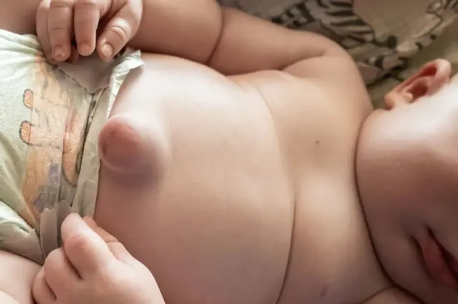
Umbilical hernia manifests as a protrusion at the umbilicus. At the first several months of life, it is a typical finding during routine well-baby checks. When they notice a bulge in their infant's belly button, new parents who are unfamiliar with this condition are often concerned.
On the one hand, parents may be afraid that their kid may experience major difficulties as a result of an umbilical hernia, and they may question whether there are any precautions they can take to avoid issues. To answer these concerns and choose when to submit the patient for surgical assessment, it is critical to understand the normal embryology and genesis of the umbilical hernia.
In children, umbilical hernia is caused by an inadequate closure of the fascia of the umbilical ring, allowing intraabdominal contents to protrude. After the umbilical cord is severed, the ring normally closes spontaneously due to the expansion of the rectus muscles and the union of the fascial layers. An umbilical hernia develops as a result of a failure or delay in this process. The actual cause is uncertain, however, it generally happens through the ring's umbilical vein component.
Umbilical hernias are frequent in children, affecting 10-30% of all white children at birth and reducing to 2-10% after one year, with males and girls affected equally. For unknown causes, umbilical hernias are very frequent in African-American neonates, with a frequency as high as 26.6 percent observed. It is also more prevalent in preterm and low-birth-weight kids, with an 84 percent prevalence in newborn infants weighing 1000 to 1500 grams and a 20.5 percent incidence in those weighing 2000 to 2500 grams.
During a well-child care visit, parents' histories may include a bulge of the belly button that worsens when the infant is screaming, coughing, or straining. The magnitude of the umbilical hernia defect, its reducibility, and the existence of symptoms of incarceration or strangling should all be determined. Abdominal discomfort, nausea, and vomiting are common symptoms of an incarcerated or strangulated umbilical hernia. The physical examination will be important for abdominal pain, distension, and erythema of the skin.
Umbilical hernias, in the vast majority of cases, have no medical consequences. A thorough physical exam is adequate to make the diagnosis and explain the usual course of the ailment with worried parents; no testing are necessary. Although pediatric umbilical hernias are frequent in healthy newborns, they are also related with a number of diseases that the pediatrician or pediatric surgeon should be aware of when examining a patient.
Pediatric umbilical hernias are more prevalent in common autosomal trisomies (for example, Trisomy 21 and 18), metabolic abnormalities (for example, hypothyroidism, mucopolysaccharidoses), and several dysmorphic syndromes (e.g., Beckwith-Wiedemann syndrome, Marfan syndrome). As a result, it is critical to separate healthy patients with an isolated umbilical hernia from patients with an umbilical hernia and additional syndromic symptoms, such as macroglossia or hypotonia, with the latter group requiring further investigation.
Repair of umbilical hernias in neonates is frequently postponed due to a low complication rate, and the majority of umbilical abnormalities resolve naturally after 2 years. The size of the hernial ring can be used to predict spontaneous closure. Expectant therapy of asymptomatic umbilical hernias is both safe and the standard of care for many pediatricians.
Conclusion
In clinical practice, umbilical hernias are prevalent. The majority of patients are examined by their primary care physician or an emergency room physician initially. Because these hernias are associated with a significant probability of imprisonment, surgery is advised for all patients. To avoid the morbidity of an umbilical hernia, an interdisciplinary team approach is required.
A preoperative workup is required in patients with concomitant comorbidities to reduce post-surgery complications. The nurse anesthetist must ensure that the patient is physically and mentally fit for surgery. Patients with cirrhosis and/or ascites should be extensively evaluated before undergoing surgery, as complications are prevalent.
Following the operation, the nurse should educate the patient on the need of decreasing weight, quitting smoking, and avoiding excessive lifting. The patient must be taught on the importance of eating a nutritious diet. When the patient's recuperation is complete, he or she should be encouraged to increase physical activity and reduce weight. This is critical for avoiding a recurrence.
Nurse practitioners who examine children should inform parents that tiny hernias will resolve on their own, but if they are still open by the age of 5, surgery is indicated.
Most patients with an umbilical hernia have a satisfactory prognosis, although recurrences do occur in around 1-3 percent of cases, despite advancements in mesh therapy.